
Pseudopyrochroa depressa (Pic)
|
publication ID |
https://doi.org/ 10.11646/zootaxa.4175.2.7 |
|
publication LSID |
lsid:zoobank.org:pub:46F1E733-8136-498B-A987-DA2D09A63841 |
|
DOI |
https://doi.org/10.5281/zenodo.6088043 |
|
persistent identifier |
https://treatment.plazi.org/id/A23C1F41-CB68-FFF6-FF61-FC430868EB90 |
|
treatment provided by |
Plazi |
|
scientific name |
Pseudopyrochroa depressa (Pic) |
| status |
|
Pseudopyrochroa depressa (Pic)
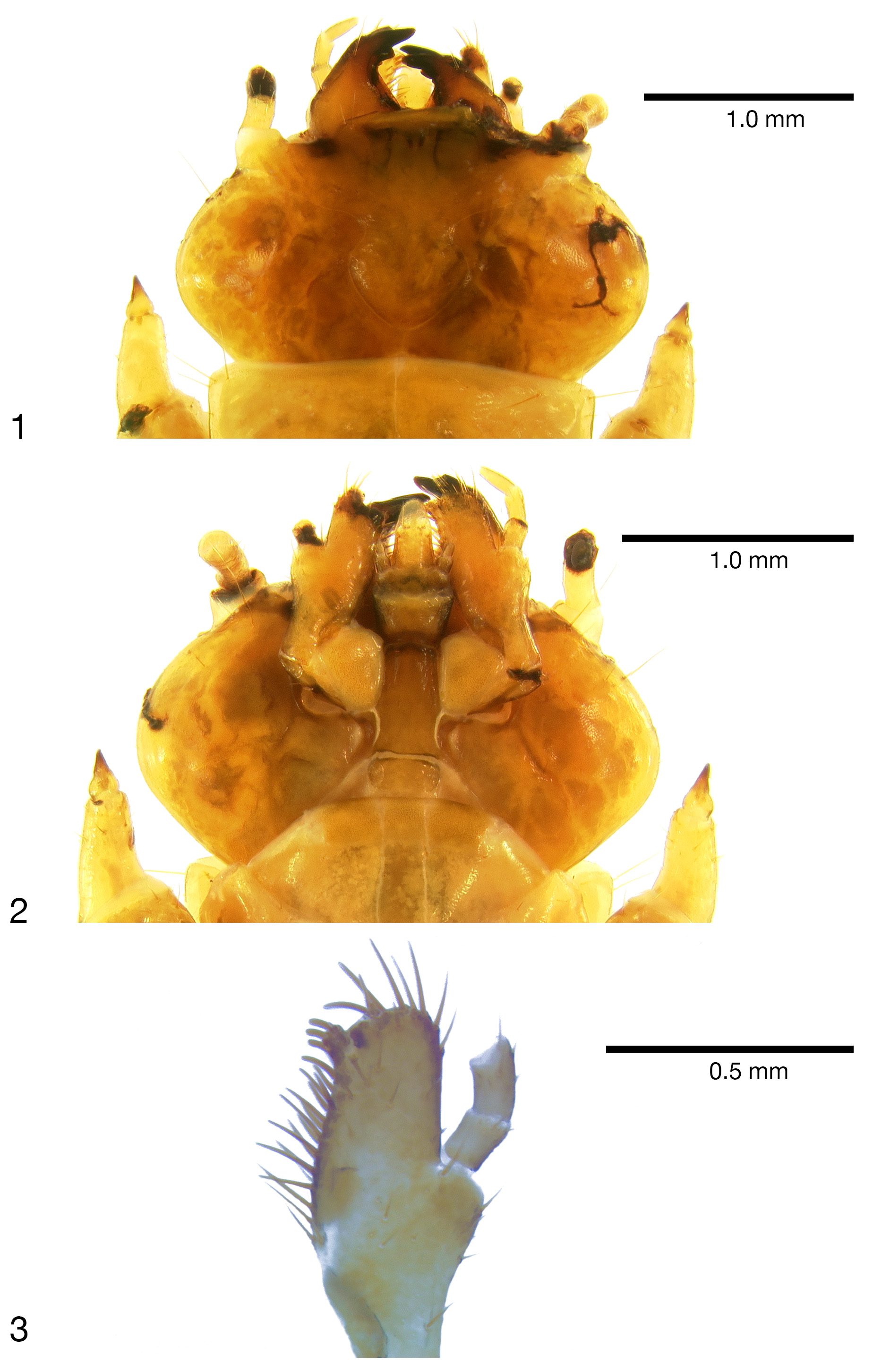
( Figures 1–6 View FIGURES 1 – 6 )
Description of mature larva. The single, relatively intact mature larva (probably on the small side of the average based on reared adults males) measures 17.5 mm (mesal labral apex to apices of urogomphi) and width (across widest portion of eighth abdominal segment) of 2.5 mm. Body orthosomatic with sides subparallel; moderately sclerotized except much of cranium, mandibles, and urogomphal plate more heavily sclerotized; body vestiture consisting of short to moderately elongate, scattered setae. Thoracic and abdominal terga 1–9 lacking distinct parabasal ridges. Head and body creamy-yellowish to amber, melanization much darker in areas of heavy sclerotization such as tips of mandibles, urogomphi, urogomphal lip and urogomphal pits.
Head: Prognathous, flattened, exerted from prothorax. Epicranial suture lyriform ( Fig. 1 View FIGURES 1 – 6 ) with stem short, frontal arms complete nearly to antennal insertions; endocarinae absent. Symmetrical labrum anterad fused frontoclypeal region. Four stemmata on each side of head in 2 groups of 2, lower pair parallel with antennal insertions. Antennal insertions fully exposed, antennae presumably filiform, 3-segmented ( Figs. 1-2 View FIGURES 1 – 6 ; incomplete in single larva; not intact on two exuviae). Mouthparts ( Fig. 2 View FIGURES 1 – 6 ) retracted. Mandibles heavily sclerotized, movable, asymmetrical, molar area of mandibles well-developed, left mandible bearing a prominent molar tooth; apex of right mandible distinctly tridentate, left mandible bidentate, both bearing 1-2 smaller subapical teeth. Maxillae each with 1-segmented cardo which is diagonally folded anteriorly upon itself toward the stipes and thus appearing 2-segmented; a well-developed, undivided, pad-like maxillary articulating area ( Fig. 2 View FIGURES 1 – 6 ); ventral surface of stipes with dense, largely double row of stout setae mesad palpifer along adoral margin ( Figs. 2–3 View FIGURES 1 – 6 ); galea and lacinia fused to form maxillary mala; mala bearing stout apical and adoral setae and spinose-dentiform uncus at apicoadoral margin ( Fig. 3 View FIGURES 1 – 6 ), three, stout, elongate-spatulate setae associated with uncus; 3-segmented, filiform maxillary palpus [3rd palpomere missing in Fig. 3 View FIGURES 1 – 6 ], 2nd palpomere about 1.5X length of first, 3rd palpomere 0.8X length of 2nd, 3rd palpomere tapering distally, acutely rounded apically. Labium ( Fig. 2 View FIGURES 1 – 6 ) with mentum ovate-subquadrate, submentum elongate with sides shallowly sinuate basally, apical margin slightly more heavily sclerotized, convexly rounded; well-developed, elongate ligula; each labial palpus short, 2-segmented. Hypopharyngeal sclerome well-developed, heavily sclerotized, molar-like, transverse; proximal region of hypopharynx with setal brushes. Hypostomal rods ( Fig. 2 View FIGURES 1 – 6 ) well developed, divergent; gular sutures separate.
Thorax and Abdomen: Thoracic segmentation well developed, sides parallel; cervicosternum divided into three plates. Legs well-developed, moderately short, 5-segmented including tarsungulus, vestiture consisting of sparse, short setae; coxae large, separated by 2–3 coxal diameters. Abdomen flattened, moderately sclerotized, tergites 1–7 Fig. 2 View FIGURES 1 – 6 : cranium, ventral view
Fig. 3 View FIGURES 1 – 6 : distal left maxilla, ventral view
FIGURES 1–6 View FIGURES 1 – 6 . (Continued)
Fig. 4 View FIGURES 1 – 6 : abdominal segments 8–9, dorsal view Fig. 5 View FIGURES 1 – 6 : abdominal segments 8–9, ventral view Fig. 6 View FIGURES 1 – 6 : distal urogomphal plate: urogomphal pits, ventral view.
subequal in length and width; 8th tergite approximately 2X length of 7th. Sternite 8 emarginate apically. Tergite 9 ( Fig. 4 View FIGURES 1 – 6 ) hinged, capable of considerable dorso-longitudinal movement, extending ventrally, thus forming the urogomphal plate ( Figs. 4–5 View FIGURES 1 – 6 ), widest basally where it forms well developed lateral lobes, lobes acutely rounded apically; surface of urogomphal plate bearing numerous, well-developed callosities and several larger, dorsal and lateral setigerous calli on the dorsal and lateral urogomphal surfaces; urogomphi heavily sclerotized, long, slender, subparallel, tapering and acuminate apically; ventral surface of urogomphal plate shallowly, but sharply excavate basally at articulation with 9th sternite, excavation narrowing distally to bases of urogomphi. Urogomphal plate possessing a heavily sclerotized, mesally acutely produced urogomphal lip ventrally, between the two, heavily sclerotized, shallow urogomphal pits ( Fig. 6 View FIGURES 1 – 6 ), which, in turn, arise distally between the heavily sclerotized, fixed urogomphi; urogomphal pits bearing characteristic, parallel rugae ( Fig. 6 View FIGURES 1 – 6 ). Sternite 9 ( Fig. 6 View FIGURES 1 – 6 ) broadly, transversely U-shaped, divided mesally by weak, desclerotized line, partially recessed into shallow emargination of 8th sternite, possessing continuous semicircular arch of approximately 28–30 well-developed asperites along anterior margin. Tenth segment reduced, crescentiform, basal margin rounded, recessed into emargination of 9th sternite, visible ventrally.
Spiracles: One pair of well-developed, ovate thoracic spiracles, situated ventro-laterally on laterotergite along anterior margin of mesothorax. Paired, ovate abdominal spiracles, subequal in size, located on dorsolateral margin of 1st abdominal tergite and ventrolateral margins of abdominal laterotergites 2–7; paired spiracles of abdominal laterotergite 8 ( Fig. 6 View FIGURES 1 – 6 ) slightly larger, annular-ovate, located ventrolaterally at distal 1/3 of its length.
Material Examined. [2 adult ♂♂]: TAIWAN: Miaoli Hsien // Syuejian ; ca. 1900m // 24.424625°N / 121.013511°E // 29 January 2015 // Yun Hsiao, leg.; [2nd label]: Reared from larva ex: // beneath bark (exposed // surfaces) of decaying logs; // humidity moderate; [3rd label]: Pseudopyrochroa // depressa ♂ // Pic // det. D. K. Young GoogleMaps . [2 vials, one each for exuviae from last larval instars of reared adults]: [data as above but on single, EtOHformatted label]; [additional data on EtOH label in vials]: ASSOCIATED ADULT IN // PINNED COLLECTION; [2nd label]: Pseudopyrochroa // depressa ♂ // Pic // det. D. K. Young. [1 mature larva]: [same data as EtOHformatted data labels, above]; [additional label in vial]: Pseudopyrochroa // depressa // Pic // det. D. K. Young GoogleMaps .
Diagnosis. The structure of the apico-adoral surface of the maxillae has been shown to be somewhat diagnostic at the species level in larvae of Pseudopyrochroa ( Hayashi 1969, Young 1996b, 2001). The three, stout, elongatespatulate setae associated with the uncus ( Fig. 3 View FIGURES 1 – 6 ) are unique among the three currently known Pseudopyrochroa larvae from Taiwan.
Also diagnostic are structures associated with the ninth abdominal segment. The structure of the urogomphal plate has consistently been very useful and reliable for species recognition of a wide range of pyrochroid larvae (e.g., Young 1975, 1991, 1996b, Young et al. 2016, Hayashi 1969). The most diagnostically useful characters of P. depressa larvae are the elongate, slender, subparallel urogomphi ( Figs. 4–5 View FIGURES 1 – 6 ), the mesally acutely produced urogomphal lip, and shallow urogomphal pits with characteristic parallel rows of rugae ( Figs. 4–6 View FIGURES 1 – 6 ).
Among other known Taiwanese pyrochroid larvae ( Young et al. 2016), those of P. fainanensis also have a prominent urogomphal lip ( Fig. 7 View FIGURE 7 ), but it is broadly rounded, the sub-ovate urogomphal pits are more developed, and the urogomphi are strongly incurved with recurved apices. The heavily sclerotized, long, slender, subparallel, tapering, and apically acuminate urogomphi of P. carinifrons (Fig. 8) are quite similar to those of P. depressa . However, the acutely produced urogomphal lip and shallow urogomphal pits of P. depressa ( Figs. 5–6 View FIGURES 1 – 6 ) easily separate larvae of the two species.
Natural history. On 29 January 2015, three larvae of P. depressa , two live and one dead, were collected (YH) at an elevation of 1900 meters in Miaoli Hsien (24.424625°N / 121.013511°E). The larvae were found beneath bark of dead logs, from exposed surfaces of logs. The humidity of the site was relatively moderate, drier than that associated with collections of P. carinifrons and P. fainanensis larvae ( Young et al. 2016). GoogleMaps
The two live larvae were successfully reared to the adult stage; one emerged on 16 February 2015.

No known copyright restrictions apply. See Agosti, D., Egloff, W., 2009. Taxonomic information exchange and copyright: the Plazi approach. BMC Research Notes 2009, 2:53 for further explanation.
|
Kingdom |
|
|
Phylum |
|
|
Class |
|
|
Order |
|
|
Family |
|
|
Genus |