
Polycarpa
|
publication ID |
https://doi.org/ 10.11646/zootaxa.4114.3.1 |
|
publication LSID |
lsid:zoobank.org:pub:6EA59057-0E05-4AA5-8B84-327CBDB32E5B |
|
DOI |
https://doi.org/10.5281/zenodo.6068917 |
|
persistent identifier |
https://treatment.plazi.org/id/A25D4D00-D650-763A-7BF3-F9037CF2FF33 |
|
treatment provided by |
Plazi |
|
scientific name |
Polycarpa |
| status |
|
Polycarpa sp.
Figure 24 View FIGURE 24
Station. CP 4381.
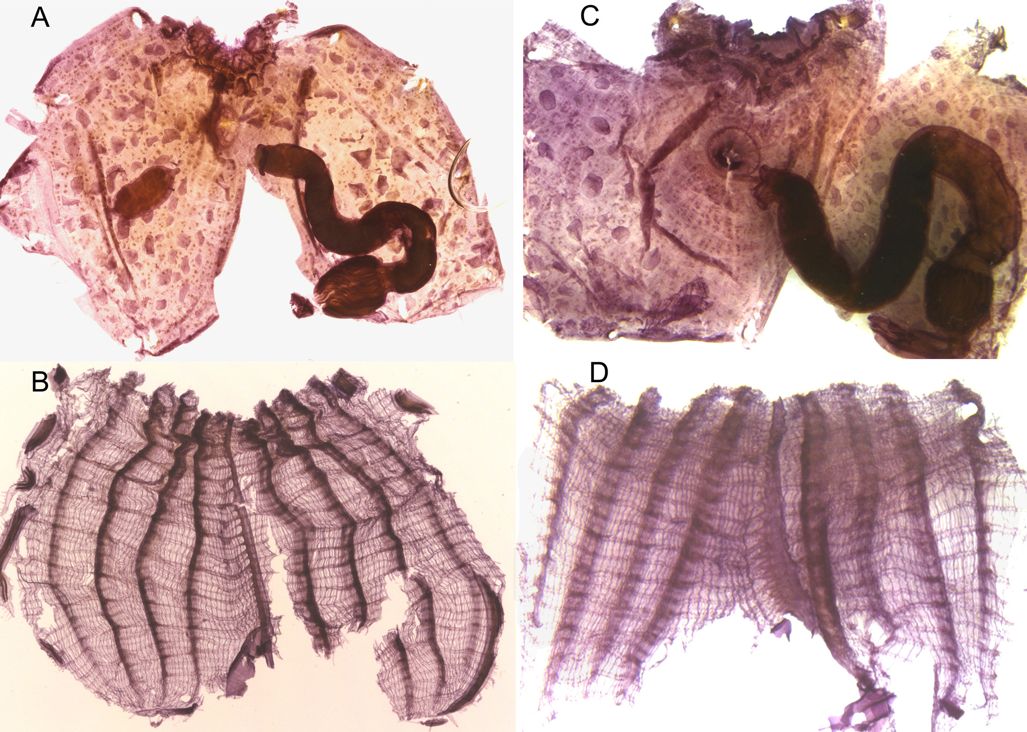
The largest of the 3 specimens collected at 114m are partially incrusted with sand with a few root-like basal expansions of the tunic. The largest is 9 x 7 mm. The siphons at a short distance from each other are not protruding. The tunic is thin but opaque. Without tunic the body is light brown with four darker lines on the siphons. The body wall musculature is weak in a felting of thin fibres. The internal layer of the body wall is spotted with groups of darker cells ( Fig. 24 View FIGURE 24 A,C) and the area near the siphons has a swollen pavement. Round endocarps are scattered everywhere ( Fig. 24 View FIGURE 24 A,C). There is an oral velum. About 50 oral tentacles are alternated in 3 orders of size, the largest thick and twisted like a corkscrew. The dorsal tubercle is C-shaped or a vertical slit. The space between the tentacles and the branchial sac is particularly narrow. The dorsal lamina is a low blade. The branchial sac has 4 spaced folds becoming thinner posteriorly ( Fig. 24 View FIGURE 24 B,D) Formulae of the right side in 2 specimens are:
RE- 6(11)8(12)7(12)7(12)6-DL
RE- 6(10)10(9)10(10)10(10)9-DL
The folds are low and it is often subjective to decide whether a vessel belongs to a fold ( Fig. 24 View FIGURE 24 B,D). There are 1 or 2 stigmata in a mesh between the folds and parastigmatic vessels. The digestive tract is loosely attached to the body wall. It occupies the dorso-ventral part of the left side ( Fig. 24 View FIGURE 24 A,C). The stomach has 8 to 9 longitudinal ridges on the mesial side. The caecum is button-shaped. The rectum is long in a deep secondary gut loop ( Fig. 24 View FIGURE 24 A,C) with the anus in 2 low lobes. One specimen has 2 polycarps on each side, the anterior one smaller; another specimen has 2 gonads only on the right side; the third one has no gonads. The polycarps are oval very loosely attached to the body wall and to the branchial sac by thin tissue strips. Male and female papillae are joined. There is a ring of filiform processes at the atrial aperture
Polycarpa species having 4 folds/side and several vessels between the folds, and numerous endocarps but no more than 2 polycarps on each side are not common. P. fibrosa ( Stimpson, 1852) from the northern Atlantic has in common with the Guiana P. s p. a coating of filaments with sand on the tunic, a similar branchial sac but with fewer longitudinal vessels between the folds, many endocarps and a punctuate internal layer of the body wall. It differs in having many more polycarps which are less elongated and the anus with large petaloid lobes. P. porculus Monniot C. & F. 1979 from 250m depth in Norway has similar tentacles, 2 or 3 polycarps on each side but it has a different shape of the branchial folds and no longitudinal vessels between them.
The deep Polycarpa species described here is likely a new species, but the variability observed among only 3 specimens does not allow to create a new species.
No known copyright restrictions apply. See Agosti, D., Egloff, W., 2009. Taxonomic information exchange and copyright: the Plazi approach. BMC Research Notes 2009, 2:53 for further explanation.
|
Kingdom |
|
|
Phylum |
|
|
SubPhylum |
Tunicata |
|
Class |
|
|
Order |
|
|
Family |
Polycarpa
| Monniot, Françoise 2016 |
P. fibrosa (
| Stimpson 1852 |