
Polycarpa itapoa Rocha & Moreno, 2000
|
publication ID |
https://doi.org/ 10.11646/zootaxa.4114.3.1 |
|
publication LSID |
lsid:zoobank.org:pub:6EA59057-0E05-4AA5-8B84-327CBDB32E5B |
|
DOI |
https://doi.org/10.5281/zenodo.6068915 |
|
persistent identifier |
https://treatment.plazi.org/id/A25D4D00-D656-7639-7BF3-FBD57B31FBBE |
|
treatment provided by |
Plazi |
|
scientific name |
Polycarpa itapoa Rocha & Moreno, 2000 |
| status |
|
Polycarpa itapoa Rocha & Moreno, 2000
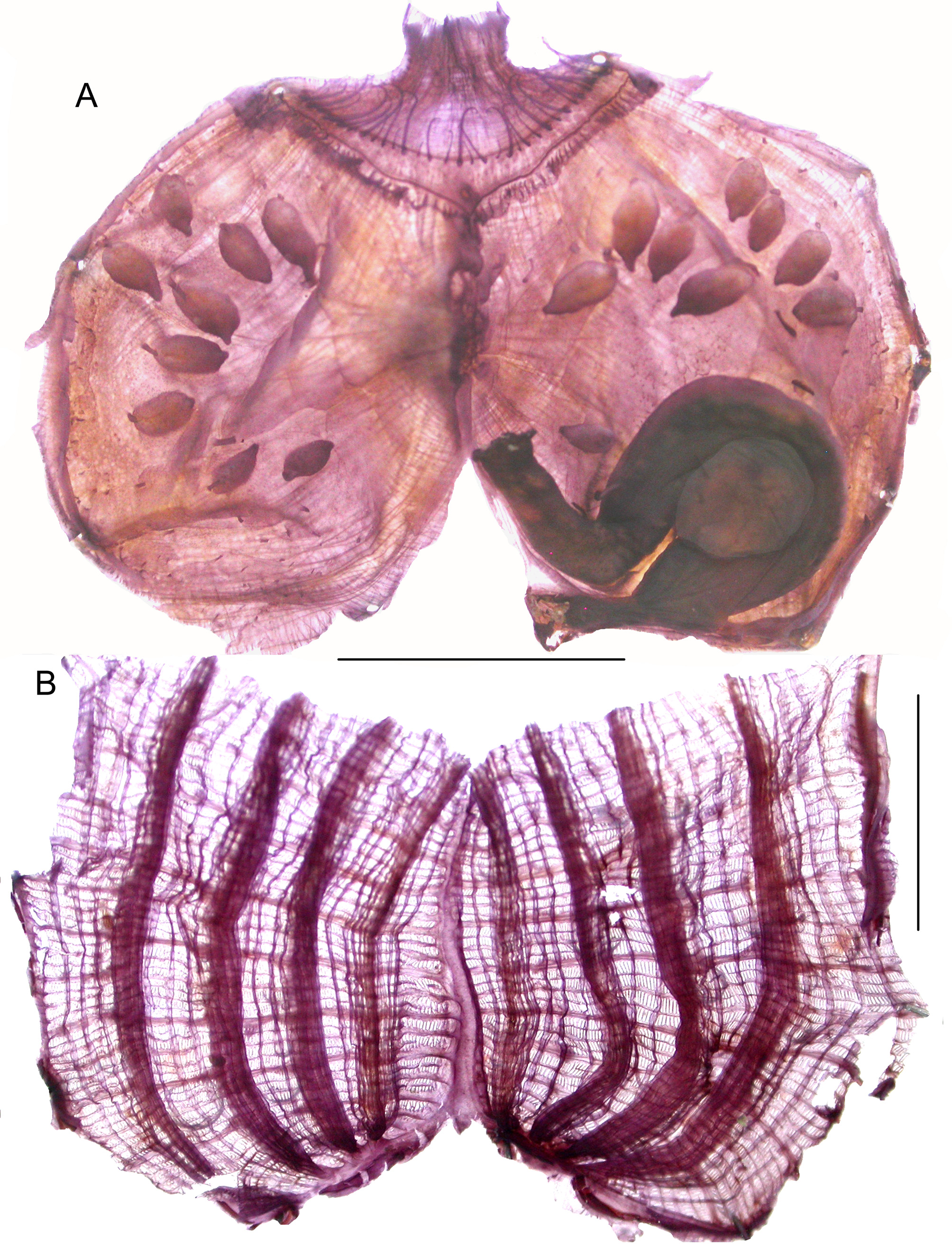
Figure 21 View FIGURE 21 .
Stations. SC7; SS10; SS12.
Either solitary or aggregated the specimens are erect with a maximum size of 1 cm
and fixed by their posterior side ( Fig. 21 View FIGURE 21 A). The siphons are well separated and protruding, the atrial one more dorsal. The tunic is thick but soft, rough with sand particles limited to the basal part of the body ( Fig. 21 View FIGURE 21 A) the tunic is thick but soft. The body wall is opaque with a weak musculature. The oral tentacles are numerous behind a high velum. The C-shaped dorsal tubercle opens anteriorly. The dorsal lamina has a smooth edge. There are 4 branchial folds on each side not recovering each other ( Fig. 21 View FIGURE 21 B) and one or 2 longitudinal vessels lie between the folds. One branchial formula is:
RE-2(5)2(8)1(10)1(12) DL (12)2(8)2(8)2 (7)2 LE
There are no parastigmatic vessels. The digestive tract occupies a half of the left body side ( Fig. 21 View FIGURE 21 C). Its diameter is almost the same along its whole length. The stomach does not show folds externally and has no caecum. The primary loop is open and the rectum follows the dorsal body line. The anus has 2 rounded lobes. Numerous round polycarps ( Fig. 21 View FIGURE 21 C) are distributed over both body sides, 35 of them were counted on the right side of a specimen. They protrude from the body wall with the ovary enclosed inside the testis lobes, the papillae are small and joined. Numerous endocarps are irregularly located between the polycarps. A ring of filiform tentacles encircles the base of the atrial siphon.This description corresponds to the original Brazilian material and in Guiana too the individuals were often mixed with polycitorid colonies of Eudistoma .
Polycarpa salutis n. sp. Figures: 22, 23.
Stations. CP4380; CP4381 (Type MNHN S1 POL.B 569); CP 4383 and 16°29.7’N – 61°31.4’W, 82 m (coll. Bouchet, 2015).
Etymology. named after Iles du Salut.
The body is globular ( Fig. 22 View FIGURE 22 A) with sessile siphons. The ventral side is incrusted with sediment and wears some tunic filaments; the upper part is naked and mammillated with small button-like papillae. The tunic is thick but soft. The largest specimen is 22 mm in diameter. The siphonal apertures are 4-lobed. Extracted from the tunic the body wall is light brown, opaque, darker at the edge of the siphons. The sphincters around the siphons are large.
The remaining of the musculature is made of a felting of dense regularly crossed fibres in a thick tissue. An oral velum is present. The oral tentacles are thin, 32 counted in one specimen in two sizes with smaller ones intercalated ( Fig. 23 View FIGURE 23 A). The pre-pharyngeal band is dorsally curved around a dorsal tubercle opened in a C. Papillae are present on the pre-pharyngeal space and the same papillae invade the whole internal layer of the body wall. There are no endocarps on the body sides except inside the gut loop ( Fig. 22 View FIGURE 22 B,23A). The branchial tissue is brown when fixed. The dorsal lamina has a plain edge and increases in height posteriorly ( Fig. 23 View FIGURE 23 B). The 4 branchial folds on each side are well spaced ( Fig. 23 View FIGURE 23 B). Formulae of 2 specimens are:
RE 3 (11) 3(13) 4(12) 4 (13) DL (10) 3 (14) 4 (13) 4 (11) 4 LE
RE 3 (10) 4 (10) 3 (9) 2 (10) 2 DL 1 (8) 2 (11) 3 (10) 4 (9) 3 LE
The branchial folds are ventrally united to a horizontal membrane without terminal papillae of the longitudinal vessels ( Fig. 23 View FIGURE 23 B). Parastigmatic vessels are present. There are 8 to 12 stigmata in a mesh between the folds. The digestive tract occupies a little less than half of the left body side ( Figs 22 View FIGURE 22 B, 23A). The oesophagus is short. The olive-shaped stomach has internal folds which do not appear externally. The primary gut loop is closed with an isodiametric intestine curved in a short rectum, the anus is rimmed by numerous round lobes. There is a caecum and a strip of tissue unites both limbs of the intestinal loop. One large or 2 foliated endocarps fill the primary gut loop ( Fig. 22 View FIGURE 22 A; 23B) but none were found elsewhere. The gonads are bottle-shaped polycarps protruding from the body wall but covered by the same papillated transparent thin layer of tissue lining the internal side of the atrial cavity. This structure is hardly seen without staining. In each polycarp the testis vesicles are on each side of the central ovary. The sperm duct opens close to the female papilla. Nine to 12 scattered polycarps were counted on the right side and 6 to 9 on the left in specimens 2cm in size. They are not neatly arranged in lines ( Figs. 22 View FIGURE 22 B; 23A). The atrial siphon has a high circular membrane with filifom papillae and in addition a large internal velum.
This deep living species differs from other Polycarpa known from the Western Atlantic (listed in Rocha & Moreno 2000) by a lobed anus, the absence of endocarps except in the gut loop, and the internal layer of the body wall covered with small papillae. The external aspect is also original with sand incrustation limited to the basal hemisphere of the body leaving naked the upper siphonal side.
The external aspect of P. salutis reminds that of P. n i v o s a (Sluiter, 1898) redescribed by Sloot (1969) which differs in having a thin body wall no pyloric caecum a bilobed anus and no endocarps in the gut loop.


No known copyright restrictions apply. See Agosti, D., Egloff, W., 2009. Taxonomic information exchange and copyright: the Plazi approach. BMC Research Notes 2009, 2:53 for further explanation.
|
Kingdom |
|
|
Phylum |
|
|
SubPhylum |
Tunicata |
|
Class |
|
|
Order |
|
|
Family |
|
|
Genus |