
Ascidia curvata ( Traustedt, 1882 )
|
publication ID |
https://doi.org/10.11646/zootaxa.4114.3.1 |
|
publication LSID |
lsid:zoobank.org:pub:6EA59057-0E05-4AA5-8B84-327CBDB32E5B |
|
DOI |
https://doi.org/10.5281/zenodo.6068901 |
|
persistent identifier |
https://treatment.plazi.org/id/A25D4D00-D65E-7637-7BF3-FBC47FA8F816 |
|
treatment provided by |
Plazi |
|
scientific name |
Ascidia curvata ( Traustedt, 1882 ) |
| status |
|
Ascidia curvata ( Traustedt, 1882)
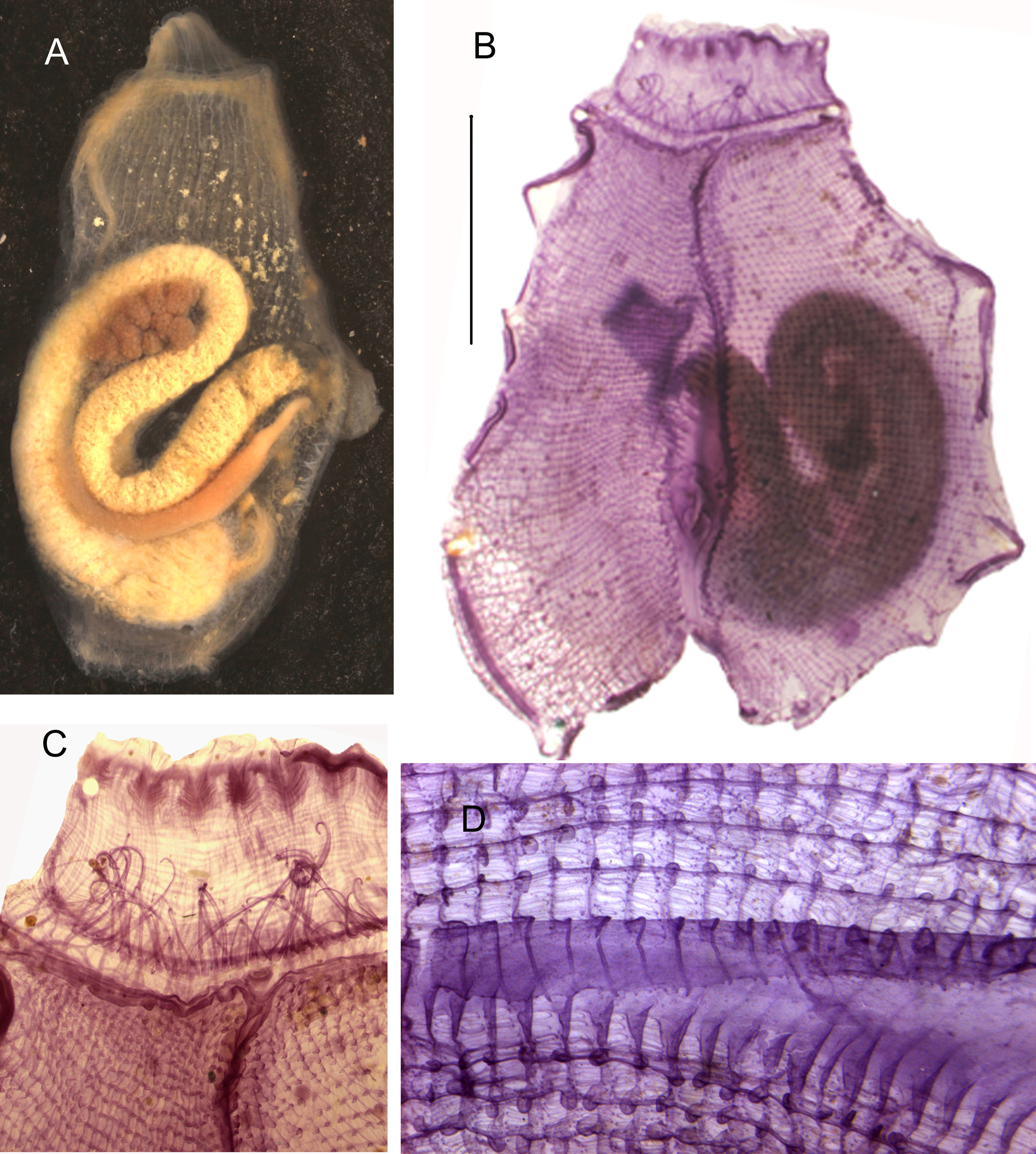
Figure 14 View FIGURE 14 .
Stations. CP4357; CP4388.
The largest of the two specimens is 19 mm in length and 11mm in width ( Fig. 14 View FIGURE 14 A). The body was attached by its left side. The tunic is vitreous and naked. The oral siphon has 8 lobes and the atrial siphon 6 lobes, both with red spots. The body wall is thin, especially on the left side which is almost devoid of musculature. A network of vessels is conspicuous in the posterior part of the body wall. Both siphons have regularly spaced circular muscles in a sphincter. The longitudinal muscle fibres issued from the oral siphon do not extend beyond the level of the third row of stigmata on the left, but extend a little more posteriorly on the right. Transverse fibres cover the whole right body side and become thicker near the dorsal line. Sixty oral tentacles were counted on one specimen and 55 in the other ( Fig. 14 View FIGURE 14 C). The pre-pharyngeal band is slightly curved dorsally. The neural gland lies at some distance from the button-like c-shaped dorsal tubercle which opens anteriorly. The space between the pre-pharyngeal band and the branchial tissue is particularly narrow. The dorsal lamina is long with high ribs on the left side and numerous denticles on the edge ( Fig. 14 View FIGURE 14 D). It extends farther than the oesophagus entrance on the left and on the right side the transverse vessels end in papillae ( Fig. 14 View FIGURE 14 B). The branchial tissue is flat and lies beyond the gut loop ( Fig. 14 View FIGURE 14 B). The longitudinal vessels are entire, 36 on the left side and 44 on the right in the largest specimen, but 32 and 36 in the smaller. They bear round papillae. There are no intermediate papillae and no parastigmatic vessels ( Fig. 14 View FIGURE 14 D). Three to 4 stigmata occur in a mesh. The digestive tract occupies 2/3 of the left body side. The oesophagus is short; the stomach has only inconspicuous internal folds. The intestine is not inflated with a long closed primary loop and a deeply curved secondary bend ( Fig. 14 View FIGURE 14 A). The plain-edged anus opens posteriorly to the top of the primary gut loop. The testis lobes are scattered above the intestine and the ovary comprises several round joined lobes included inside the primary gut loop ( Fig. 14 View FIGURE 14 A). The large oviduct and the sperm duct accompany the rectum and both open against the anus. Many oocytes were found freely in the atrial cavity.
A. curvata is a common species of the western Atlantic recorded from Panama ( Bonnet & Rocha 2011a), northern Brazil ( Bonnet & Rocha 2011 b), the Caribbean area ( Van Name 1902, 1945; Monniot C. 1983a) and Bermuda (Berrill 1932)
No known copyright restrictions apply. See Agosti, D., Egloff, W., 2009. Taxonomic information exchange and copyright: the Plazi approach. BMC Research Notes 2009, 2:53 for further explanation.
|
Kingdom |
|
|
Phylum |
|
|
SubPhylum |
Tunicata |
|
Class |
|
|
Order |
|
|
Family |
|
|
Genus |