
Enicospilus, Stephens, 1835
|
publication ID |
https://doi.org/ 10.5852/ejt.2018.483 |
|
publication LSID |
lsid:zoobank.org:pub:72738E88-9179-4758-B127-ADF33D9D3207 |
|
DOI |
https://doi.org/10.5281/zenodo.3845873 |
|
persistent identifier |
https://treatment.plazi.org/id/A30D87CA-FF8F-FFDB-F0BB-FB49CB6FFE29 |
|
treatment provided by |
Valdenar |
|
scientific name |
Enicospilus |
| status |
|
Identification key to the Swedish species of Enicospilus View in CoL View at ENA
1. Fore wing lacking sclerites in glabrous area of discosubmarginal cell ( Fig. 13C View Fig ); vein Rs+2 r conspicuously curved before junction with pterostigma; large species, wing length about 20 mm .................................................................................................. E. inflexus (Ratzeburg, 1844) View in CoL [For separation between E. inflexus (Ratzeburg, 1844) View in CoL and E. undulatus (Gravenhorst, 1829) View in CoL , see Broad & Shaw 2016.]
– Fore wing with at least one distinct sclerite in discosubmarginal cell ( Figs 4 View Fig A–D, 13A–B); vein Rs+2 r slightly sinuate before junction with pterostigma; smaller species, wing length usually less than 16 mm ....................................................................................................................................... 2
2. Fore wing lacking any trace of central sclerite; distal sclerite very weak or absent ( Fig. 13A View Fig ); clypeus in lateral view flattened ( Fig. 14B View Fig ) ................................................. E. repentinus (Holmgren, 1860) View in CoL
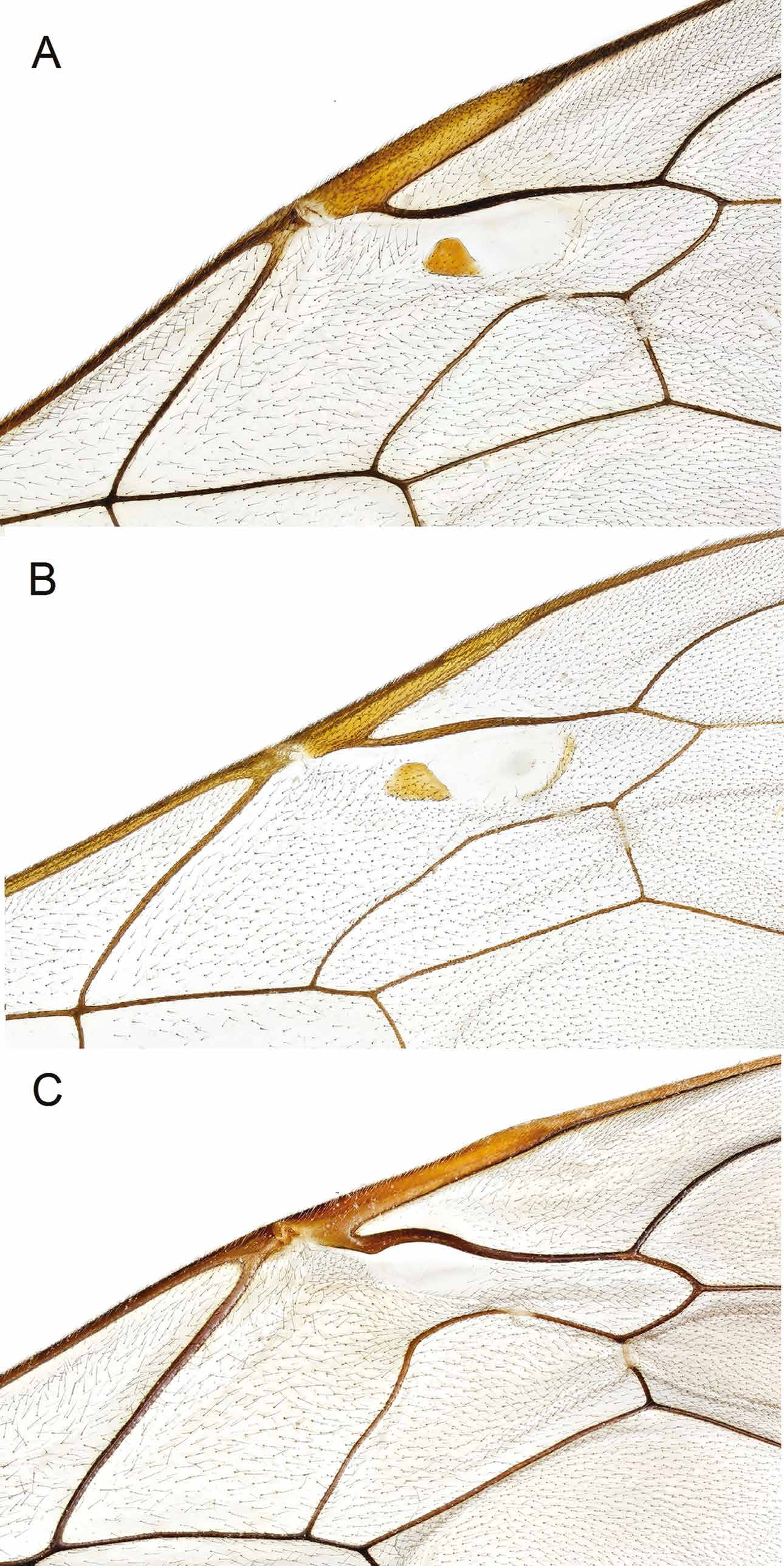
– Fore wing with central sclerite present, but sometimes completely translucent ( Figs 4 View Fig A–D, 13B); clypeus in lateral view distinctly convex ( Fig. 14A View Fig ) ( E. ramidulus View in CoL species group) ......................... 3
3. Fore wing with central sclerite completely translucent ( Fig. 13B View Fig ) ..................................................... ..................................................................................................... E. merdarius (Gravenhorst, 1829) View in CoL – Fore wing with central sclerite distinctly pigmented ( Fig. 4 View Fig A–D) ................................................... 4
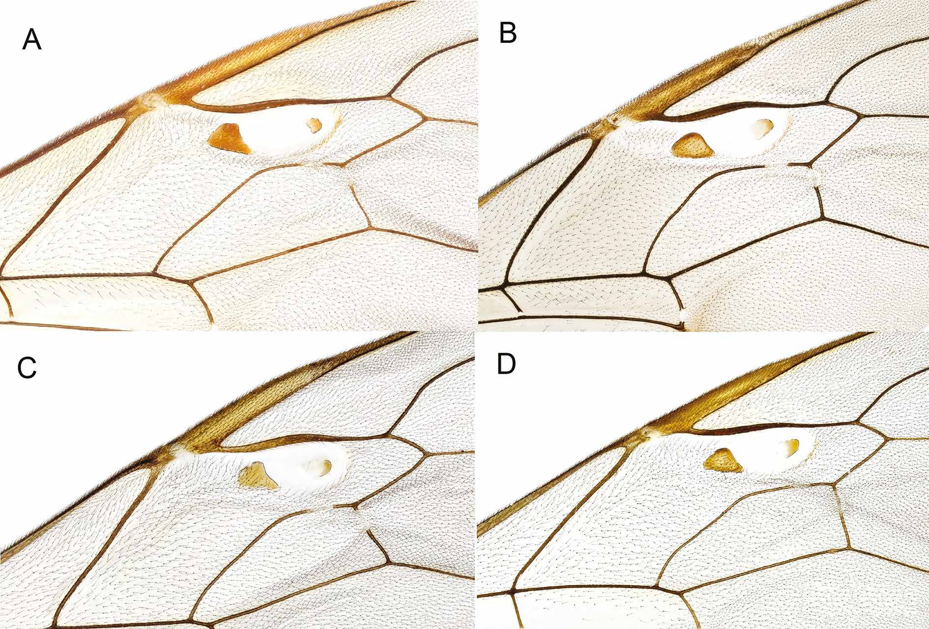
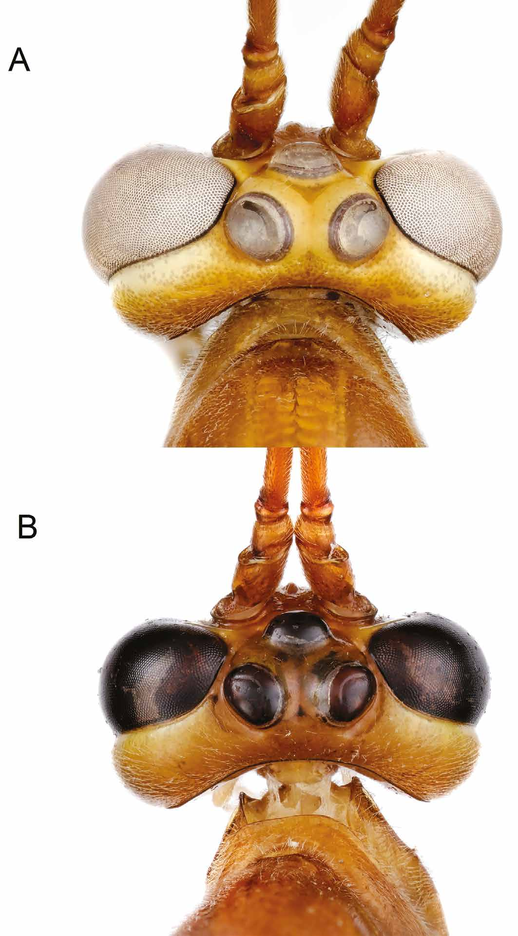
4. Antenna with central and apical flagellomeres very slender with central and apical segments more than 2 times as long as wide ( Fig. 3 View Fig G–H); number of flagellomeres 53–56; occipital carina only slightly curved before indicated junction with hypostomal carina ( Fig. 6B View Fig ); epicnemial carina between mesopleural angles and sternal part almost straight ( Fig. 12A View Fig ); central sclerite in glabrous area in fore wing usually more circular in shape, largely unpigmented; central and proximal sclerite closer to each other ( Fig. 4C View Fig ). Small species, fore wing length 12–13 mm ....... E. ryrholmi sp. nov.
– Antenna with central and apical flagellomeres at most 1.8 times as long as wide ( Fig. 3 View Fig A–F); occipital carina distinctly curved before indicated junction with hypostomal carina, ( Fig. 6A View Fig ); epicnemial carina between mesopleural angles and sternal part sinuous ( Fig. 12B View Fig ); central sclerite in glabrous area in fore wing more semi-ovoid in shape, often elongate; central and proximal sclerites further apart ( Fig. 4 View Fig A–B, D) ......................................................................................................................... 5
5. Mesosoma usually with extensive dark brown patches; central sclerite in fore wing mostly narrow, at most as long as wide ( Fig. 4A View Fig ); distance between central and proximal sclerites usually distinctly longer than basal side of proximal sclerite; central sclerite mostly entirely pigmented .................................................................................... E. combustus (Gravenhorst, 1829) View in CoL
– Mesosoma lacking distinct dark patches, uniformly testaceous; central sclerite usually longer than wide ( Fig. 4 View Fig B–C); distance between central and proximal sclerites almost equal to basal side of proximal sclerite; central sclerite mostly largely unpigmented proximally ..................................... 6
6. Antenna with central and preapical flagellomeres shorter, at most 1.5 times as long as wide ( Fig. 3 View Fig A–D) ....................................................................................................................................... 7
– Antenna with central and preapical flagellomeres longer, at least 1.7 times as long as wide ( Fig. 3 View Fig E–F) ........................................................................................................................................ 8
7. Antenna with 51–56 flagellomeres; temples very strongly narrowed behind eyes, head with no gap between eye and lateral ocelli ( Fig. 5A View Fig ); central and apical flagellomeres about 1.3 times as long as wide ( Fig. 3 View Fig A–B) ..................................................................................... E. cerebrator Aubert, 1966 View in CoL
– Antenna with 59–62 flagellomeres; temples strongly buccate, head with distinct gap between ocelli and eye ( Fig. 5B View Fig ); central and apical flagellomeres about 1.5 times as long as wide ( Fig. 3 View Fig C–D) ...................................................................................................... E. cederbergi sp. nov.
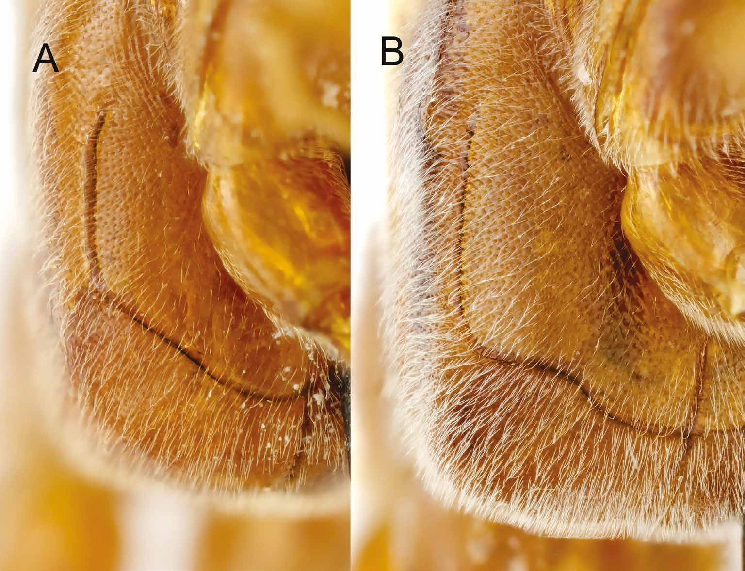
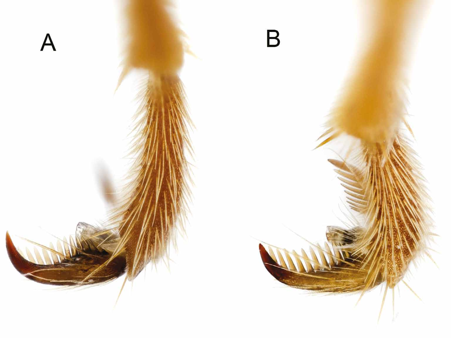
8. Metasoma in female abruptly black-tipped from the 5th (in females) or 6th (in males) tergite; hind tarsal claws in female conspicuously curved ( Fig. 15A View Fig ).................... E. ramidulus (Linnaeus, 1758) View in CoL
– Metasoma never abruptly black-tipped; hind tarsal claws in female not conspicuously curved ( Fig. 15B View Fig ) .......................................................................................................................................... 9
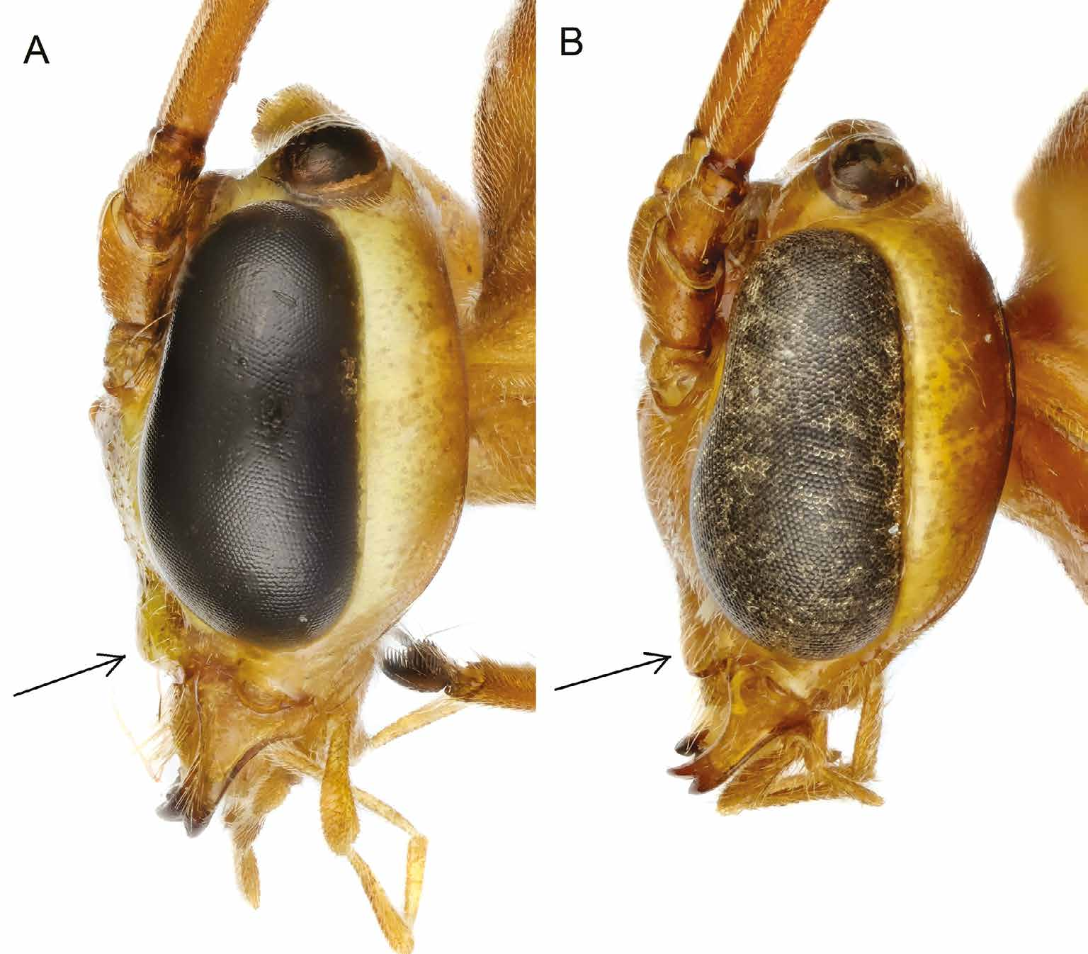
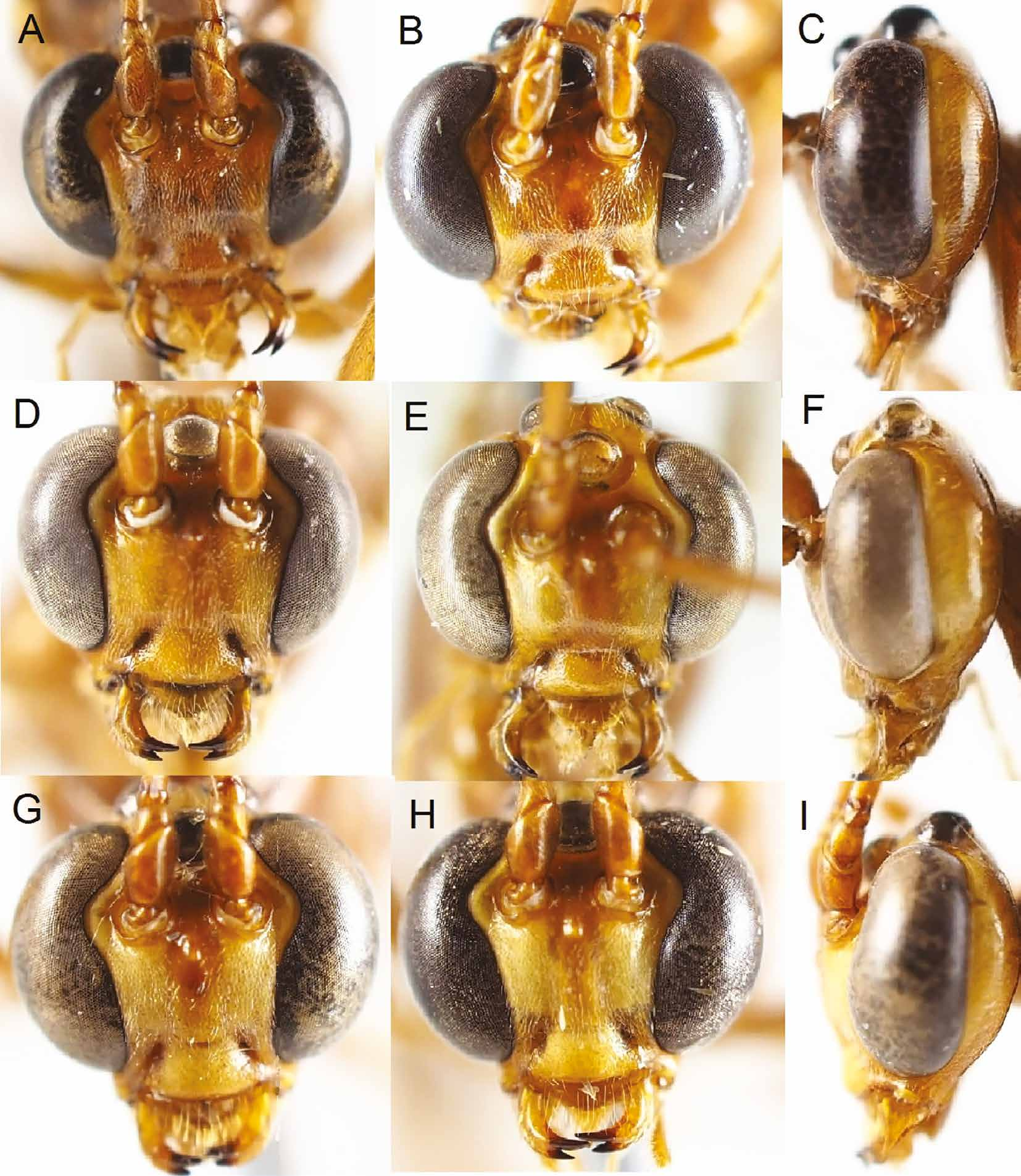
9. Head in lateral view with temples narrow, at most about 0.4 times the width of compound eye ( Fig. 9I View Fig ); face generally with extensive yellow markings ( Fig. 9 View Fig G–H); face narrow in anterior view ( Fig. 9 View Fig G–H); lateral ocellus touching compound eye ................................. E. adustus (Haller, 1885) View in CoL
– Head in lateral view with temples wide, about 0.7 times the width of compound eye ( Fig. 9C, F View Fig ); face generally more testaceous; face wider in anterior view ( Fig. 9 View Fig A–B, D–E); lateral ocelli often with more or less distinct gap between lateral ocellus and compound eye .................................... 10
10. Number of flagellomeres 56–59; head in anterior view more rounded ( Fig. 9 View Fig D–E); ocelli small, gap between lateral ocelli and inner margin of compound eye wide, about 0.2–0.3 times the diameter of ocellus ............................................................................................. E. myricae Broad & Shaw, 2016
– Number of flagellomeres 62–67; head in anterior view distinctly transverse ( Fig. 9 View Fig A–B); ocelli large, gap between lateral ocelli and inner margin of compound eye narrow, about 0.1 times the diameter of ocellus .......................................................................................... E. intermedius sp. nov


No known copyright restrictions apply. See Agosti, D., Egloff, W., 2009. Taxonomic information exchange and copyright: the Plazi approach. BMC Research Notes 2009, 2:53 for further explanation.