
Pediopsoides (Pediopsoides) alba Li, Dai & Li
|
publication ID |
https://doi.org/ 10.11646/zootaxa.4150.3.5 |
|
publication LSID |
lsid:zoobank.org:pub:9A230549-A9DF-47E6-BD92-6B599F0ACAFE |
|
DOI |
https://doi.org/10.5281/zenodo.6087710 |
|
persistent identifier |
https://treatment.plazi.org/id/A333EE78-F85B-FFA8-26FF-194CD773F93F |
|
treatment provided by |
Plazi |
|
scientific name |
Pediopsoides (Pediopsoides) alba Li, Dai & Li |
| status |
sp. nov. |
Pediopsoides (Pediopsoides) alba Li, Dai & Li View in CoL sp. nov.
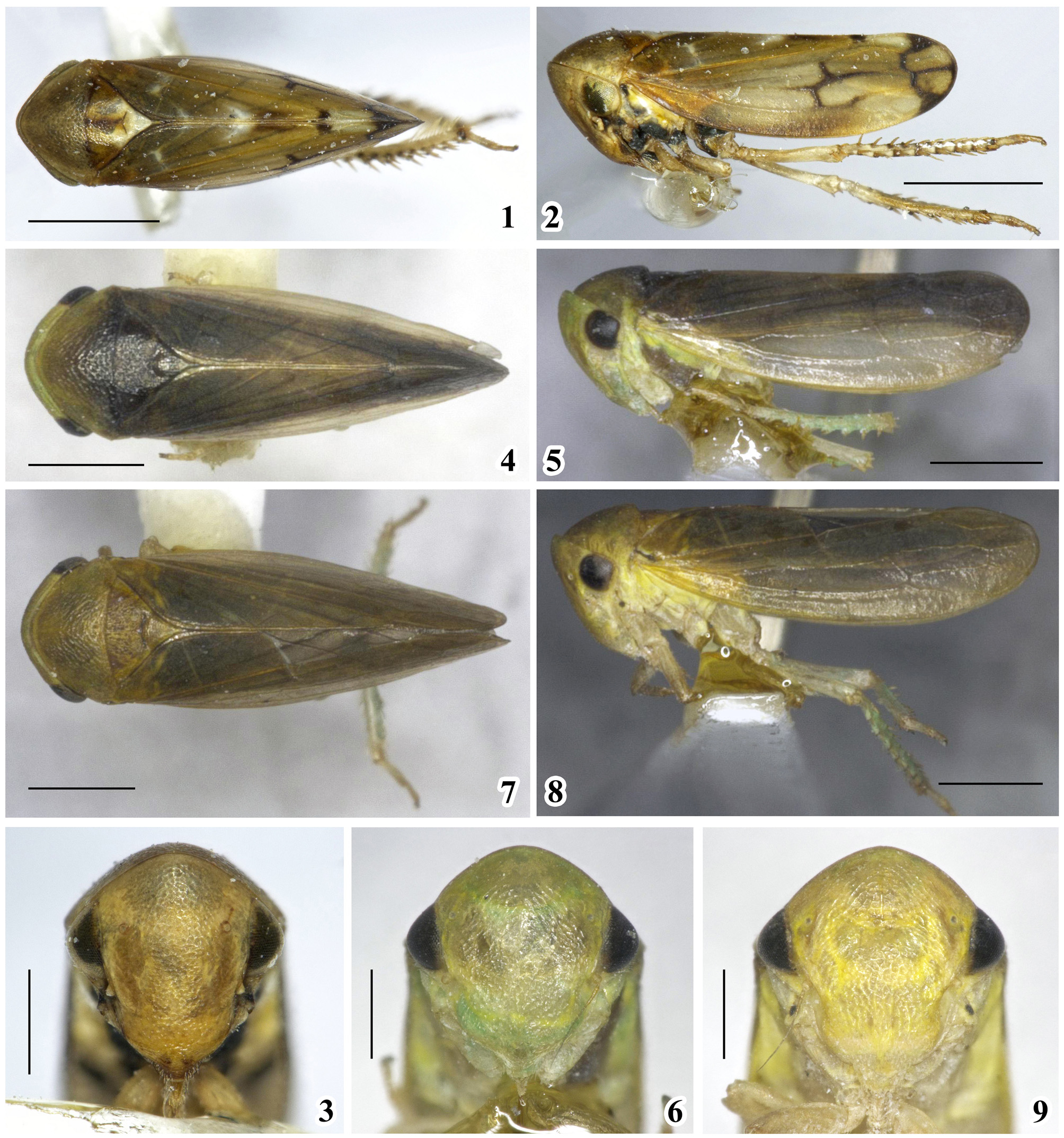
( Figs 1–3 View FIGURES 1 – 9 , 10–19 View FIGURES 10 – 19 , 36)
Body length (including tegmina). ♂, 3.25–3.30 mm.
Holotype description. Head and thorax (color). Background color ( Figs 1–3 View FIGURES 1 – 9 ) yellow brown. Vertex yellowish brown; face yellowish; eyes dark brown in front aspect and pale brown in dorsal aspect; postclypeus yellow, anteclypeus yellowish with brown tip; scape and pedicel of antenna yellowish, antennal fossa black. Proepisternum with black spot. Pronotum brown with central part dark brown, darker on sides of midline, anterior margins near eyes yellowish. Mesonotum yellowish brown, with middle line and lateral angles dark brown. Scutellum milk white, scutellar suture dark brown. Forewing clavus dark brown, tips of veins 1A and 2A, and cross veins between claval veins whitish, claval sutures yellow, veins surrounded apical cells and cross veins black. Legs yellowish, bases of macrosetae dark brown.
Morphology. Body appearance ( Figs 1–2 View FIGURES 1 – 9 ) typical of genus; head, face, pronotum, and scutellum obviously striated. Head strongly angularly produced in dorsal view, inverted V-shaped, including eyes as broad as pronotum; crown parallel-margined, and strongly shortened and bandlike. Face ( Fig. 3 View FIGURES 1 – 9 ) across eyes as wide as long; ocelli between eyes with distance of about 5 × longer than that of ocellus to adjacent eye; sutures between lora and frons almost invisible; anteclypeus projected beyond lora and gena; transclypeal suture obsolete. Pronotum ( Fig. 1 View FIGURES 1 – 9 ) 2.0 × broader than long, with inconspicuous longitudinal medial carina, and oblique and reticular striations; anterior margin strongly produced, slightly depressed near eyes, posterior margin clearly concave medially. Mesonotum and scutellum ( Fig. 1 View FIGURES 1 – 9 ) 1.4 × wider than long, nearly triangular, striated except bilateral corners; scutellar suture depressed and arched onward. Forewings ( Fig. 2 View FIGURES 1 – 9 ) opaque, venation prominent. Hind tibia with 10–11 macrosetae on PD row, 7 on AD row, 4 on AV row.
Male genitalia. Pygofer ( Fig. 10 View FIGURES 10 – 19 ) broad basally, in lateral view, dorsal margin depressed and sloping caudally, caudal margin prolonged on dorsal part, ventral margin tapered to end with several fine setae, apex with two processes extended dorsomesad with acute tips of caudal one pointed inward and anterior one pointed dorsad ( Fig. 11 View FIGURES 10 – 19 ), basally with several scattered fine setae. Subgenital plate ( Fig. 10 View FIGURES 10 – 19 ) slender, elongate and slightly longer than pygofer, dorsoventral margins nearly parallel, end slightly tumid, surface except basal 1/3 with scattered setae and apex with tuft of longer setae. Style ( Fig. 13 View FIGURES 10 – 19 ) slender, nearly angled on basal 1/3, clearly inflated subapically, then suddenly narrowed with blunt tip directed dorsad, stem outer margin with row of setae. Dorsal connective composed of dorsal and ventral portions; dorsal portion paired, with front margin strongly depressed and hind margin bearing three spines ( Fig. 12 View FIGURES 10 – 19 ) freely attached with ventral portion and segment X; ventral portion ( Figs 14– 15 View FIGURES 10 – 19 ) unpaired and unarmed. Aedeagus ( Figs 16–17 View FIGURES 10 – 19 ), relatively robust, shaft with paired spines basally on lateral margins half as long as shaft; in lateral view, aedeagal shaft broad, curved dorsally, ventral margin slightly serrated below gonopore, dorsal margin sinuate, apex blunt, dorsal apodeme weakly developed; in ventral view, aedeagus broad basally, shaft compressed, apex subacute; gonopore apical. Connective ( Figs 18–19 View FIGURES 10 – 19 ), in dorsal aspect, with posterior margin and both lateral margins strongly excavated medially, anterior margin as wide as posterior margin, with fingerlike process in middle of anterior margin longer than lateral arms, both lateral arms slender and bent dorsally.
Female. Unknown.
Material examined. Holotype: ♂, CHINA: Yunnan Province, Dehong Autonomous Prefecture , Lianghe County, Mengyang Township , 25. VII. 2013, collected by Fan Zhi-Hua . Paratypes: 1 ♂, same data as holotype, except collected on 27. VII. 2013 by Yang Wei-Cheng.
Distribution. China (Yunnan Province) (Fig. 36).
Remarks. The new species is similar to P. (P.) kodaiana Viraktamath, 1996 from India but differs from the latter by the oblique striated pronotum, the aedeagal shaft with a pair of basal spines on the lateral margins, and by the different shapes of the pygofer ventral processes and dorsal connective.
Etymology. The new species name is derived from the Latin words “ albus ”, refers to the white scutellum.

No known copyright restrictions apply. See Agosti, D., Egloff, W., 2009. Taxonomic information exchange and copyright: the Plazi approach. BMC Research Notes 2009, 2:53 for further explanation.