
Trouessartia capensis, Berla, 1959
|
publication ID |
https://doi.org/10.11646/zootaxa.4860.1.1 |
|
publication LSID |
lsid:zoobank.org:pub:A64165A5-D29A-4CCD-B0B1-2A78497B9A6F |
|
DOI |
https://doi.org/10.5281/zenodo.4536010 |
|
persistent identifier |
https://treatment.plazi.org/id/A34C1E77-FFDE-FF84-FF42-8DBAFA02ADCA |
|
treatment provided by |
Plazi |
|
scientific name |
Trouessartia capensis |
| status |
|
Key to New World species of the capensis group
(Males and females)
1. In both sexes, setae d1 present........................................................................... 2
- In both sexes, setae d1 absent........................................................................... 12
2. In females, setae h1 filiform............................................................................. 3
- In females, setae h1 narrowly lanceolate................................................................... 4
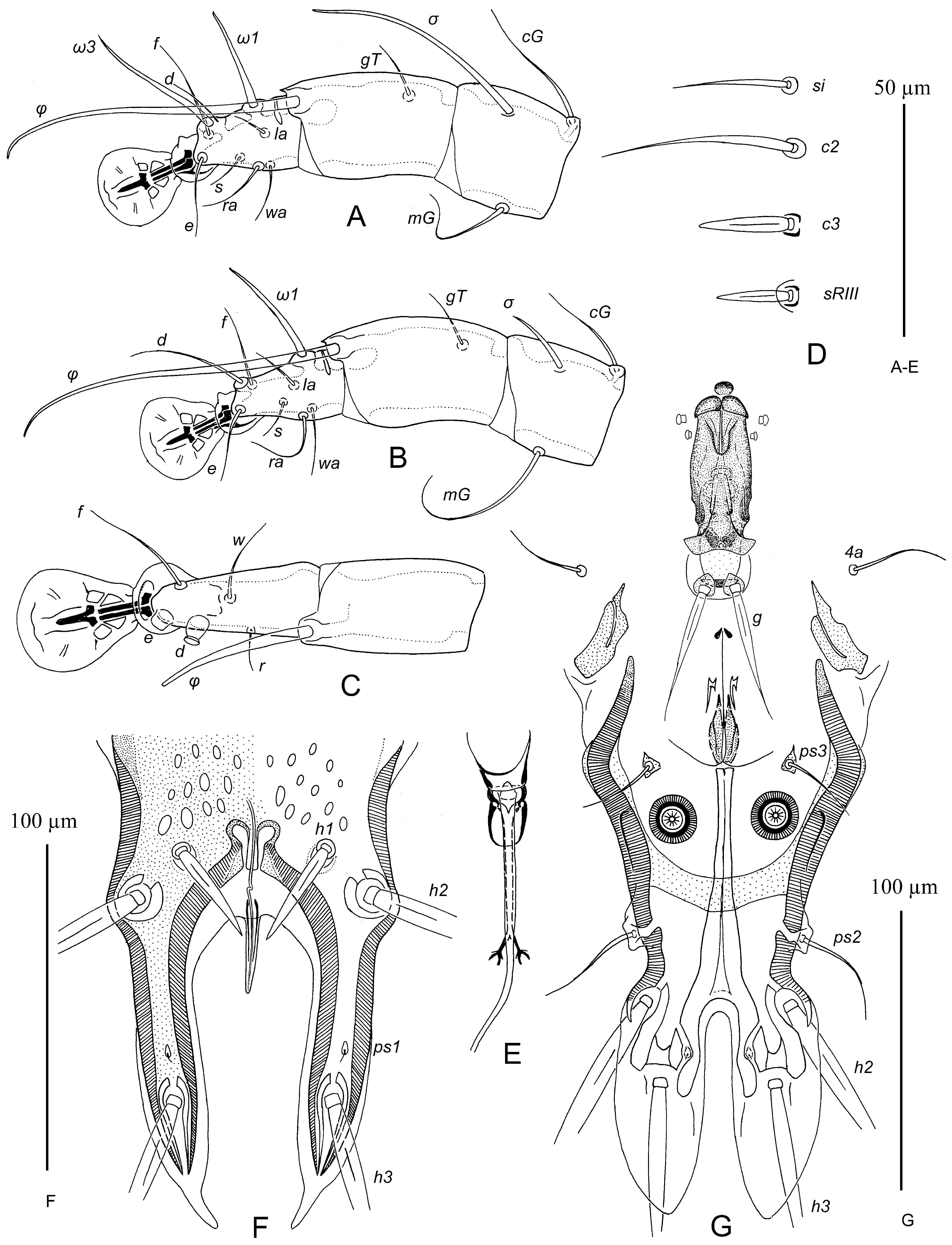
3. In both sexes, anterolateral extensions of prodorsal shield angular, extending to epimerites Ia. In males, bases of setae g distinctly separated, postgenital plaque horseshoe-shaped, setae 4b situated posterior to 3a, rudimentary sclerites rEpIIa large ovate, 15–20 in diameter. In females, external copulatory tube finger-like, 20–22 long, anterior part of hysteronotal shield with large ovate lacunae along lateral margins........................................... T. mangaratibensis Berla, 1959
- In both sexes, anterolateral extensions of prodorsal shield rounded, not extending to epimerites Ia. In males, bases of setae g almost touching, postgenital plaque small trapezoidal or bow-shaped, setae 4b anterior to 3a, and rudimentary sclerites rEpIIa small ovate ( Figs. 5B View FIGURE 5 , 7G View FIGURE 7 ). In females, external copulatory tube short conical, 7–10 long, posterior part of hysteronotal shield with small ovate lacunae, anterior part without any lacunae ( Figs. 6A View FIGURE 6 , 7F View FIGURE 7 )............................... T. ciris sp. n.
4. In females, external copulatory tube extending beyond level of setae h3.......................................... 5
- In females, external copulatory tube extending maximally to midlength between levels of setae h2 and h3............... 6
5. In females, external copulatory tube 70–80 long and extending to midlength between levels of setae h3 and posterior tips of hysteronotal shield ( Fig. 4E View FIGURE 4 ). In males, setae g setiform, situated at level of setae 4a or slightly anterior; adanal shields shaped as small sclerites of irregular form with long blade-like extension directed anterior............................................................................................ T. geospiza OConnor, Foufopoulos and Lipton, 2005
- In females, external copulatory tube about 50–60 long and extending slightly beyond level of setae h3. In males, setae g strongly widened and flattened in basal 2/3, about 5 in width, situated distinctly anterior to setae 4a; adanal shields shaped as small circles around bases of setae ps3............................................... T. sicaliae Hernandes, 2014
6. In males, genital shield crescent-shaped, apophyses of adanal apodemes with rectangular ledge at anterior end ( Fig. 4A View FIGURE 4 ). In females, external copulatory tube short conical, 9–15 long ( Fig. 4C View FIGURE 4 ).............................. T. passerinae sp. n.
- In males, genital shield of another shape or absent, apophyses of adanal apodemes of another structure. In females, external copulatory tube stylet- or finger-like, 30–60 long ( Figs. 10F View FIGURE 10 , 13F View FIGURE 13 )............................................... 7
7. In males, setae g strongly widened and flattened in basal 2/3, about 5 in width ( Fig. 10G View FIGURE 10 ). In females, setae ps1 approximately equidistant from levels of setae h2 and h3 ( Fig. 10F View FIGURE 10 )......................................................... 8
- In males, setae g moderately thickened but not flattened ( Fig. 13G View FIGURE 13 ). In females, setae ps1 closer to level of setae h3 ( Fig. 13F View FIGURE 13 ) ................................................................................................... 9
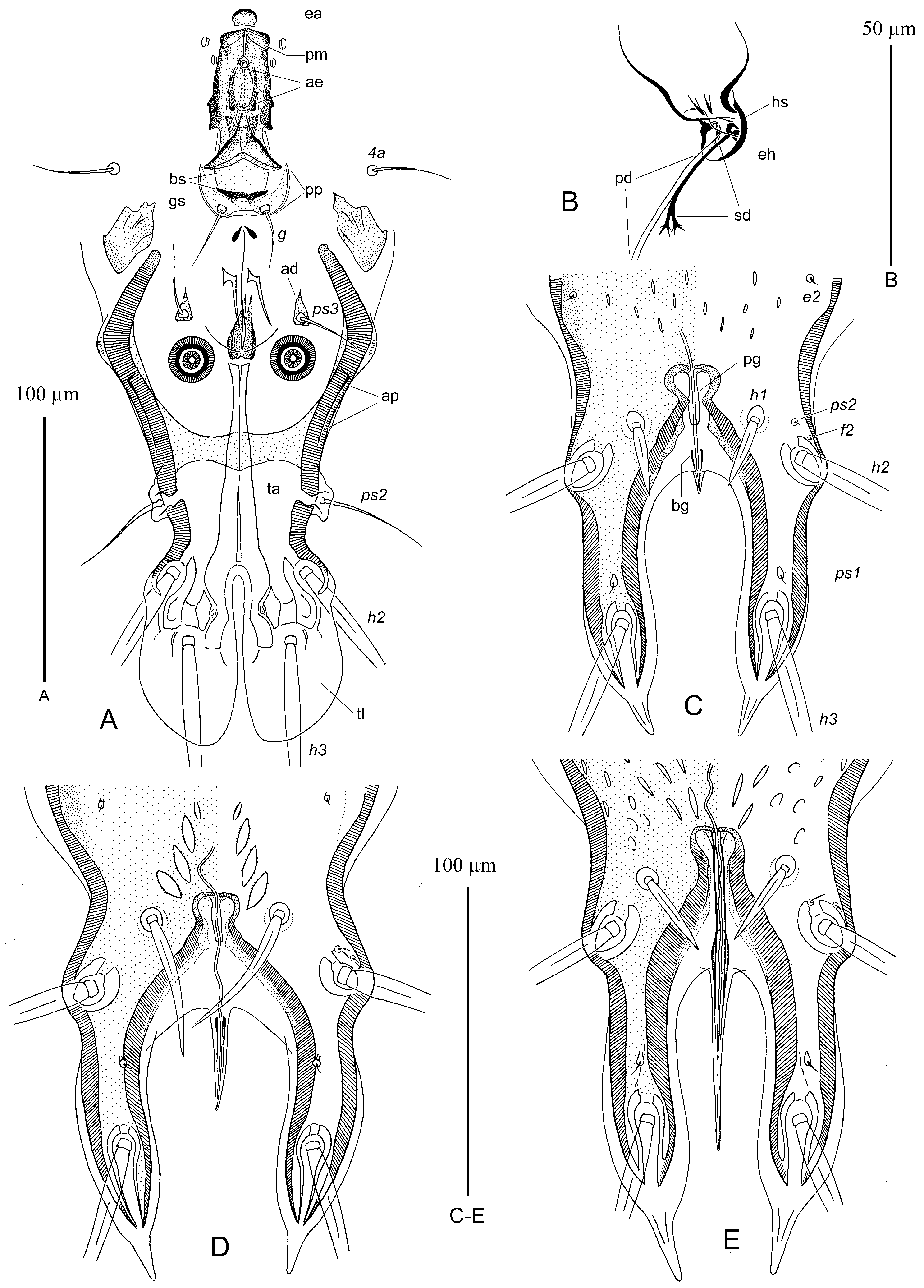
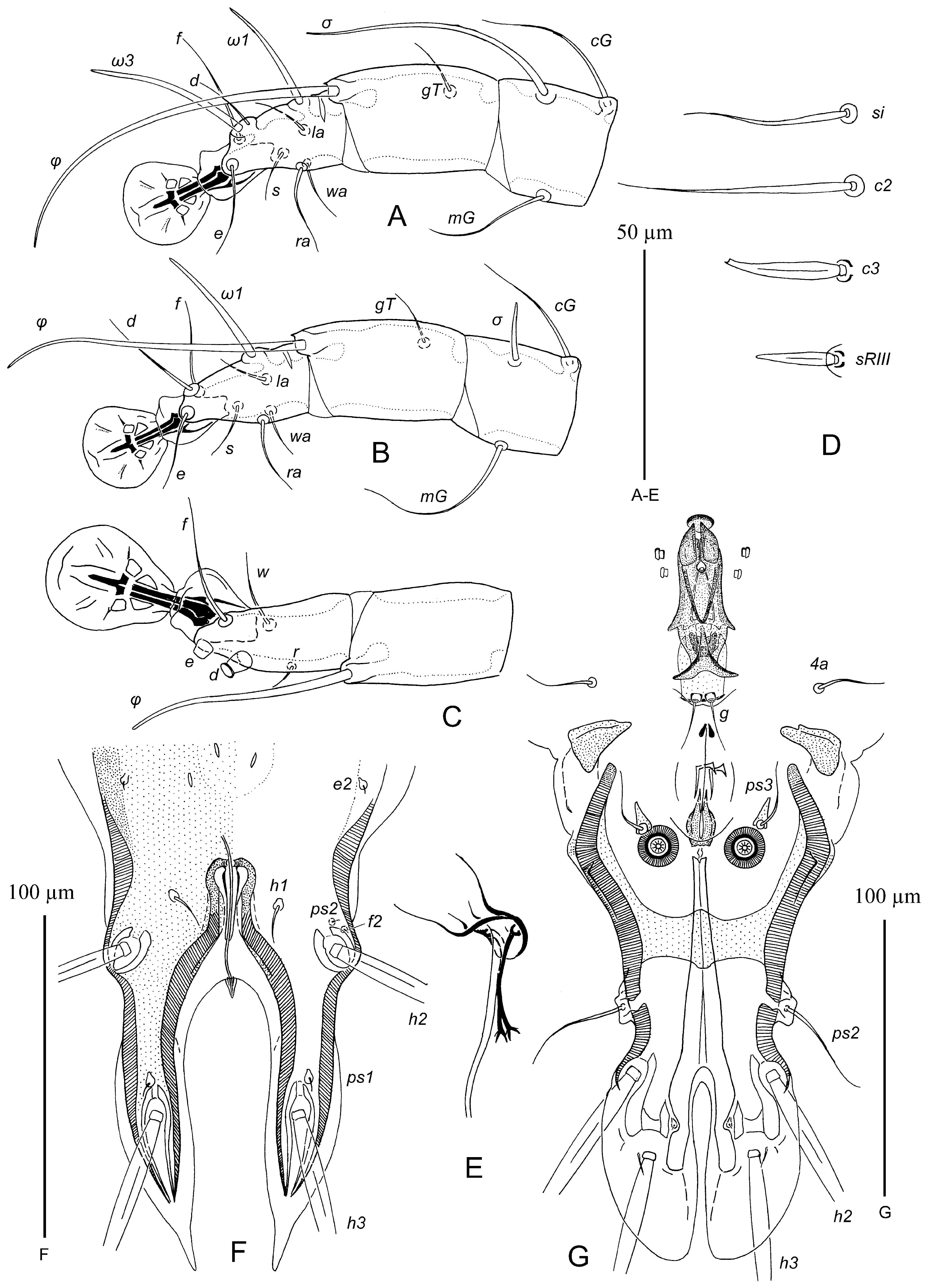
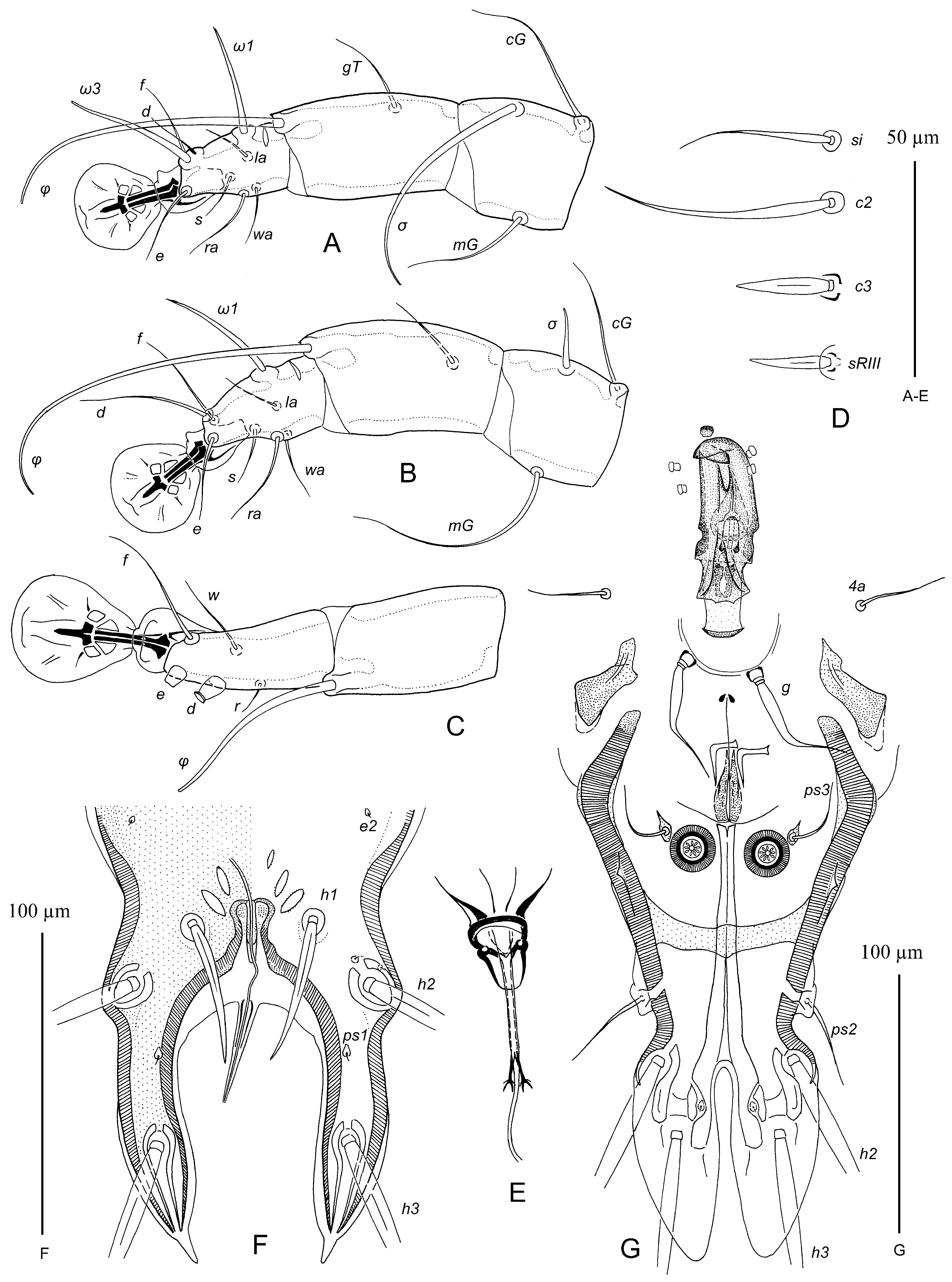
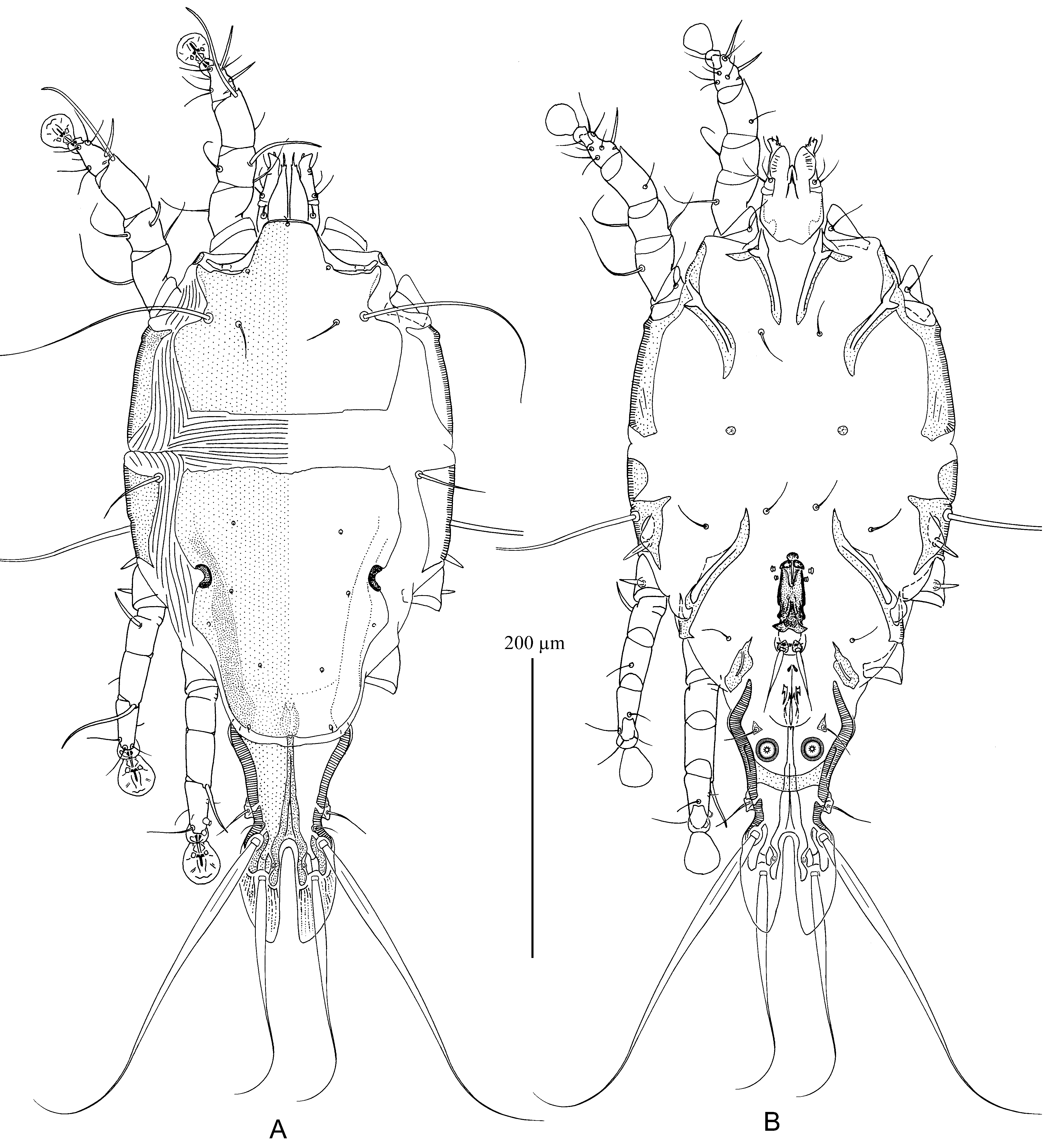
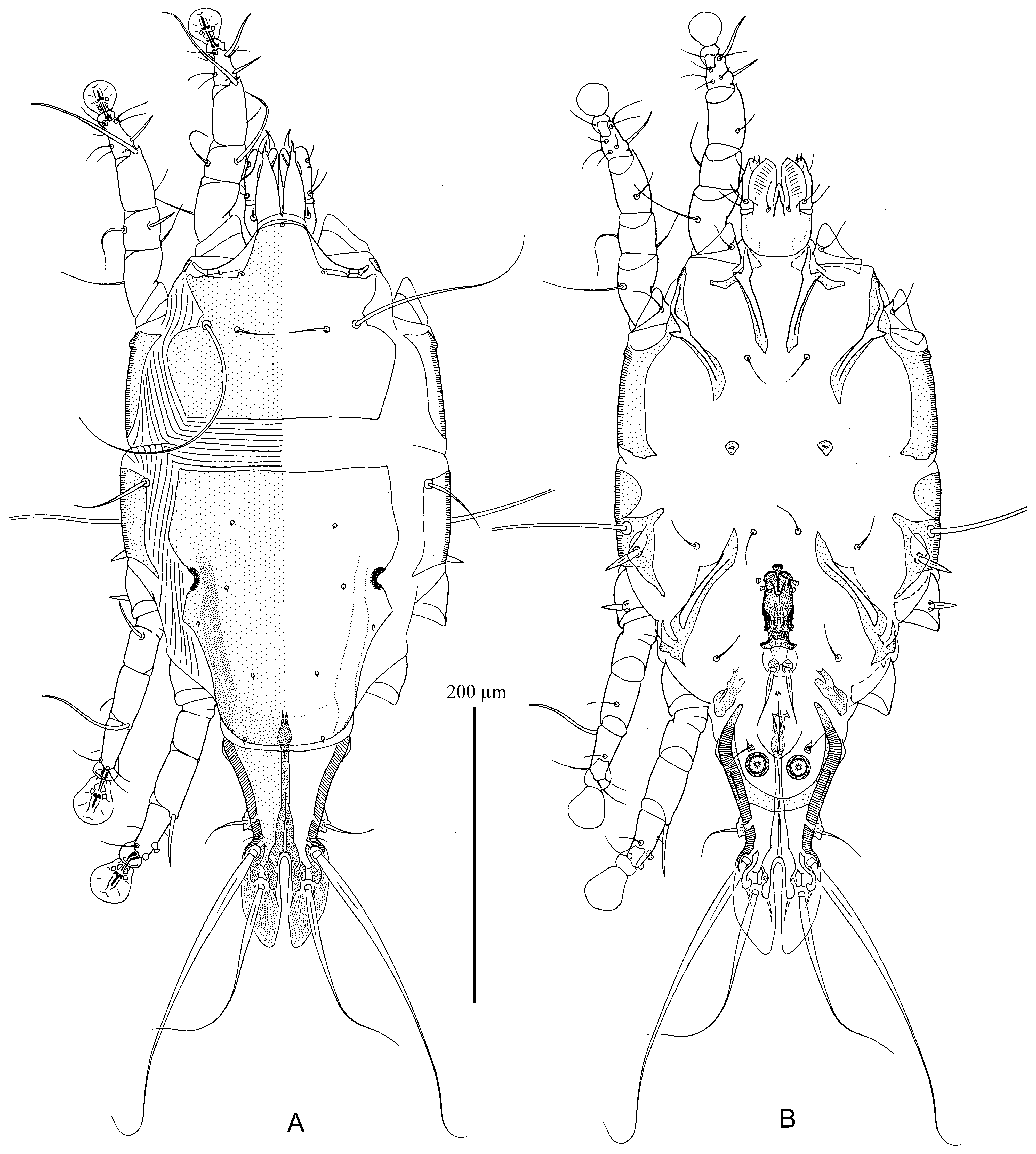
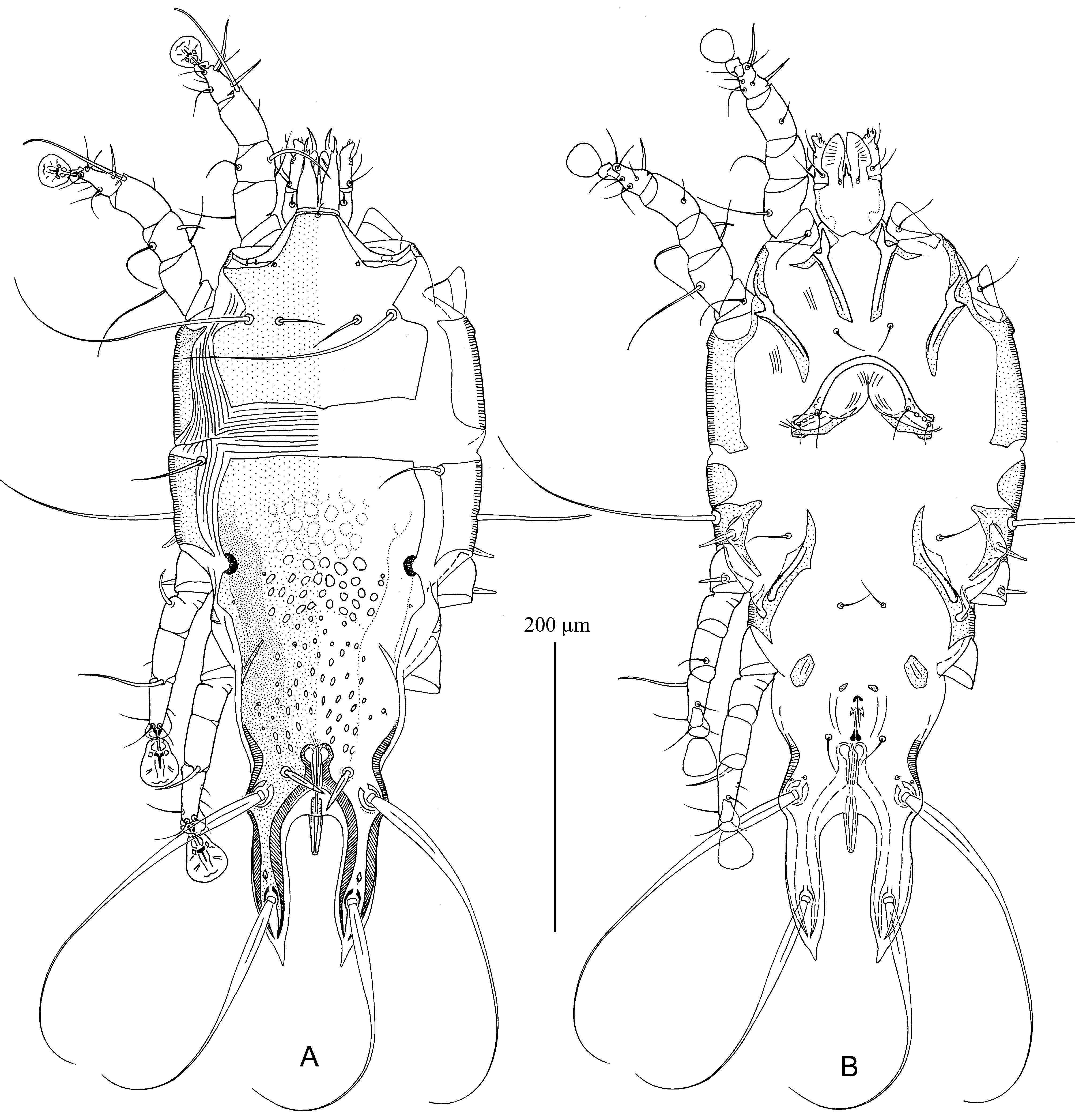
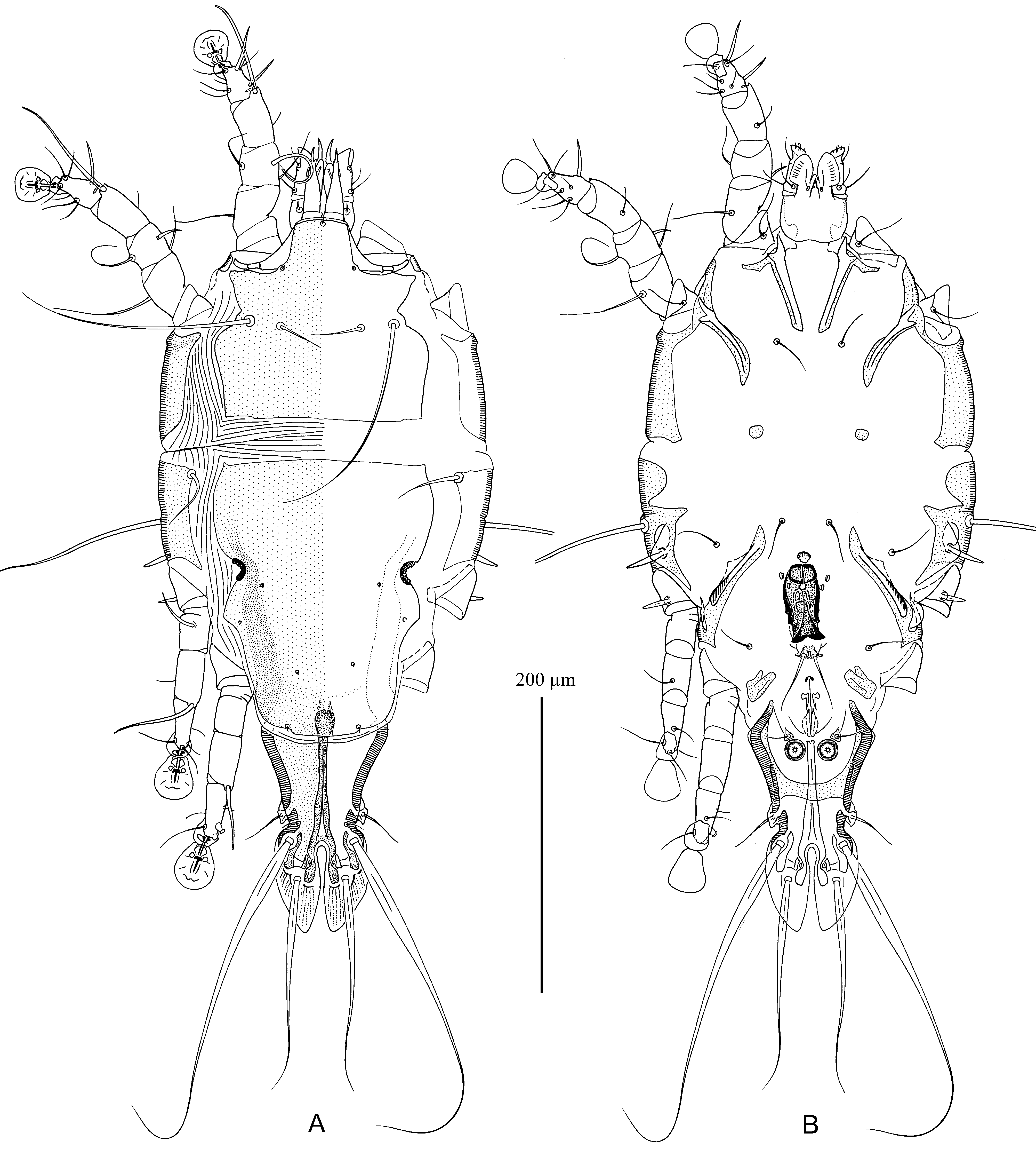
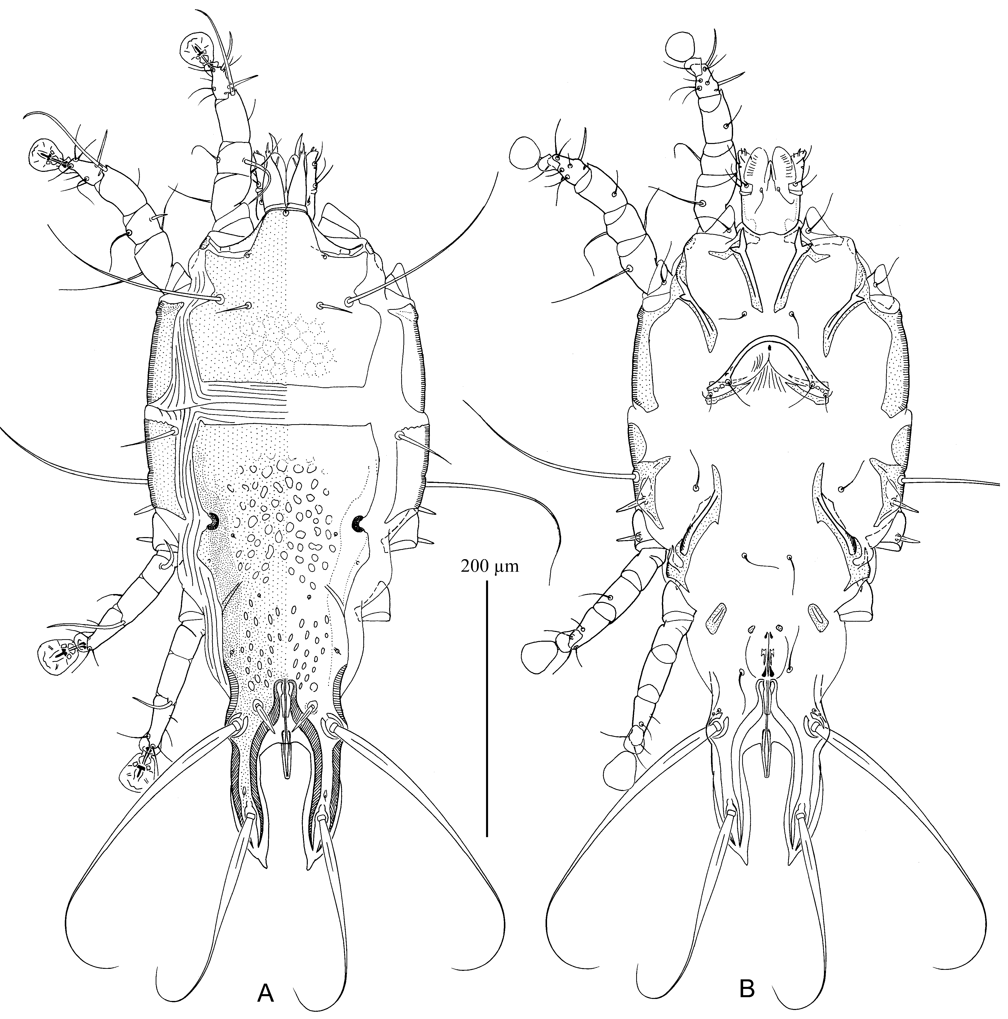
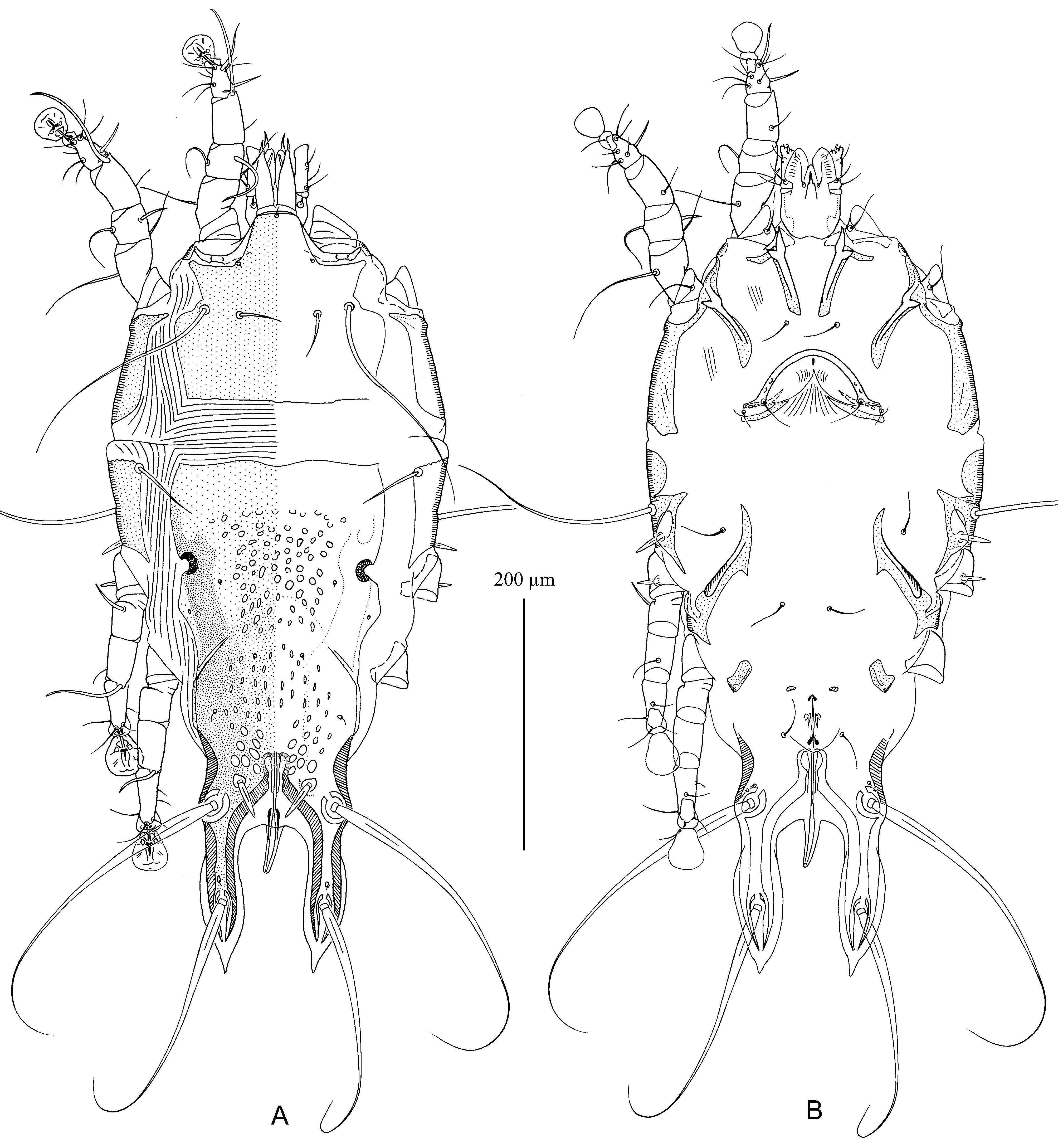
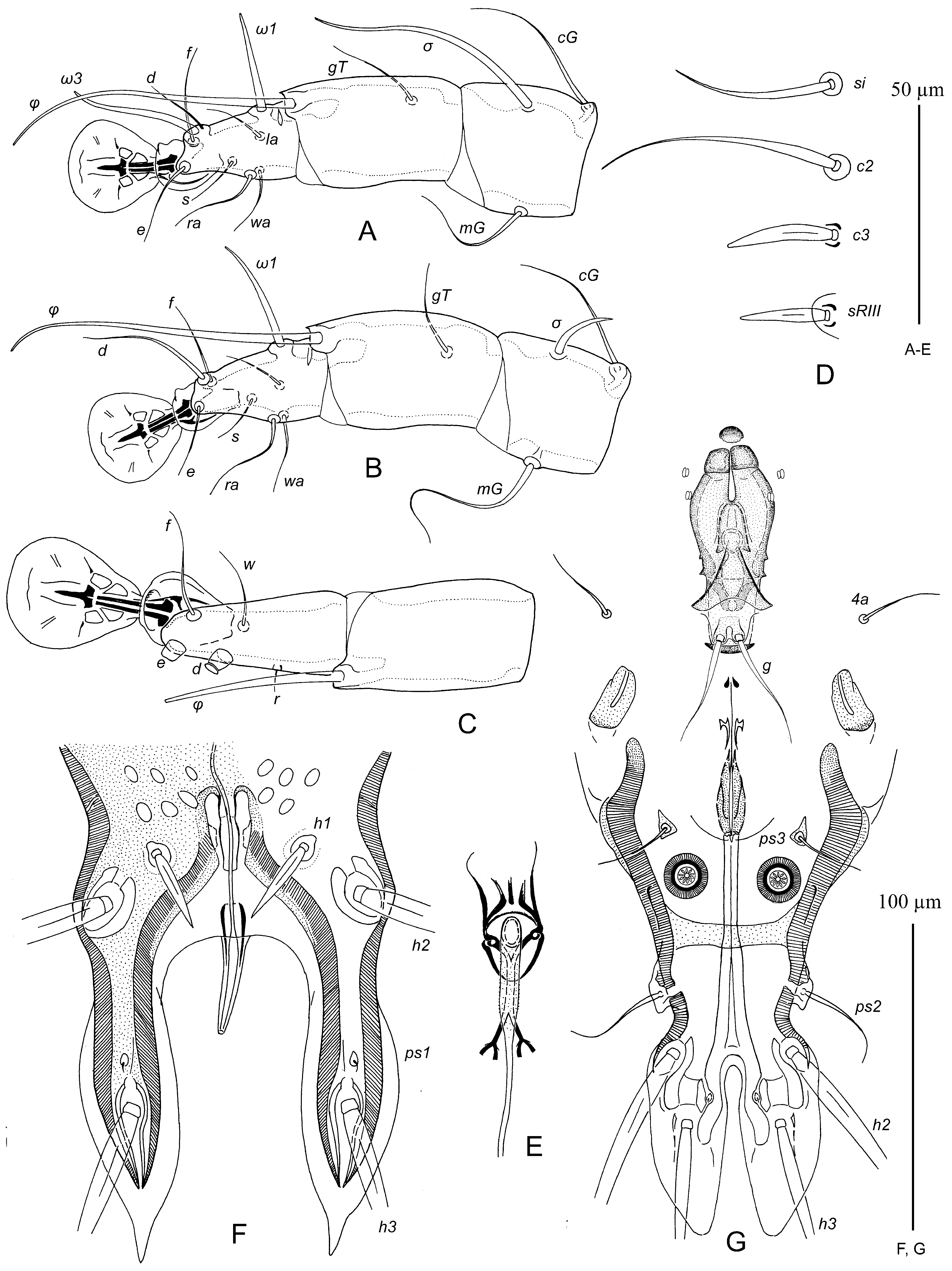
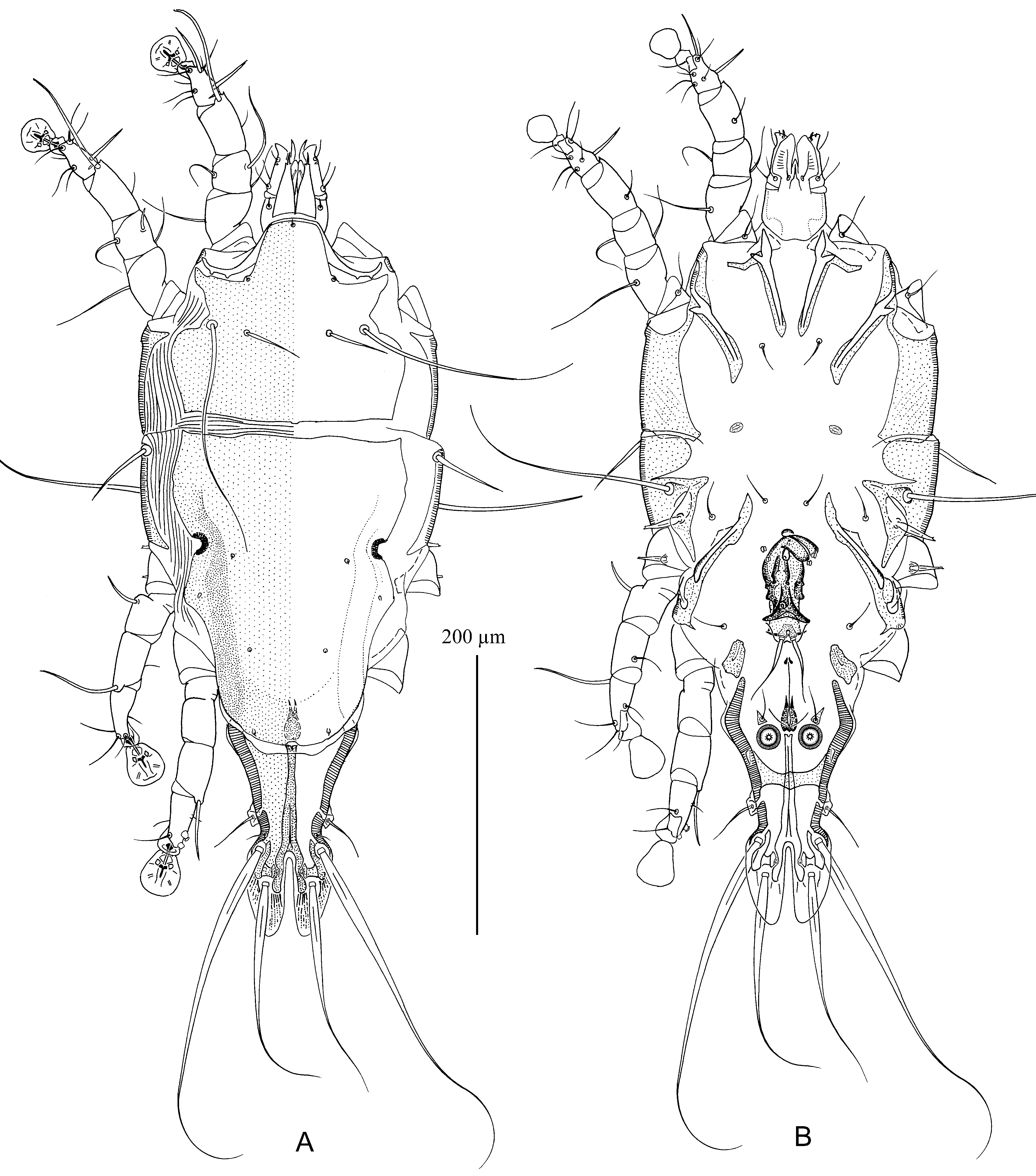
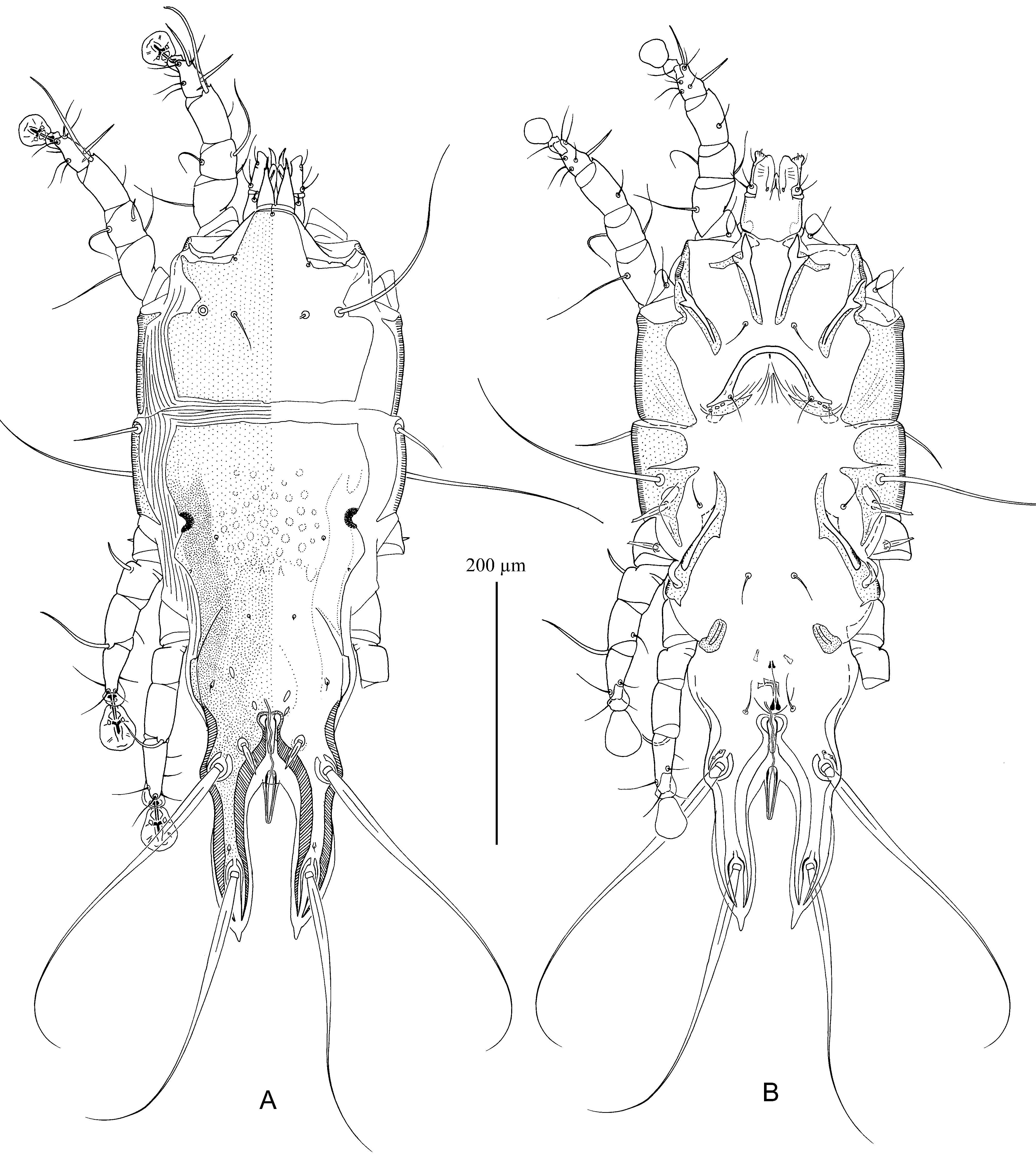
8. In males, setae g situated in area of postgenital plaque. In females, anterior part of hysteronotal shield with poorly distinct ornamentation of ovate and irregularly shaped lacunae, median area of posterior part with obliquely orientated ovate and lanceolate lacunae, setae h1 almost extending to level of setae ps1 ( Fig. 4D View FIGURE 4 )............................. T. capensis Berla, 1959 In males, setae g situated off postgenital plaque ( Fig. 10G View FIGURE 10 ). In females, anterior part of hysteronotal shield with reticulate ornamentation, posterior part with oblique ovate lacunae, setae h1 extending slightly beyond level of setae ps1 ( Fig. 10F View FIGURE 10 )............................................................................................. T. spizellae sp. n.
9. In males, anterior genital papillae larger than posterior genital papillae ( Figs. 13G View FIGURE 13 , 16G View FIGURE 16 ). In females, distal end of primary spermaduct before entering in external copulatory tube with S-like bend ( Figs. 13F View FIGURE 13 , 16F View FIGURE 16 )........................... 10
- In males, anterior and posterior genital papillae similar in size ( Fig. 19G View FIGURE 19 ). In females, distal end of primary spermaduct straight or slightly sinuous ( Fig. 19F View FIGURE 19 )........................................................................... 11
10. In males, sclerotized areas around bases of setae g not connected, rudimentary sclerites rEpIIa circular ( Figs. 14B View FIGURE 14 , 16G View FIGURE 16 ). In females, setae h1 32–38 long, slightly extending beyond free margin of interlobal membrane ( Fig. 16F View FIGURE 16 )....................................................................................................... T. americana sp. n. In males, sclerotized areas around bases of setae g connected by narrow bridge, rudimentary sclerites rEpIIa teardrop-shaped ( Figs. 11B View FIGURE 11 , 13G View FIGURE 13 ). In females, setae h1 28–30 long, extending to free margin of interlobal membrane ( Fig. 13F View FIGURE 13 )................................................................................................... T. seiurus sp. n.
11. In males, bases of setae g surrounded by small sclerotized areas, anterior ends of epimerites IVa usually bifurcate and almost extending to level of setae g ( Figs. 17B View FIGURE 17 , 19G View FIGURE 19 ). In females, central part of hysteronotal shield with poorly pronounced small ovate lacunae, posterior part with dash-shaped lacunae; tips of setae h1 extending distinctly beyond free margin of interlobar membrane ( Figs. 18A View FIGURE 18 , 19F View FIGURE 19 ).............................................................. T. helmitheros sp. n.
- In males, bases of setae g without sclerotized areas, anterior ends of epimerites IVa simple and distinctly not reaching level of setae g. In females, central and posterior parts of hysteronotal shield with well pronounced small ovate and dash-shaped lacunae, setae h1 barely extending to free margin of interlobar membrane..................... T. basieuteri Hernandes, 2014
12. Length of idiosoma in males 470–490 and in females 540–590. In males, rudimentary sclerites rEpIIa large ovate, each with a small lacuna ( Fig. 26B View FIGURE 26 ). In female, posterior half of hysteronotal shield with two different types of lacunae: small narrowly ovate lacunae between levels of setae e1 and e2, and with large ovate lacunae between setae e2 and h1 ( Figs. 27A View FIGURE 27 , 28F View FIGURE 28 )............................................................................................. T. tigrina View in CoL sp. n.
- Length of idiosoma in males 410–450 and in females 490–535. In males, rudimentary sclerites rEpIIa small ovate, without lacuna ( Fig. 23B View FIGURE 23 ). In female, posterior half of hysteronotal shield with uniform lacunae ( Figs. 24A View FIGURE 24 , 25F View FIGURE 25 )............... 13
13. In both sexes, almost half of coxal fields II heavily sclerotized ( Figs. 29B View FIGURE 29 , 30B View FIGURE 30 ). In males, setae 4b posterior to level of setae cp; epimerites IVa extending to level of setae g; apophyses of adanal apodemes represented by narrow membranes with convex free margin ( Fig. 31G View FIGURE 31 ). In females, hysteronotal shields 345–350 long, posterior part of hysteronotal shield usually with several (4–10) narrowly lanceolate lacunae ( Figs. 30A View FIGURE 30 , 31F View FIGURE 31 )......................................... T. pensylvanica View in CoL sp. n.
- In both sexes, only narrow lateral area of coxal fields II heavily sclerotized ( Fig. 23B View FIGURE 23 , 24B View FIGURE 24 ). In males, epimerites IVa not extending to level of setae g, apophyses of adanal apodemes represented by longitudinal ridges with rounded tubercle at anterior end. In females, hysteronotal shield 315–335 long, posterior part with numerous small ovate lacunae.................. 14
14. In both sexes, setae c3 with acute apices ( Fig. 25D View FIGURE 25 ). In males, inner margins of terminal lamellae slightly convex and almost touching, setae g slightly thickened in basal part ( Fig. 25G View FIGURE 25 ). In females, posterior part of prodorsal shield with faint reticulate ornamentation ( Fig. 24A View FIGURE 24 )................................................................... T. ruticilla sp. n.
- In both sexes, setae c3 with truncate or bidentate apices ( Fig. 22D View FIGURE 22 ). In males, inner margins of terminal lamellae almost parallel-sided, setae g long filiform ( Fig. 22G View FIGURE 22 ). In females, prodorsal shield without ornamentation ( Fig. 21A View FIGURE 21 )...................................................................................................... T. mniotilta sp. n.


No known copyright restrictions apply. See Agosti, D., Egloff, W., 2009. Taxonomic information exchange and copyright: the Plazi approach. BMC Research Notes 2009, 2:53 for further explanation.
|
Kingdom |
|
|
Phylum |
|
|
Class |
|
|
Order |
|
|
Family |
|
|
Genus |