
Ditiola haasii Oberw., Zeitschrift
|
publication ID |
https://doi.org/ 10.11646/phytotaxa.522.2.4 |
|
DOI |
https://doi.org/10.5281/zenodo.5556257 |
|
persistent identifier |
https://treatment.plazi.org/id/A34C87AF-FF86-3323-FF44-CC06FBEFF872 |
|
treatment provided by |
Plazi |
|
scientific name |
Ditiola haasii Oberw., Zeitschrift |
| status |
|
Ditiola haasii Oberw., Zeitschrift View in CoL für Mykologie 55(2): 205, 1989 Figs. 1–4 View FIGURE 1 View FIGURE 2 View FIGURE 3 View FIGURE 4
Holotype. Germany, Bavaria, Allgäu, Oberjoch , elev. 1200 m, leg. L. Kisimova-Horovitz & F. Oberwinkler, collection FO 31799, 20 Sep 1981 (herbarium M; not studied by us).
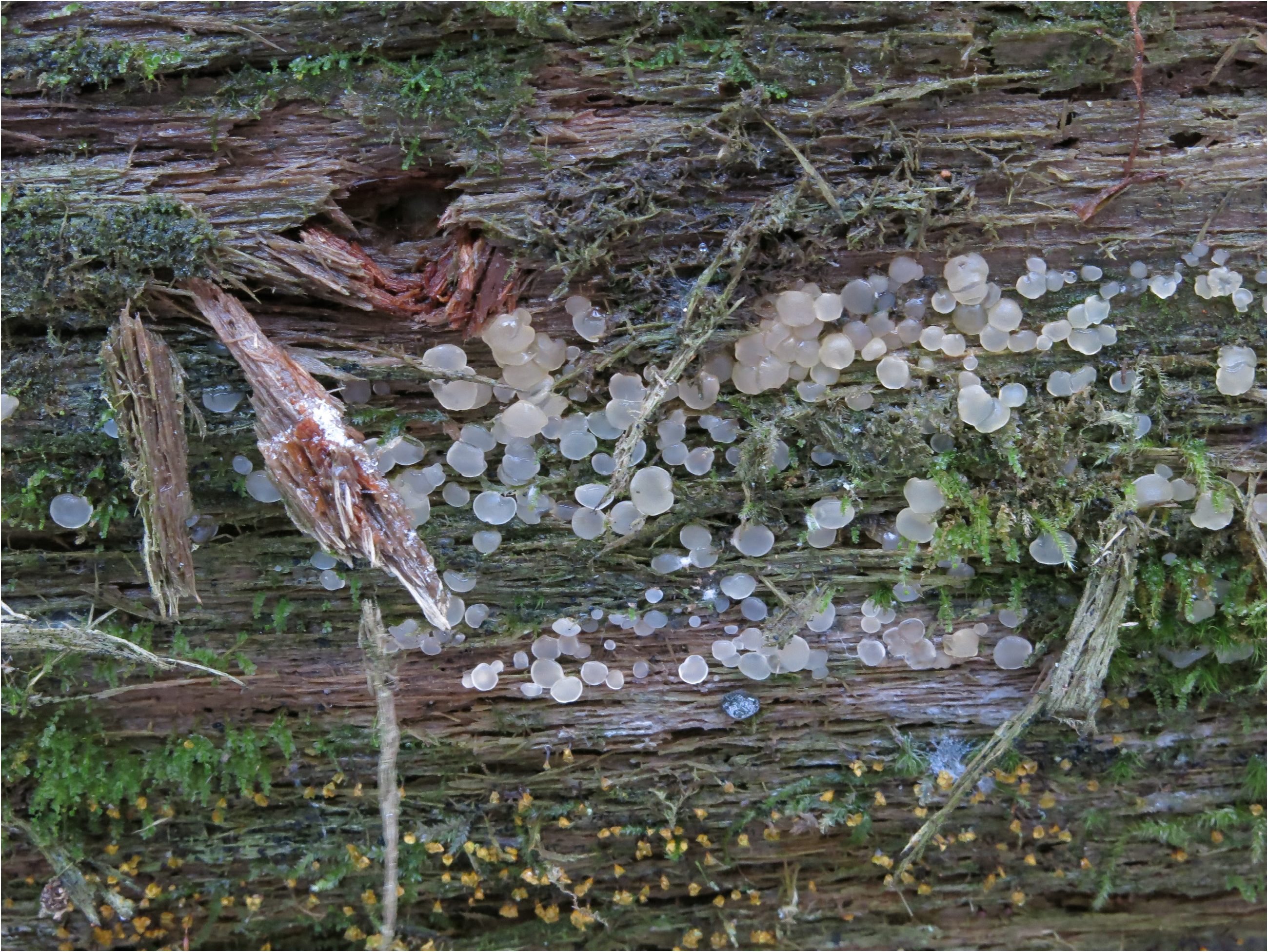
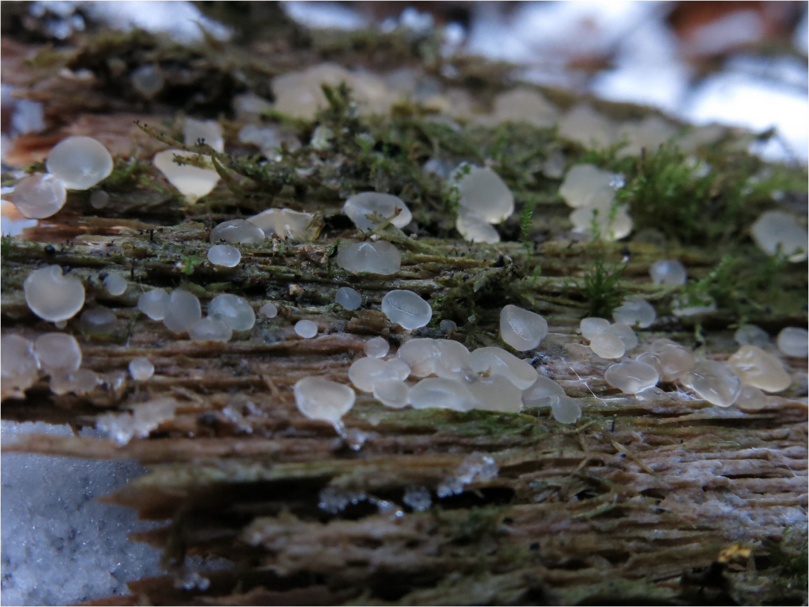
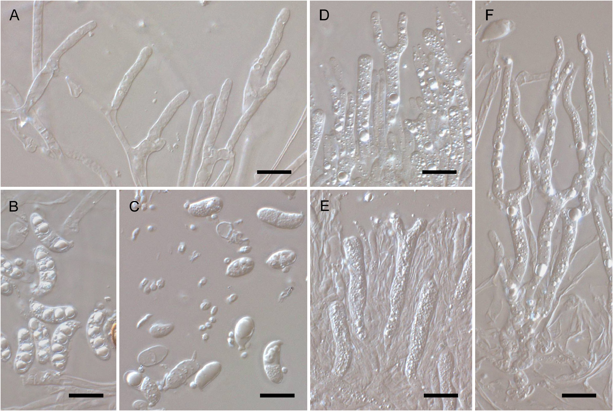
Basidiomata turbinate to ± discoid or cushion-shaped when hydrated, sessile to stalked, centrally attached to the substrate by a rooting cord that may not be very conspicuous, ca. 1–4.5 mm in diameter and 1–3 mm high (estimates derived from re-hydrated material), whitish to pale yellow when fresh, strongly gelatinous, with a shiny, slightly roughened, well-delimited cortex, and a pruinose hymenium, collapsing onto the substrate when dried as a thin, brittle, pale yellowish to orangish layer. Cortical and marginal hyphae thin-walled, clamped, with ± cylindrical terminal cells, 2.5–4 µm in diameter, simple to sparsely branched. Internal hyphae mostly thin-walled, with smooth walls and clamped, but some hyphae becoming thick-walled towards the base. True hyphidia probably absent, only some thin-walled, unbranched hyphidia-like cells observed among the basidia, probably representing young basidial growths. Basidia cylindric to slightly attenuated towards the base, thin-walled and clamped, becoming bifurcate by the development of two sterigmata. Mature basidial body 31–45 × 4–5(5.5) µm, sterigmata 18–55 × 2.5–3.5 µm. Basidium apex frequently U-shaped, without or with a barely developed apical protuberance. Basidiospores narrowly ellipsoid-allantoid to cylindric-allantoid, (10.5)11–16 × 5–6.2 µm, thin-walled, inamyloid, becoming 0–3 septate at maturity. Microconidia ovoid, ca. 2–2.5 × 1.5–2 µm. Cell cytoplasm frequently showing very conspicuous and strongly refractive lipid drops, especially in basidia and basidiospores, which are hyaline or have a yellowish hue.

No known copyright restrictions apply. See Agosti, D., Egloff, W., 2009. Taxonomic information exchange and copyright: the Plazi approach. BMC Research Notes 2009, 2:53 for further explanation.