
Prosopocoilus yangi Fukinuki, 2004
|
publication ID |
https://doi.org/10.5281/zenodo.201939 |
|
DOI |
https://doi.org/10.5281/zenodo.6194476 |
|
persistent identifier |
https://treatment.plazi.org/id/A41687E4-FFED-C957-5FCB-5726FCCF734B |
|
treatment provided by |
Plazi |
|
scientific name |
Prosopocoilus yangi Fukinuki, 2004 |
| status |
|
Prosopocoilus yangi Fukinuki, 2004 View in CoL
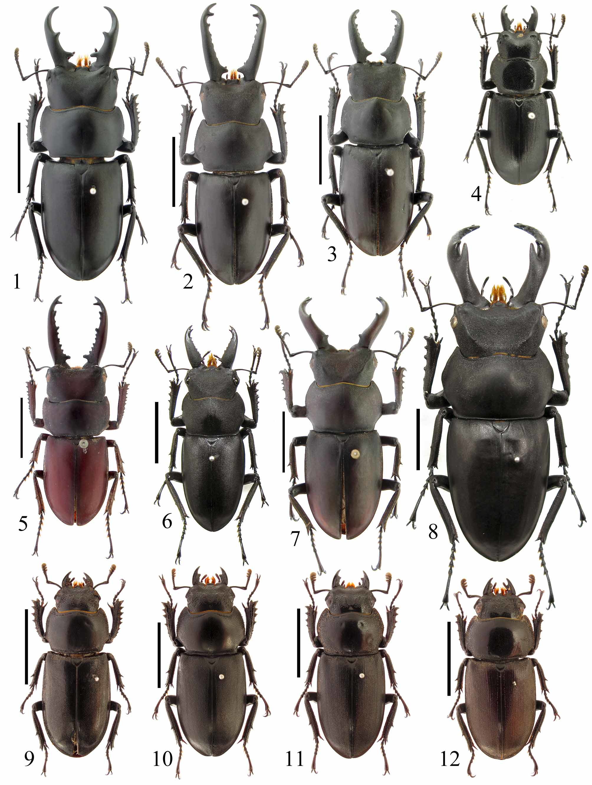
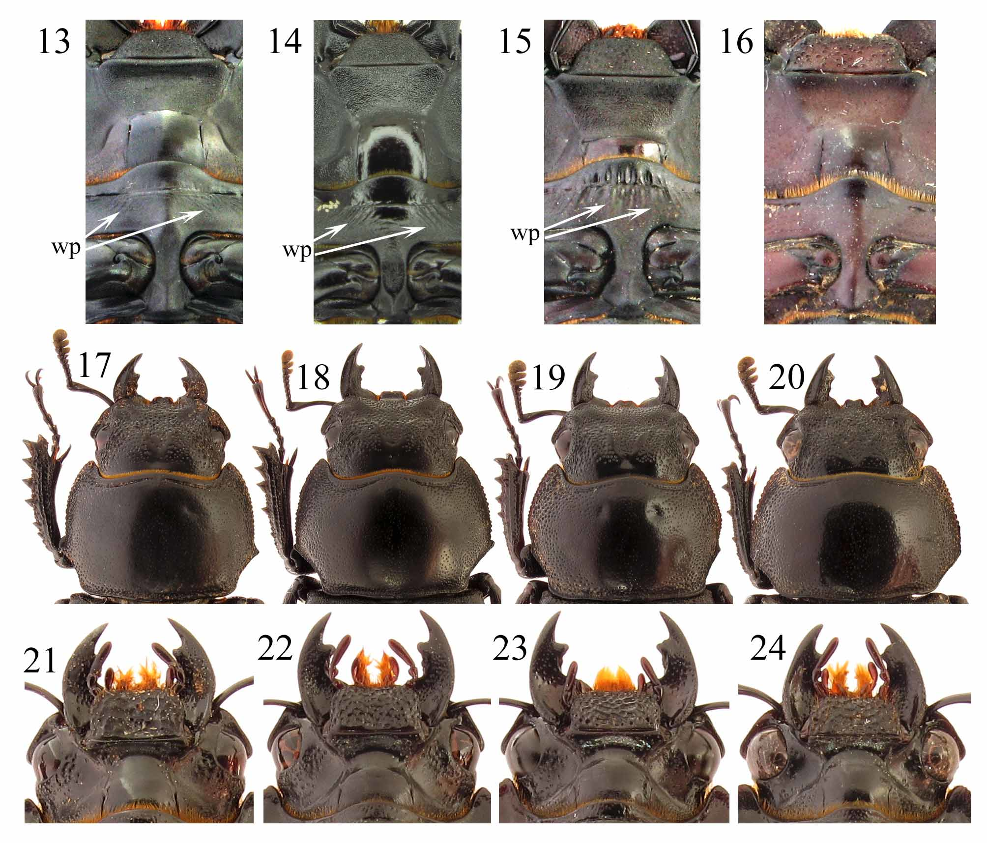
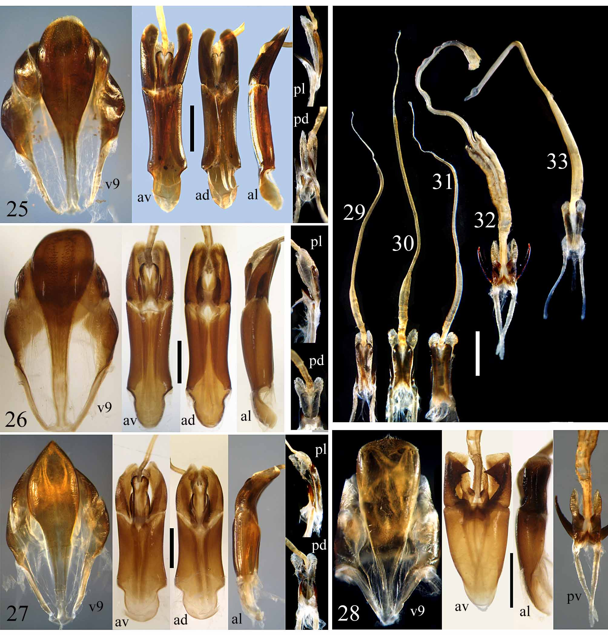
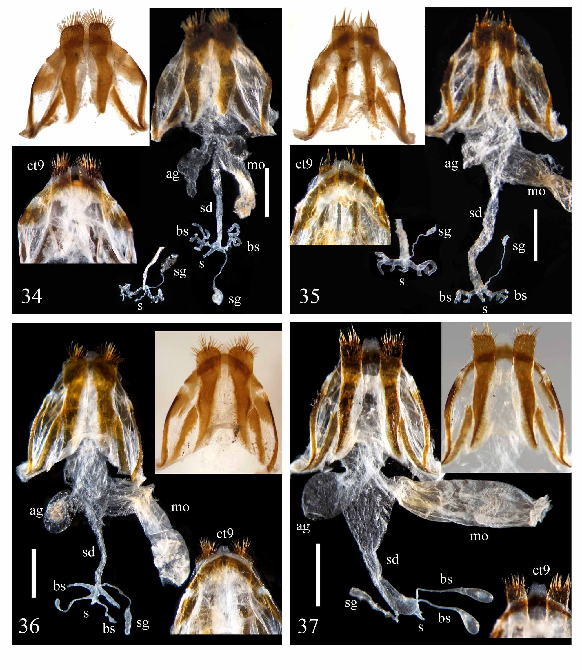
( Figs. 1, 9 View FIGURES 1 – 12 , 13, 17 View FIGURES 17 – 20 , 21, 25, 29 View FIGURES 29 – 33 , 34 View FIGURES 34 – 37 )
Prosopocoilus yangi Fukinuki, 2004: 31 View in CoL , Fig. 7 View FIGURES 1 – 12 (male holotype), “Junbon shan, Leanshin county, Jangxi ( sic) prov”
Material examined. CHINA: Chongqing: 42 3, 34 ƤƤ, Nanchuan County, Mt. Jinfoshan, 1000-1400 m, several larvae collected by He-Li Deng, Xiao-Dong Yang and Li-Jiang Wang from field in VI.2008, reared by J. Hao, emerging into adults in III.2009; most specimens reared in 2009, emerging into adults in III–IV.2010. All specimens deposited in Chang-Chin Chen’s collection ( Tianjin, China) except 13, 1Ƥ will be deposited in the Natural History Museum, London and 13, 1Ƥ will be deposited in Luca Bartolozzi’s collection (Firenze, Italy).
Description of additional males ( Figs. 1 View FIGURES 1 – 12 , 13). Body length measured from apex of mandible to terminal tip of elytra: 29–40 mm.
Color and pubescence: Both dorsal and ventral surfaces of the entire body black, opaque and glabrous.
Head finely microsculptured and smooth on dorsal surface, about 1.7 times as wide as long. Vertex depressed gradually in a triangular area defined by the anterolateral angles and the posterior end of the head. Frontal margin wave shaped, protruding and rounded medially as an intermandibular projection ( sensu Holloway 2007). Labrum defined posteriorly by a transverse labral suture, usually divided by a central split along the midline, with each lobe about 1.5–2.0 times as wide as long, and with the conjoined anterior margin of the lobes straight or convex; central split varying in length and sometimes absent in males. Canthus occupying half of the outer margin of the eye. Preocular margin straight. Anterolateral angle of the head a sharp obtuse angle. Postocular margin slightly convex. Mentum punctate, trapezoidal, with anterolateral angles rounded. Submentum clearly defined and microsculptured. Gula smooth. Maxilla with the tip of the lacinia not hooked. Labium with the ligula deeply bifurcate and setose. General shape of the mandible not different between the large-sized specimens and the small-sized specimens; mandible about 1.3–1.5 times as long as the head, straight from the base to the anterior 1/4 point and gently incurved at apex, with a longer subbasal inner tooth at the basal 1/5 or 1/6 point, and with a shorter median inner tooth in the middle or just beyond the middle; no subapical tooth; no tooth or denticle appeared between the median and subbasal teeth; the inner margin between the apex and the median tooth smooth in most specimens, but with one or two small denticles in a few specimens as in holotype. Antennal club with three pubescent antennomeres; antennomere 7 with width slightly greater than that of antennomere 6 and sharply pointed at tip, not lamellate as antennomeres 8–10.
Pronotum micropunctate and smooth on the surface, slightly longer and wider than head, widest at about the anterior 1/3 point; lateral margin evenly rounded at the anterior 2/3, abruptly pointed into a sharp lateral angle at the posterior 1/4 point, and concave behind the lateral angle; posterior angle rounded.
Elytra micropunctate and smooth on the surface, with no depression or large punctures, 1.7–1.8 times as long as wide, and almost as wide as the pronotum.
Legs: Protibia with 4–8 ( 4 in left protibia and 5 in right protibia of holotype) distinct teeth along the lateral margin. Mesotibia with a distinct lateral spine. Metatibia with 0–1 lateral spine.
Male genitalia ( Figs. 25, 29 View FIGURES 29 – 33 ): Ventral plate of the 9th abdominal segment with the basal part almost parallel sided, and without a longitudinal membranous stripe along the midline of the posterior expansion. Aedeagus in dorsal view about 5 times as long as wide. Basal piece in dorsal view rather oblong and elongate, nearly twice as long as parameres, with dorsal plates well marked; caudal margin of the ventral surface flat and not protruding medially, with the caudal margin of the sclerotized portion slightly concave. Paramere triangular and sharply pointed at apex in lateral view, lamellate and rounded at apex in dorsal or ventral view, without a basal process on the ventral surface. Penis (not counting the basal struts and the caudal membranous pouches) elongate, about 2/3 times as long as paramere, membranous along the middle and sclerotized along the lateral margins ventrally; membranous dorsal surface forming a pair of caudal pouches but not forming any dorsal pouch when being fully inflated; pair of processes on the dorsal end of the cross bar about half as long as penis; flagellum (permanently everted internal sac) a little longer than aedeagus, broader and belt-like at basal 1/4, narrower and belt-like from the basal 1/4 point to the apical 1/5 point, and thread-like at the apical 1/5.
Description of females ( Figs. 9 View FIGURES 1 – 12 , 17 View FIGURES 17 – 20 , 21). Body length measured from apex of mandible to terminal tip of elytra: 23–26 mm.
Color and pubescence: Both dorsal and ventral surfaces of the entire body black, opaque and glabrous.
Head punctate on dorsal surface, punctures on the posterior surface markedly smaller than on the anterior surface, punctures around the eye denser and partly fused. Vertex with a pair of large and poorly-defined lateral bulges. Frontal margin concave with the intermandibular projection weakly marked. Labrum defined posteriorly by a transverse labral suture, about 4 times as wide as long and flat at tip. Canthus occupying 2/3 of the outer margin of the eye. Preocular margin nearly straight. Anterolateral angle of the head obtuse and poorly defined. Postocular margin slightly convex. Mentum densely punctate, rectangular, about 1.5 times as wide as long, and rounded at anterolateral angles. Submentum clearly defined and sparsely punctate. Gula smooth. Maxilla with the tip of the lacinia hooked. Labium with the ligula deeply bifurcate and setose. Mandible half as long as head, evenly incurved, and with a median inner tooth. Antennal club with three pubescent antennomeres; antennomere 7 with width slightly greater than that of the antennomere 6 and sharply pointed at tip, not lamellate as antennomeres 8–10.
Pronotum densely punctate on the surface; without central depression; about 1.6 times as wide as long, widest at the posterior 1/3 point forming a clear lateral angle; lateral margin minutely crenulate, weakly convex from the anterior angle to the lateral angle, concave at the posterior 1/3, and rounded at the posterior angle.
Elytra densely micropunctate and opaque on the surface except for the sutural area, sparsely punctate and weakly shiny on the sutural area, about 1.7 times as long as wide, and almost as wide as the pronotum.
Legs: Protibia strongly incurved, continuously serrate with 5–8 distinct teeth along the lateral margin; apex deeply bifurcate with the branches widely separated and pointed at tip. Mesotibia and metatibia straight, each with a distinct lateral spine.
Female genitalia ( Fig. 34 View FIGURES 34 – 37 ): Last abdominal tergite semicircular, with a broad membranous area along the midline. Last abdominal ventrite with a large membranous area at middle. Hemisternite with the inner apex protruding a little beyond the outer apex. Spermatheca membranous, with a pair of lateral branches near apex; each lateral branch coral-like and irregular. Spermathecal duct nearly parallel sided. Spermathecal gland and its duct nearly half as long as spermathecal duct.
Distribution. Jiangxi (Junboshan, the type locality), southern Chongqing (Mt. Jinfoshan).
Remarks. An examination of a large number of specimens shows that the shape of the male mandible does not vary with the size of the specimen. Therefore P. yangi is characterized by the following combination of male mandible characters: mandible about 1.4–1.6 times as long as head; subbasal tooth of the mandible clearly marked and longer than all other inner teeth; median inner tooth of the mandible situated nearly in the middle and a little shorter than the subbasal tooth; no tooth or denticle between the subbasal and median teeth of the mandible; no subapical tooth behind the apex of the mandible. Fukinuki (2004) was correct in regarding P. b o re l i (Boileau, 1904) from India as the most similar species to P. yangi , using the characters of mandibles and pronotum. In addition, P. passaloides (Hope, 1845) from Java is also a similar species, but can be distinguished by the different shape of the male pronotum and the different position of the major inner tooth of the male mandible. A comparison in male and female genitalia showed P. yangi to be quite similar to P. simianshanus and P. cyclommatoides . The male and female genitalia of P. b o re l i have not yet been examined.
No known copyright restrictions apply. See Agosti, D., Egloff, W., 2009. Taxonomic information exchange and copyright: the Plazi approach. BMC Research Notes 2009, 2:53 for further explanation.
|
Kingdom |
|
|
Phylum |
|
|
Class |
|
|
Order |
|
|
SuperFamily |
Scarabaeoidea |
|
Family |
|
|
Genus |
Prosopocoilus yangi Fukinuki, 2004
| Huang, Hao & Chen, Chang-Chin 2011 |
Prosopocoilus yangi
| Fukinuki 2004: 31 |