
Ascetoaxinus quatsinoensis, Oliver & Frey, 2014
|
publication ID |
https://doi.org/ 10.11646/zootaxa.3869.4.8 |
|
publication LSID |
lsid:zoobank.org:pub:EE91053B-A15A-4A1F-866E-D23BA7F79D58 |
|
DOI |
https://doi.org/10.5281/zenodo.5120034 |
|
persistent identifier |
https://treatment.plazi.org/id/A462AF16-B97E-4C10-CBBB-B0786018119B |
|
treatment provided by |
Felipe |
|
scientific name |
Ascetoaxinus quatsinoensis |
| status |
sp. nov. |
Ascetoaxinus quatsinoensis View in CoL sp. nov.
Figs. 2–6 View FIGURE 2 View FIGURES 3–4. 3 View FIGURE 5 View FIGURE 6
Type material. Holotype, single specimen, off Quatsino Sound , Vancouver Island, British Columbia, Canada, 50°15.482´N, 128°26.400´W to 50°14.519´N, 128°26.567´W, 1086–1318m. Coll. J. Boutillier, Fisheries and Oceans Canada, 02 September 2004. RBCM 010-00221 View Materials - 005 View Materials .
Measurements.
Description. Shell ( Fig. 2 View FIGURE 2 ). 31.3mm in length. Thin, brittle. Equivalve. Moderately tumid. Strongly inequilateral, prosogyrous beaks close to the anterior margin. Outline obliquely oval; anterior margin almost straight bounding a large excavated lunule; ventral margin long, almost straight; posterior margin broad, sulcate with a distinct posterior dorsal sinus. Posterior sulcus sharply defined, relatively narrow; submarginal sulcus sharply defining a flattened escutcheon. Posterior ventral slope anterior of the posterior sulcus a little flattened, creating a weak secondary ridge. Hinge teeth lacking, ligament partially sunken, relatively short being less than half the length of the escutcheon. Sculpture of well-defined growth lines; edge of lunule drawn out into five rounded projections. Muscle scars indistinct; anterior adductor scar elongate and parallel with ventral margin. Shell colour white.
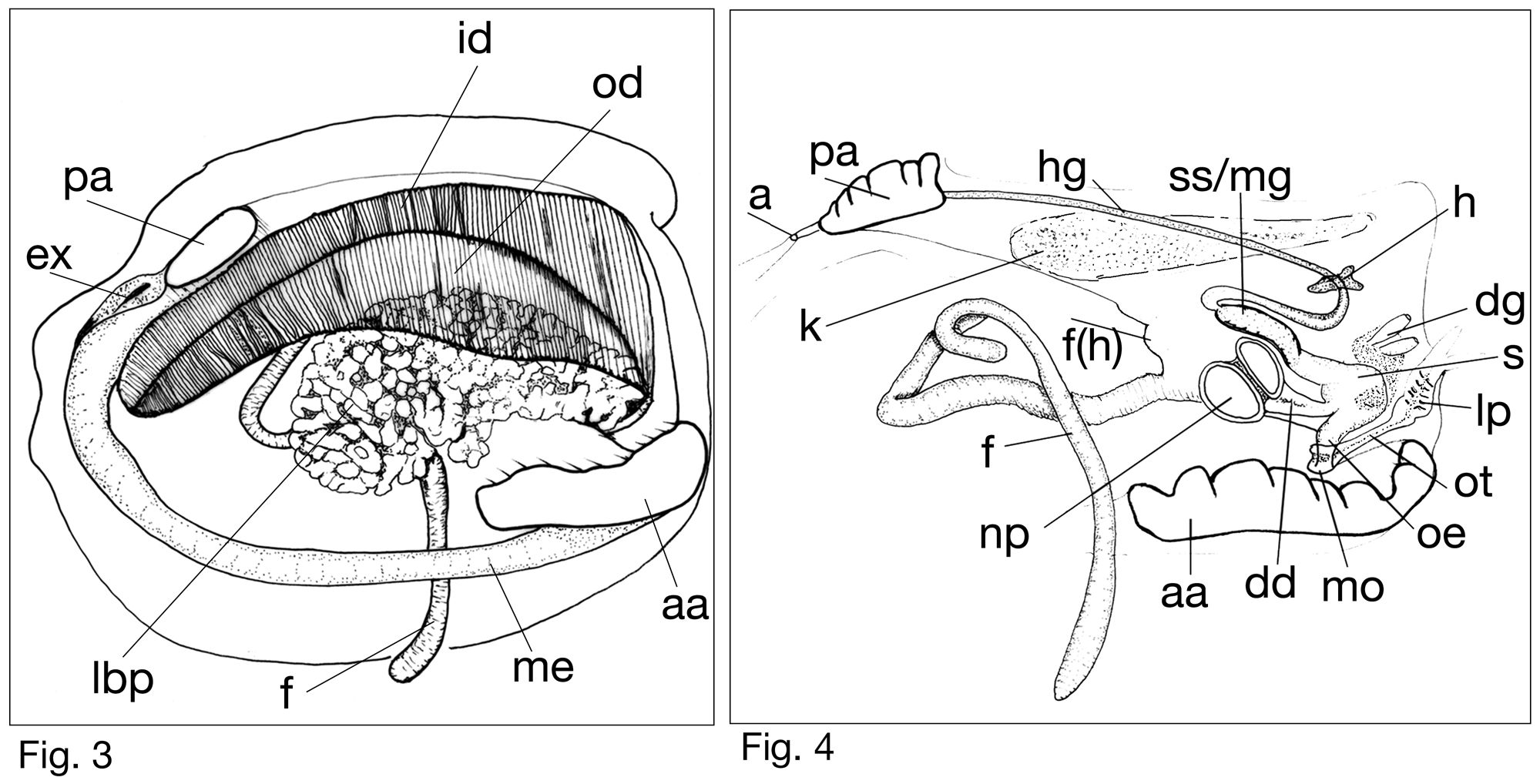
Anatomy ( Figs. 3–6 View FIGURES 3–4. 3 View FIGURE 5 View FIGURE 6 ). Mantle is thin; unfused except for the formation of a small exhalant aperture ( Fig. 3 View FIGURES 3–4. 3 , ex). Anterior adductor scar elongate and approximately 4 times longer than posterior adductor scar; free from mantle edge for about one-fifth of its ventral edge; posterior adductor muscle oval. Foot vermiform ( Fig. 3 View FIGURES 3–4. 3 ), very long, tip not noticeably expanded, heel obsolete, protractor muscles very slender. Labial palps small, narrow with a distinctly grooved dorsal zone and a long tubular portion leading to the mouth ( Fig. 5A, B View FIGURE 5 ). Alimentary system ( Fig. 4 View FIGURES 3–4. 3 ) with short oesophagus leading to a relatively small stomach; large digestive ducts leading to the lateral body pouches open into the ventral anterior face ( Fig. 5D View FIGURE 5 , dd, np), smaller ducts open dorsally on the left side and lead to a pair of outgrowths ( Fig. 5B View FIGURE 5 , dg); walls of the combined style sac and mid gut thickened ( Fig. 5D View FIGURE 5 , ss/mg), remainder of mid gut coiling back over style sac, passing through heart and coiling towards the posterior as the hind gut; anus opening into exhalant aperture ( Fig. 4 View FIGURES 3–4. 3 ). Lateral body pouches large, arborescent, terminations cloven or single, blunt ( Fig. 5C View FIGURE 5 ). Kidney large and packed with golden coloured granules. Ctenidium large, both demibranchs with fully reflected filaments; outer demibranch about half the depth of the inner demibranch ( Fig. 3 View FIGURES 3–4. 3 , od, id). Filaments laminar ( Fig. 6A, C View FIGURE 6 ), frontal zone narrow, abfrontal zone extended and fused across the ascending and descending arms, creating junctions between the ascending and descending lamellae ( Fig. 6A View FIGURE 6 , abs). Ventrally every filament is fused. The frontal surface ( Fig. 6B View FIGURE 6 ) is ciliated with distinct lateral cilia (lc) and laterofrontal cirri (lfc); frontal cilia not apparent, instead adjacent frontal zone appears wide and smooth (sfz). Abfrontal surfaces lined with bacteriocytes that are domed and roundly polygonal with a glycocalyx ( Fig. 6C View FIGURE 6 , bc). Bacilli bacteria also present in defined bundles, measuring 1.1 um in length ( Fig. 6D View FIGURE 6 , bct).


No known copyright restrictions apply. See Agosti, D., Egloff, W., 2009. Taxonomic information exchange and copyright: the Plazi approach. BMC Research Notes 2009, 2:53 for further explanation.
|
Kingdom |
|
|
Phylum |
|
|
Class |
|
|
SubClass |
Heterodonta |
|
Order |
|
|
SuperFamily |
Thyasiroidea |
|
Family |
|
|
Genus |