
Buenia lombartei Kovačić, Ordines & Schliewen, 2018
|
publication ID |
https://doi.org/ 10.11646/zootaxa.4392.2.3 |
|
publication LSID |
lsid:zoobank.org:pub:DB0BAC2A-70C6-4C81-8340-4C054A379B7D |
|
DOI |
https://doi.org/10.5281/zenodo.5960626 |
|
persistent identifier |
https://treatment.plazi.org/id/EAEB514C-842C-4223-95BD-9CE7ED2C3B94 |
|
taxon LSID |
lsid:zoobank.org:act:EAEB514C-842C-4223-95BD-9CE7ED2C3B94 |
|
treatment provided by |
Plazi |
|
scientific name |
Buenia lombartei Kovačić, Ordines & Schliewen |
| status |
sp. nov. |
Buenia lombartei Kovačić, Ordines & Schliewen , sp. nov.
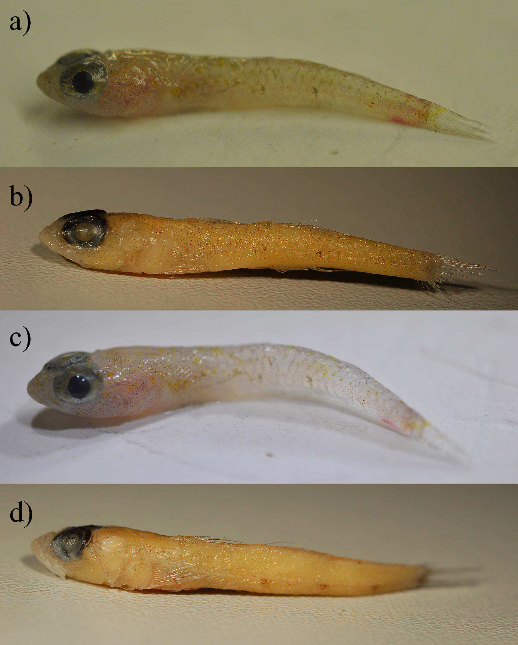
Holotype. PMR VP4108 View Materials , adult male, 27.5 mm SL, caudal fin damaged, Spain, Balearic Islands, Migjorn Mallorca ( Badia ), 39.297ºN, 2.4128ºE, mud, 343 m depth, coll. Ordines, Lombarte & Kovačić, IEO-COB, 19 June 2016, beam trawl ( Figs. 2a, 2b View FIGURE 2 and 3b View FIGURE 3 ). GoogleMaps
Paratype. ZSM 46818 adult female, 26.0 mm SL, caudal fin damaged, Spain, Balearic Islands, Ponent Mallorca ( Dragonera ), 39.7465ºN, 2.3138ºE, mud, 375 m depth, coll. Ordines, Lombarte & Kovačić, IEO-COB, 18 June 2016, bottom trawl ( Figs. 2c, 2d View FIGURE 2 and 3a View FIGURE 3 ). GoogleMaps
Diagnosis. (1) anterior oculoscapular canal semi-closed with pores σ, λ, Κ, α, ρ and additional pores and open furrows; (2) suborbital row c of 6 papillae; (3) A I/7–I/8; (4) LL 25–27; (5) TR 6; (6) P 18; (7) D 1 II, the longest spine of D1, backwards reaching to D2 middle in males when folded down, in females not reaching D 2 I; (8) V anterior membrane reduced to less than 1/6 of spinous ray in midline depth; (9) tongue well developed, bilobed; (10) head length 33.1–35.0 of SL; (11) eye 32.9–33.5% of head length; (12) anal fin base 11.7–12.2% of SL; (13) pelvic to anus 19.4–20.4% of SL; (14) snout 72.1–75.0% of eye diameter; (15) pectoral fin 20.2% of SL; (16) cheek depth 15.4–15.7% of head length; (17) colouration of fresh material reduced to rarely scattered yellow dots and miniature melanophores densely distributed on head and body, only forming three small marks visible at lateral midline; (18) colouration of preserved material reduced to scattered miniature melanophores on head and body, forming only three small marks visible at lateral midline.
Description (all morphometric values and meristics in the text are presented as holotype first and paratype in parentheses). General morphology ( Fig. 2 View FIGURE 2 ). Body proportions are given in Table I. Body moderately elongate, laterally compressed towards caudal fin. Head large, 33.1 (35.0) % of SL, moderately depressed and narrow, its greatest width being 55.5 (51.6) % of its length, with a nearly horizontal predorsal profile. Snout short, just 72.1 (75.0) % of eye length, dorsal profile of snout gently sloping. Eyes large, dorsolateral, extending above dorsal profile in fresh material, 33.5 (32.9) % of the length of the head, size even more relatively large to body length than to head, 11.1 (11.5) % of SL. Eyes very close to each other, interorbital width just 9.8 (8.3) % of eye diameter. Anterior nostril short, tubular, erect, without process from rim; posterior nostril pore-like, near orbit, with erected rim. Mouth oblique, lower jaw projecting beyond the upper, posterior angle of jaws below anterior half of pupil. Rows of teeth in lower jaw with one row of large outer teeth, one intermediate row of smaller teeth and inner row with teeth larger than in intermediate row. Rows of teeth in upper jaw with one row of large outer teeth, one intermediate row of smaller teeth and inner row with teeth larger than in intermediate row. Teeth in outer and inner rows pointed and poorly curved towards the buccal cavity in both jaws. Tongue well developed, bilobed. Branchiostegal membrane attached to entire lateral margin of isthmus from immediately anterior to near pectoral fin base.
Fins. D 1 VI (VI); D2 I/9 (I/9); A I/7 (I/8); C damaged to count branched rays, 16 (16) segmented rays; P 18 (18), V I/5 + 5/I (I/5 + 5/I). Fin-bases and lengths in proportion to standard body length are given in Table I. D 1 II the longest, D 1 II with elongate filament in male, backwards reaching to half of D2 base when folded down, in female not reaching D 2 I. Fin membrane of D 1 VI not connected with base of D 2 I. D2 commences at vertical of urogenital papilla, with last ray over or slightly behind vertical of last A ray. A commences below first to second segmented ray of D2. C rounded. P uppermost rays within membrane. P rays all branched except uppermost and lowermost rays unbranched, ending back before D 2 I. V complete and rounded, reaching behind anus in male, not in female. V anterior membrane reduced to less than 1/6 of spinous ray in midline depth.
Squamation. Scales lost on both specimens, counts made from stained scale pouches. Predorsal area, including nape and along D1 base to D 1 VI, opercle and cheek naked. Breast scaled with cycloid scales, scales along ventral midline 3–4. Scales in lateral series 25–27, left and right side 25/26 (27/26), in transverse series 6 (6).
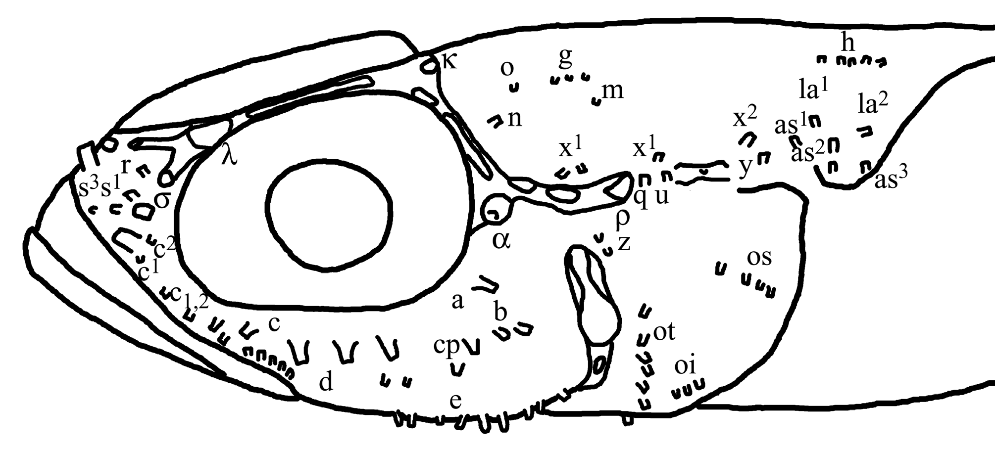
Lateral line system ( Fig. 4 View FIGURE 4 ). Head with anterior oculoscapular canal semi-closed with pores σ, λ, Κ, α, ρ and additional pores and open furrows ( Fig. 3c View FIGURE 3 ). Posterior oculoscapular canal with pores ρ 1 and ρ 2 or as open furrow. Preopercular canals with pores γ, δ, ε or as open furrow. Sensory papillae of the head lateral line system large, elongated and flattened and reduced in number in all rows ( Fig. 3d View FIGURE 3 ). Rows with number of sensory papillae in parentheses counted on left side of both specimens: (1) preorbital: snout with three rows in median preorbital series, superior row r close to pore σ as single papilla, inferior row s with two sections, s 1 (2) close to nostrils, s 3 (1) above upper lip. Lateral series c in three parts: superior c 2 (1-2) between anterior and posterior nostrils, middle c 1 (1 larger papillae) behind and below anterior nostril and inferior c 1,2 (2 larger papillae) above upper lip. (2) suborbital: rows a and c, including cp, without transverse proliferation; a longitudinal (1–2 larger papillae) below rear part of eye; c (6 larger papillae) longitudinal below frontal and middle part of eye starting anteriorly with two papillae above below each other at vertical of front border of eye, posteriorly ending before row a anteriorly starts and distant from row b beginning. Two cp larger papillae below a row, lower cp’ anterior to upper cp. Longitudinal row b short (2–3), anteriorly beginning below rear border of eye. Longitudinal row d distantly separated in supralabial (5–7) and horizontal part back on cheek (2–3). (3) preoperculo-mandibular: external row e and internal row i divided into anterior (e: 8–15, i: 8–11), and posterior sections (e: 8–13, i: 8–10); row f (1). (4) oculoscapular: anterior longitudinal row x 1 divided in anterior section (2–4) vertical to pore γ and posterior section (1) above between pore ρ and posterior oculoscapular canal, not visible in paratype, posterior longitudinal row x 2 (1–2) above row y; row z (2) behind pore γ, rows q (1) and u (1) larger papillae behind pore ρ, row y single papilla above posterior edge of opercle. Axillary rows as 1 (1–3), as 2 (2–3), as 3 (2–3), la 1 (1) and la 2 (1) present. (5) opercular: transverse row ot (8); superior longitudinal row os (4); inferior longitudinal row oi (3). Rows ot, os, oi not visible in paratype due to the damaged surface. (6) anterior dorsal: rows n and o as single papillae, longitudinal row g (3), row m (1) present and row h continuous (5-9). Rows n, o, g, and m not visible in paratype due to the damaged surface.
Colour of freshly collected material ( Figs. 2a and 2c View FIGURE 2 ). No distinct sexual dimorphism evident except for colouration of P and V and head underside. Head and body yellowish white to dusky white with xanthophores forming a few scattered yellow dots and miniature melanophores more densely distributed. Melanophores widely scattered on head, upper body, but also on ventrally on head and along A fin. Only prepelvic area and belly with no pigments. Three small marks visible along lateral midline, two below D2, one on caudal peduncle. Melanophores follows to some degree the scale margins on body. No other marks, stripes or saddles or any other pattern formed by melanophores present on body or head. Yellow pigments present on upper part of body with more dots anteriorly than posteriorly. Reddish colour from gills visible through gill cover as well as silvery peritoneum visible below skin on belly and grey vertebral column silhouette along the posterior part of body. Caudal peduncle with two reddish dispersed marks on upper and lower edge. Head with uniformly scattered miniature melanophores on predorsal area, snout, cheeks, opercle. Head underside more strongly pigmented in male than in female. Eyes green golden to brass, with dark green to black pupil. Yellow dots on head present on predorsal area, snout and upper opercle. Fins mostly transparent with more melanophores on D1 and D2, and less on C and A. P and V pigmented in male, female without melanophores on these fins. D1 and D2, P and C with yellow dots at base, V and A without.
Colour of preserved specimens in alcohol ( Figs. 2b and 2d View FIGURE 2 ) very pale yellow to white brown (buff to tan), with dark brown pattern similar to freshly collected specimens. No distinct sexual dimorphism evident except for P and V and head underside. Melanophores widely scattered on head, upper body, but also on ventrally on head and along A fin. Only prepelvic area and belly with no pigments. Three small marks still visible at lateral midline, on upper body pigments follows to some degree the scale margins. No other marks, stripes or saddles or any other pattern formed by melanophores present on body or head. Eyes black, with green reflective pupil. Head with uniformly scattered miniature melanophores on predorsal area, snout, cheeks, opercle. Head underside more pigmented in male than in female. D1, D2, C and A mostly transparent with a few melanophores. P and V pigmented in male, female without melanophores on these fins.
Vertebral column and pterygiophore insertion pattern (pty) based on paratype ZSM 46818): 12 precaudal and 18 caudal vertebrae, including urostyle; total count 30. One epural; pty 3- 122100; two prehaemal pterygiophores. Total number of caudal fin rays inserting in the hypurals 5, 3 + 4 (fused), hypural 2 + 3 (fused) and hypural 1 (parhypural): 16; fused hypural 1 + 2 and 3 + 4 separated by a large gap, which is inserted by one branched caudal ray (compare with Fig. 5 View FIGURE 5 ).
Etymology. The species is named lombartei after Dr. Antoni Lombarte, researcher of the Institut de Ciències del Mar-CSIC, Barcelona, who found the first specimen of the new species during the MEDITS_ES05_16 fieldwork, the paratype, among trawl debris despite its small size.
Ecological and geographical distribution. Both specimens of B. lombartei sp. nov. were collected from sampling stations located relatively close to each other on the upper slope off the west coast off Mallorca (geographical positions included in holotype and paratype specifications above). At these stations, at 343 and 375 m, the temperature ranged between 13.43 and 13.44 ºC, the salinity ranged between 38.53 and 38.55 ‰. Sediments collected in the respective beam trawl samples in each sampling station only recovered bathyal mud with very few individuals of epibenthic species, most belonging to the decapod crustacean genus Munida (mainly Munida intermedia ). The bottom trawl collected a higher diversity of species, including epibenthic species, such as the decapod crustaceans Munida intermedia , Nephrops norvegicus and Macropipus tuberculatus , and nektobenthic species such as the decapod crusteceans Plesionika heterocarpus and Parapennaeus longirostris , the chondrichthyan fishes Scyliorhinus canicula and Galeus melastomus and the osteichthian fishes Chlorophthalmus agassizi , Gadiculus argenteus , Coelorinchus caelorhincus , Lepidotrigla dieuzeidei , Helicolenus dactylopterus and Lepidorhombus boscii . Both sampling stations were located in the Balearic Islands upper slope bottom trawl fishery area, targeting at those depths P. longirostris and N. norvegicus . In the Balearic Islands the upper slope is the bathymetric stratum supporting a comparatively low fishing effort, which in the archipelago is mainly directed to the middle slope, targeting the decapod crustacean Aristeus antennatus , and in the continental shelf the fishes Mullus surmuletus and Merluccius merluccius among a large variety of other species including also cephalopods ( Ordines et al. 2014, Quetglas et al. 2012).
Remarks. B. lombartei sp. nov. can be clearly distinguished from B. jeffreysii by 1) morphology, 2) morphometrics, 3) size, colouration of 4) fresh and 5) preserved material, 6) geographic distribution and by 7) depth range, 8) bottom composition and the (9) COI haplotype.
1) Tongue well developed, bilobed vs. tongue reduced to short flap free from the floor of the mouth in B. jeffreysii . A I/7 (I/8) and C 16 (16) segmented rays vs. A I/9 and C 15 segmented rays in B. jeffreysii . D 1 II with elongate filament in males, backwards reaching to half of D2 base when folded down vs. backwards reaching to D 2 I when folded down in B. jeffreysii . P uppermost ray unbranched vs. in P uppermost ray branched in B. jeffreysii . V anterior membrane reduced to less than 1/6 of spinous ray in midline depth vs. V anterior membrane about 2/5 of spinous ray in midline depth in B. jeffreysii . Scales in lateral series 25–27 vs. 28–30 in studied B. jeffreysii . Scales in transverse series 6 (6) vs. 7 in B. jeffreysii . Head with anterior oculoscapular canal semi-closed with pores σ, λ, Κ, α, ρ and additional pores and open furrows vs. anterior oculoscapular canal normally closed with pores σ, λ, Κ,α, ρ and no additional pores and open furrows in B. jeffreysii . Sensory papillae of the head lateral line system elongated and flattened vs. semispheric, bubble shape in B. jeffreysii . Sensory papillae row r as single papilla, vs. r with two papillae in B. jeffreysii . Sensory papillae row s 1 with two papillae vs. s 1 as single papilla in B. jeffreysii . Sensory papillae row c with 6 papillae vs. 7-8 papillae in B. jeffreysii . Sensory papillae row z with 2 papillae vs. 3–4 papillae in B. jeffreysii . Sensory papillae row ot with 8 papillae vs. 18–20 papillae in B. jeffreysii . Sensory papillae row os with 4 papillae vs. 8–12 papillae in B. jeffreysii . Sensory papillae row oi with 3 papillae vs. 4–6 papillae in B. jeffreysii .
2) B. lombartei sp. nov. with head large, 33.1 (35.0) % of SL and narrow, its greatest width being 55.5 (51.6) % of its length vs. head length 28.5–31.0 % of SL and head width 58.5-69.4 of its length in B. jeffreysii . B. lombartei sp. nov. with snout short, just 72.1 (75.0) % of eye length vs. 78.9–94.7% in B. jeffreysii . B. lombartei sp. nov. with eyes large, 33.5 (32.9) % of the length of the head and 11.1 (11.5) % of SL vs. 28.7-30.6% and 8.2–9.1% in B. jeffreysii . B. lombartei sp. nov. with interorbital width just 9.8 (8.3) % of eye diameter vs. 20.9-26.5% in B. jeffreysii . B. lombartei sp. nov. with cheek depth narrow, 15.7 (15.4) % of the length of the head vs. 17.4–20.7% in B. jeffreysii . B. lombartei sp. nov. with anal fin base short 12.2 (11.7) % of SL vs. 14.0–16.8% in B. jeffreysii . B. lombartei sp. nov. with pectoral fin short 20.2 (20.2) % of SL vs. 22.0–23.0% in B. jeffreysii . B. lombartei sp. nov. with pelvic to anus distance short 20.4 (19.4) % of SL vs. 22.1–24.1% in B. jeffreysii .
3) Size of the B. lombartei sp. nov. adults was 26.5 and 27.0 mm SL vs. 34.2–50.0 mm SL in B. jeffreysii .
4) Colouration of fresh material of B. lombartei sp. nov. contains a few scattered yellow dots and more densely distributed miniature melanophores on head and body, forming only three small marks visible at lateral midline and to some degree the scale margins on body vs. brown and orange colouration of B. jeffreysii broad and intensive, with recognizable orange-brown marbled pattern on upper head and body and five large dark brown blotches along lateral midline.
5) Colouration of preserved material of B. lombartei sp. nov. contains scattered miniature melanophores on head and body, forming only three small marks visible at lateral midline and to some degree the scale margins on body, D1, D2, C and A mostly transparent with a few melanophores vs. body and head with dark brown markings, upper part of body with intensive brown pattern, and five large dark brown marks distributed along lateral midline, D1 and D2 with oblique rows of spots, C and A densely pigmented in B. jeffreysii .
6) B. lombartei sp. nov. has been collected in the warm temperate marine zone of the Mediterranean Sea province while B. jeffreysii is restricted to the cold temperate water zone of North European seas province (Spalding et al. 2007).
7) B. lombartei sp. nov. was collected only at depth of 343 m by beam trawl and at depth of 375 m by trawl, suggesting a very narrow depth range, because extensive beam trawls had been performed between 52 m to 375 m depth and trawl between 48 m and 746 m during the MEDITS_ES05_16 survey in June 2016 on board the R/V Miguel Oliver. Contrary to that, B. jeffreysii occurs from 5 to 330 m depth ( Miller 1986). Therefore, while the depth zone of B. lombartei sp. nov. appears strictly bathyal, B. jeffreysii is mainly littoral species, present in circalittoral and infralittoral zones.
8) B. lombartei sp. nov. was collected on pure mud bottom, contrary to B. jeffreysii that is present on various bottoms (sand, shell debris, gravels, corallines) that could be muddy, i.e. mixed with mud ( Luisy 2015), but not on the pure mud sediment.
9) see below under Genetics and phylogeny
B. lombartei sp. nov. is most easily distinguished from B. massutii by posterior oculoscapular canal present at least as open furrow vs. posterior oculoscapular canal absent; sensory papillae row c with 6 papillae vs. 5 papillae, P 18 vs. 16; D 1 II in males elongate reaching half of D2 base vs. D 1 II in males elongate reaching to posterior end of D2 base; snout short, 72.1–75.0% of eye diameter vs. 78.3-93.8%; pectoral fin short, 20.2% of SL vs. 22.6– 26.5%; body with only three small marks visible at lateral midline and no dark pigment between caudal peduncle and fin vs. body with four vertical midline stripes visible at lateral midline and fifth longer vertical mark as margin between caudal peduncle and fin.
The new species could be also clearly distinguished from B. affinis by sensory papillae row c with 6 papillae vs. 5 papillae, P 18 vs. 15–16; D 1 in males elongate with spine II reaching half of D2 base vs. D 1 II in males not elongate; V anterior membrane reduced to less than 1/6 of spinous ray in midline depth vs. V anterior membrane about 1/3–1/2 of spinous ray in midline depth; head large with length 33.1–35.0% of SL vs. 27.5–30.9%; eyes large with eye diameter 32.8–35.7% of head length vs. 23.3–30.3%.


No known copyright restrictions apply. See Agosti, D., Egloff, W., 2009. Taxonomic information exchange and copyright: the Plazi approach. BMC Research Notes 2009, 2:53 for further explanation.
|
Kingdom |
|
|
Phylum |
|
|
Class |
|
|
Order |
|
|
Family |
|
|
Genus |