
Scaphoideus albovittatus Matsumura, 1914
|
publication ID |
https://doi.org/ 10.11646/zootaxa.3904.3.2 |
|
publication LSID |
lsid:zoobank.org:pub:EA840547-EE36-43E3-9FFE-90C055112BD6 |
|
DOI |
https://doi.org/10.5281/zenodo.3510356 |
|
persistent identifier |
https://treatment.plazi.org/id/A6352B3D-CB23-9538-378D-21A4FD3A5196 |
|
treatment provided by |
Plazi |
|
scientific name |
Scaphoideus albovittatus Matsumura, 1914 |
| status |
|
Scaphoideus albovittatus Matsumura, 1914 View in CoL
( Figs. 1 View FIGURE 1 A–D, F–I, K–N, 3A, 4A, 4E, 5A–H, 6A–D, 7A–P, 14A–C, 15A–C, 16A–C)
Scaphoideus albovittatus Matsumura, 1914:224 View in CoL ; Ishihara, 1961: 252; Okada, 1977: 192; Li & Wang, 1991: 188; Cai & Shen, 1999: 243; Cai & Shen, 2002: 276; Kamitani & Hayashi, 2013:515 View Cited Treatment .
Scaphoideus albivittatus View in CoL [sic], Li & Chen, 2002: 197; Li & Wang, 2006:184; Li et al., 2007: 160; Xing & Li, 2010: 137; Li et al., 2011: 209.
Body including forewings at rest moderately elongate. Head including eyes slightly narrower than pronotum. Crown prominently produced anteriorly, longer medially than next to eyes, medial length 1.4 times as long as width between eyes, narrowly rounded to face, disc smooth with shagreened patches ( Figs. 1 View FIGURE 1 A, 3A). Eyes fairly large ( Fig. 1 View FIGURE 1 F). Ocelli situated on anterior margin of crown and separate from eye by distance less than ocellar diameter. Frontoclypeus narrow, longer than width between eyes. Clypellus widened apically. Gena slightly incised below eye ( Fig. 1 View FIGURE 1 K). Pronotum slightly shorter than head, with anterior margin roundly produced and posterior margin slightly concave. Scutellum nearly equal to length of head, suture curved ( Figs. 1 View FIGURE 1 A, 3A). Forewing semitransparent, with appendix wide, outer anteapical cell acute apically; inner anteapical cell closed basally; two reflexed cross veins between outer anteapical cell and costal margin, without reflexed vein basad of outer anteapical cell ( Fig. 1 View FIGURE 1 F).
Body pale brown with whitish longitudinal stripe from tip of vertex to near apex of forewing ( Fig. 1 View FIGURE 1 A). Vertex with pair of small black spots near apex and pair of large irregular black spots near ocelli ( Figs. 1 View FIGURE 1 A, 3A); frontoclypeus pale yellow with a narrow blackish stripe along apex margin touching spot at apex of vertex and wide blackish arc across antennal pits; clypellus and lorum pale yellow; gena pale yellow with blackish marking below antenna ( Fig. 1 View FIGURE 1 K). Pronotum brown on lateral margin, with pairs of dark brown patches. Scutellum brown on lateral margin, with 2 small orange spots at center ( Figs. 1 View FIGURE 1 A, F, 3A). Forewing transparent with brown veins, darker at base and apex ( Fig. 1 View FIGURE 1 A, F).
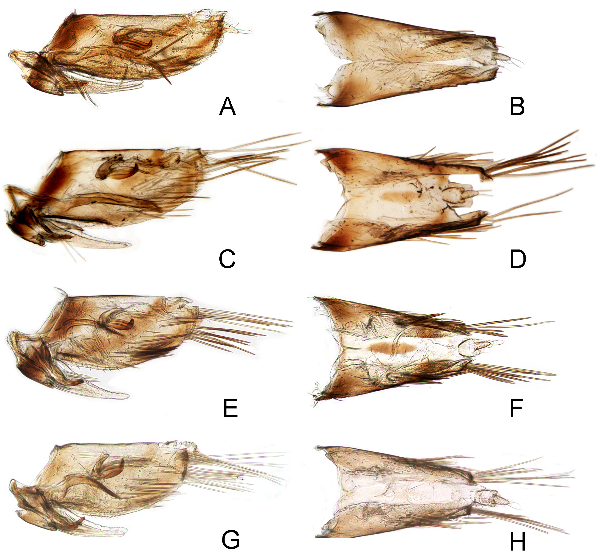
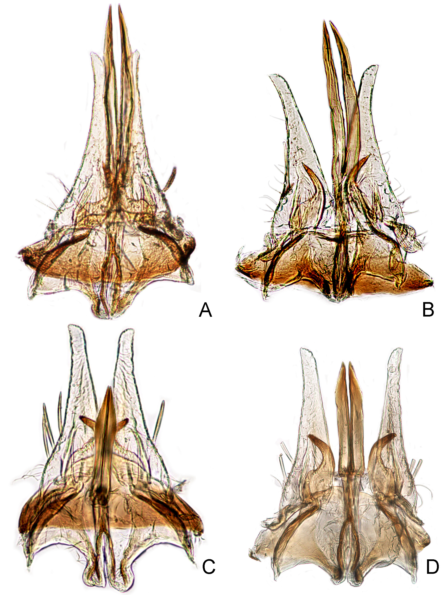
Male genitalia. Male pygofer very long and gradually tapered posteriorly, with row of very long macrosetae apically and scattered short macrosetae from subapex to middle ( Fig. 5A–H View FIGURE 5. A – H ). Subgenital plate short, extending 1/2 length of pygofer, distinctly widened basally, with uniseriate row of 2-3 macrosetae near base, and fine setae along lateral margin from base to middle ( Fig. 6A–D View FIGURE 6. A – D ). Style with broad anterior part and discernable preapical lobe; apophysis short, curved laterally ( Figs. 6A–D View FIGURE 6. A – D , 7 View FIGURE 7 B, F, J, N). Connective with anterior arms nearly parallel; posterior processes of uniform width for most of length, in distal 1/3 gradually tapered and pointed apically, in lateral aspect curved and strongly narrowed distally( Figs. 6A–D View FIGURE 6. A – D , 7 View FIGURE 7 A, E, I, M). Aedeagus robust; shaft longer than basal apodeme in lateral view, narrowed distally and curved dorsally at apex; gonopore subapical on ventral surface ( Fig. 7 View FIGURE 7 C, D, G, H, K, L, O, P).
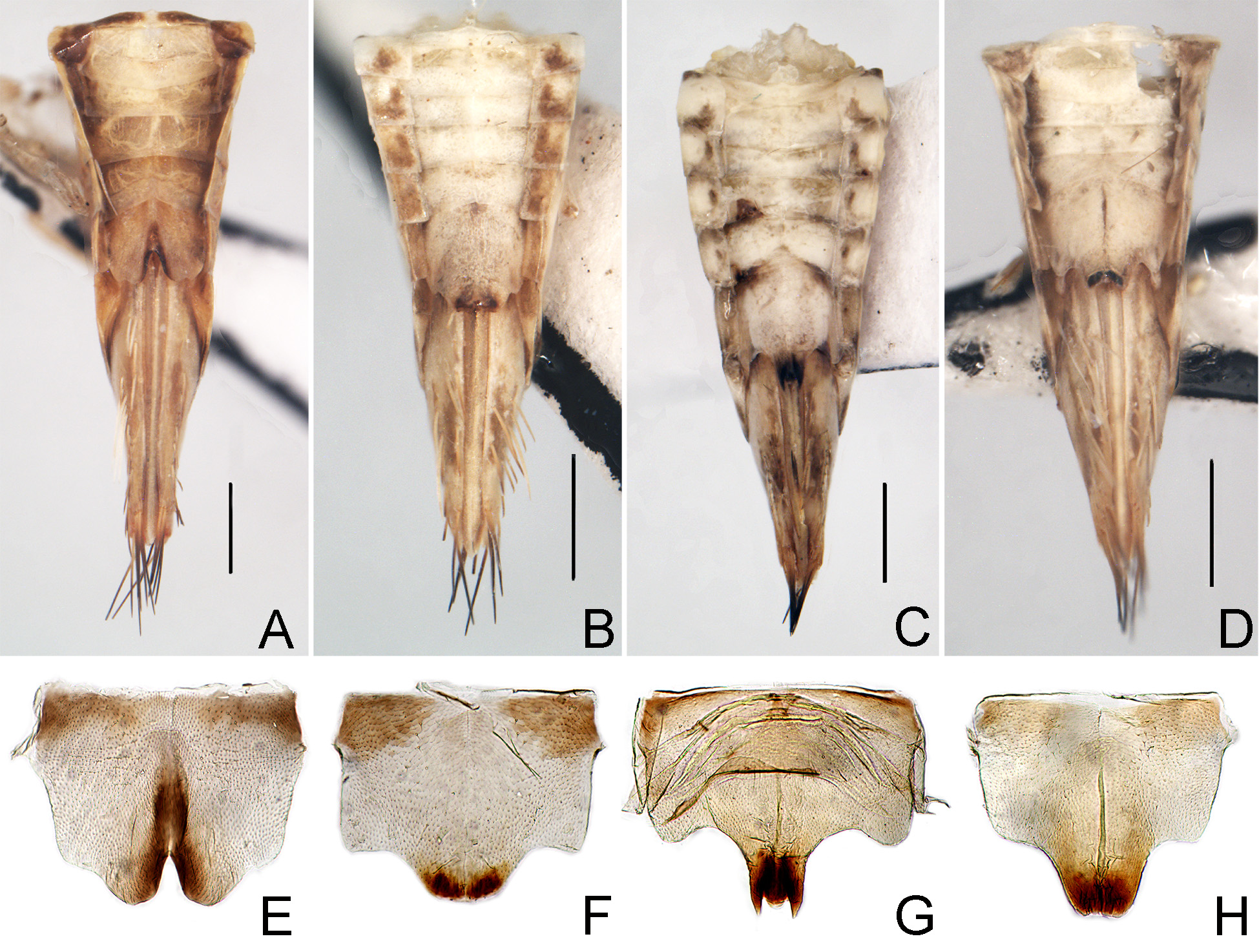
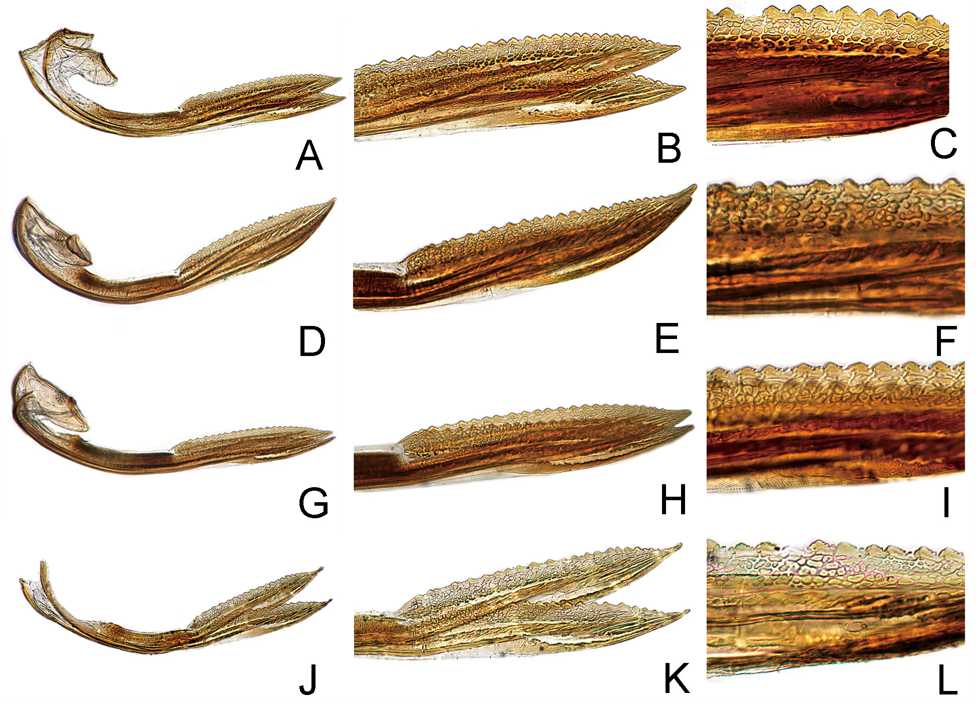
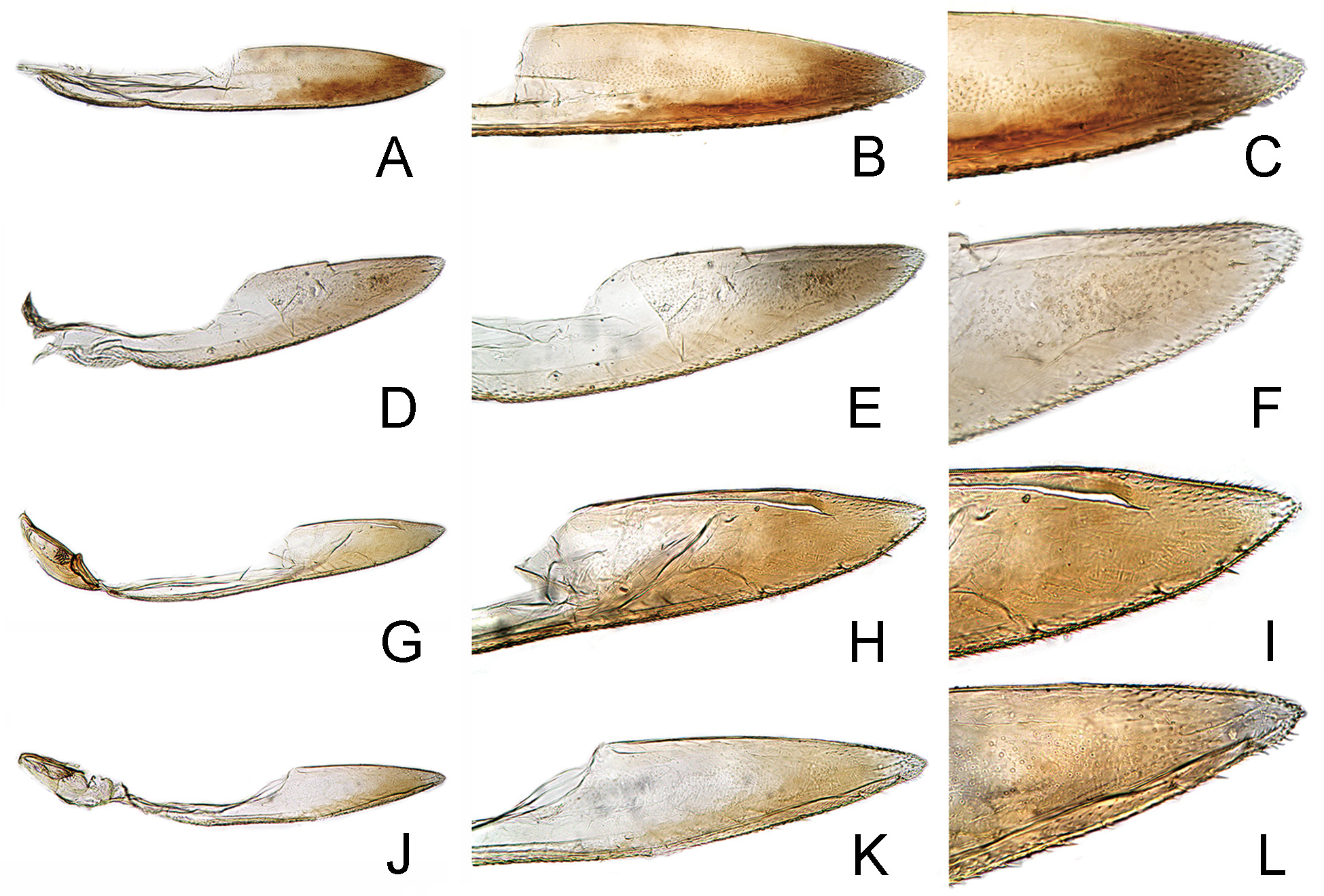
Female genitalia. Abdominal sternite VII ( Fig. 4 View FIGURE 4 A, E), in ventral view, broader than long, anterior margin straight; caudal margin deeply incised in middle, edge of incision blackish. First valvulae, in lateral view ( Fig. 14 View FIGURE 14 A–C), distinctly curved dorsally and with dorsal margin concave, dorsal reticulate sculptured area located on apical half, broadening to near apex and gradually narrowing to apex. Second valvulae ( Fig. 15 View FIGURE 15 A–C), in lateral view, distinctly expanded beyond basal curvature and abruptly narrowed near apex; apex narrowly acute; shaft bearing many triangular teeth distributed on apical half behind basal curvature, ducts extending toward dorsal margin and toward apical portion of shaft. Third valvulae ( Fig. 16 View FIGURE 16 A-C), in lateral view, with basal half narrow and apical half distinctly expanded; apex prominently produced angularly.
Measurements. Male 4.5–5.7mm long, 1.11mm wide across eyes. Female 5.5–6.1mm long, 1.1–1.3mm wide across eyes.
Material examined. 6♂, 4♀, Shandong Prov., Yantai, Kunyushan, 12.vii.2001, Qin Daozheng and Liu Zhenjiang; 2♂, Shaanxi Prov., Yangxian, 22. VIII, Wei Cong and Shang Suqin; 3♂, 1♀, Hunan Prov., Hengshan, 7.viii.1985, Zhang Yalin and Chai Yonghui; 4♀, Hunan Prov., Hengshan, 8.viii.1985, Zhang Yalin and Chai Yonghui; 1♂, 1♀, Hunan Prov., Hengshan, 8.viii.1985, Zhang Yalin and Chai Yonghui; 1♀, Henan Prov., Xixia, Huangshian Forestry Farm, 7.vii.1998, Hu Jian; 1♀, Yunnan Prov., Juanyuanxian, 4.vi. 1974, Chou Io and Yuan Feng; 1♀, Hubei Prov., Tongshanxian, Hengshi, 9.viii.2001, Huangmin; 1♂, Hubei Prov., Dabieshan, Taohuachong, 26. vii.2014, Chen Fangying; 1♂, Hainan Prov., Jianfengling, 20.viii.2009, Gao Xia; 1♂, GuangxiProv., Longzhou, Sanlian, 14.vi.2000, Yao Jian.
Distribution. China (Guangxi, Guizhou, Hainan, Hebei, Henan, Hubei, Hunan, Shaanxi, Shandong, Sichuan, Xizang, Yunnan); Japan, Korea, Russia.
Remarks. Scaphoideus albovittatus Matsumura is a very common species throughout Eastern Asia. This species shows some variation in the external coloration and male genitalia among the specimens from northern to southern China, including forms from Shandong Prov. ( Figs. 1 View FIGURE 1 A, F, K, 5A, B, 6A, 7A–D), Shaanxi Prov. ( Figs. 1 View FIGURE 1 B, G, L, 5C, D, 6B, 7E–H), Hubei Prov. ( Figs. 1 View FIGURE 1 C, H, M, 5E, F, 6C, 7I –L) and Guangxi Prov. ( Figs. 1 View FIGURE 1 D, I, N, 5G, H, 6D, 7M–P). In the Shandong form, the shaft of the aedeagus is more slender ( Fig. 7 View FIGURE 7 D) and the connective processes are less sinuate and straighter ( Figs. 6A View FIGURE 6. A – D , 7 View FIGURE 7 A). Those from Shaanxi Prov. are similar to those from Shandong except the ventral margin of the aedeagal shaft is slightly convex ( Fig. 7 View FIGURE 7 H). However, in the specimens from Hubei Prov., the aedeagus is robust and the connective processes are slightly curved ventrally ( Fig. 7 View FIGURE 7 I-L) compared to those from northern Shandong and Shaanxi, and the aedeagus is more slender than those from southern Guangxi ( Fig. 7 View FIGURE 7 P). From north to south, the pygofer becomes more slender, the shaft of aedeagus becomes stouter, and the connective processes become more curved ventrad. These differences are regarded as intraspecific variations, which can be expected in widely distributed species.


No known copyright restrictions apply. See Agosti, D., Egloff, W., 2009. Taxonomic information exchange and copyright: the Plazi approach. BMC Research Notes 2009, 2:53 for further explanation.
|
Kingdom |
|
|
Phylum |
|
|
Class |
|
|
Order |
|
|
Family |
|
|
Genus |
Scaphoideus albovittatus Matsumura, 1914
| Chen, Fangying, Dai, Wu & Zhang, Yalin 2015 |
Scaphoideus albivittatus
| Li 2011: 209 |
| Xing 2010: 137 |
| Li 2007: 160 |
| Li 2006: 184 |
| Li 2002: 197 |
Scaphoideus albovittatus
| Kamitani 2013: 515 |
| Cai 2002: 276 |
| Li 1991: 188 |
| Okada 1977: 192 |
| Ishihara 1961: 252 |
| Matsumura 1914: 224 |