
Aphelenchoides paradalianensis, Cui, Ruqiang, Zhuo, Kan, Wang, Honghong & Liao, Jinling, 2011
|
publication ID |
https://doi.org/ 10.5281/zenodo.201986 |
|
DOI |
https://doi.org/10.5281/zenodo.5664073 |
|
persistent identifier |
https://treatment.plazi.org/id/A64387F0-FFBE-193D-F294-C4A0FBA05C08 |
|
treatment provided by |
Plazi |
|
scientific name |
Aphelenchoides paradalianensis |
| status |
sp. nov. |
Aphelenchoides paradalianensis n. sp.
Figs. 1–3 View FIGURE 1 View FIGURE 2 View FIGURE 3
= Aphelenchoides sp. HR 3 in Zhuo et al. 2010
Measurements. See Table 1 View TABLE 1 .
Female Male
Material examined. Type material: Holotype female, 20 paratype females and 10 paratype males are deposited in the USDA Nematode Collection, Beltsville, Maryland; two paratype females in the University of California Nematode Collection, Riverside, California; two paratype females in the Canadian National Nematode Collection, Ottawa, Canada and two paratype females at CABI Bioscience, UK Centre, Surrey, UK. Other voucher specimens and cultures are available at the Plant Nematode Research Laboratory, College of Resources and Environmental Sciences, South China Agricultural University, Guangzhou, China.
Description. Female. Body slender and ventrally curved when heat-relaxed, occasionally dorsally curved; annules fine. Lateral fields with four incisures in mid-body. Lip region rounded in lateral view, slightly offset, bearing 6 annules. Stylet 10–12.5 μm long, with small basal swellings, stylet cone comprising ca. 45% of total stylet. Procorpus cylindrical, ca. 4–5 stylet lengths. Metacorpus oval, with conspicuous valve situated centrally. Nerve ring posterior to metacorpus. Excretory pore level with nerve ring or opposite the posterior level of the nerve ring, the position varying from 1/2 to 2/3 metacorpus length behind metacorpus. Hemizonid invisible. Pharyngo-intestinal junction immediately posterior to metacorpus. Pharyngeal glands overlapping intestine dorsally for ca. 3–5 body diameters. Monodelphic, ovary outstretched, locating left of intestine, occupying 30–50% of body length, oocytes in two rows except in anterior part. Oviduct connecting ovary and spermatheca. Spermatheca oval, sperm present in some individuals. Crustaformeria ovate-oblong, posterior to spermatheca, visible in some individuals. Uterus with thick wall, posterior to crustaformeria. Vagina inclined anteriorly at ca. 45° to body axis, both anterior and posterior vulval lips slightly protruding, vulval flap absent. Postuterine sac short, 17.5–27.5 μm long, extending 12.9–19.3% of vulva-anus distance, ca. 1–1.5 vulval body widths or 2–2.5 anal body widths long, sperm sometimes present. Tail conoid, terminus consisting of a short trunk bearing two finely rounded and smooth processes of unequal length.
Male. Much less common than females; the ratio of males to females was about one to ten thousand. Body slender, posterior region curved ventrally when heat-relaxed. Anterior region and cuticle similar to female. Testis single, anteriorly outstretched, locating left of intestine, occupying 40–50% of body length. Spicules smoothly curved, rosethorn-shaped, apex small, rounded. Rostrum short, rounded. Gubernaculum absent. Tail conoid, terminus with a small ventral papillae-like mucronate structure or a short trunk with two fine processes. Three pairs of subventral caudal papillae observed: one pair adanal, the second in the middle of tail, and the third subterminal. Bursal flap absent.
Diagnosis and relationships. Aphelenchoides paradalianensis n. sp. is characterised by an unusual tail terminus (the female tail terminus possesses a short trunk with two processes, while the male tail terminus bears a small mucronate structure or just the same as the female), the slightly offset lip region with 6 annules, four incisures in the lateral field, short postuterine sac (extending ca. 13–20% of vulva-anus distance), medium-sized spicules (14.1–18.6 μm), three pairs of caudal papillae.
The new species appears morphologically most similar to A. dalianensis Cheng, Hao & Lin, 2009 because of female tail terminus shape, but the new species is distinguished from A. dalianensis by the size and shape of the male spicule (14.1–18.6 μm vs. 10–12.9 μm; slender vs. thick), the shape of male tail terminus (simple without any mucronate structure in A. dalianensis ), shorter postuterine sac (extending for 12.9–19.3% vs. ca. 25% of vulvaanus distance, ca. 1–1.5 vulval body widths vs. ca. 2 vulval body widths), smaller female c ratio (14.6–17.7 vs. 17– 20.7).
In addition, A. paradalianensis n. sp. is closely related to A. kungradensis Karimova, 1957 , A. lilium Yokoo, 1964 and A. variacaudatus Ibrahim & Hooper, 1994 in having four lateral incisures in the lateral field and two mucronate structures on tail terminus of the female. The new species differs from A. kungradensis by the shape of female tail terminus (central section depressed in A. kungradensis ), shorter postuterine sac (extending for 12.9– 19.3% vs. ca. 43% of vulva-anus distance; ca. 1–1.5 vulval body widths vs. ca. 3 vulval body widths; 2–2.5 anal body widths vs. ca. 5 anal body widths) and the presence of males. From A. lilium by the position of the excretory pore (level with nerve ring vs. obviously posterior to nerve ring), higher b ratio (6.2–7.9 vs. 3.5–4.2 in the female; 5.7–6.8 vs. 4.3–5.6 in the male), smaller T ratio (41.6–55.4 vs. 55.2–67.7) and the shorter body length (485–683 μm vs. 640–750 μm in the female; 393–514 μm vs. 600–800 μm in the male). From A. variacaudatus by the shape of female tail terminus (central section depressed in A. variacaudatus ), position of the excretory pore (level with nerve ring, posterior to metacorpus vs. anterior to nerve ring, level with the metacorpus base), higher female a ratio (31.1–46.7 vs. 27.7–32.7), smaller female b ratio (6.2–7.9 vs. 8.6–10.9), smaller stylet length (10–12.5 μm vs. 12.7–14.6) and the presence of males. A. paradalianensis n. sp. is also close to A. goodeyi Siddiqi & Franklin, 1967 , A. brevistylus Jain & Singh, 1984 , A. parabicaudatus Shavrov, 1967 , A. bimucronatus Nesterov, 1985 , A. wallacei Singh, 1977 and A. bicaudatus (Imamura, 1931) Filipjev & Schuurmans Stekhoven, 1941 , but the new species can be easily differentiated from these Aphelenchoides species. It differs from A. goodeyi by the shape of female tail terminus (with a short trunk bearing four fine hair-like spikes and somewhat spreading in A. goodeyi ), shorter postuterine sac (ca. 1–1.5 vulval body widths vs. ca. 3 vulval body widths), and the presence of males and of sperm in the postuterine sac (both species were cultured on agar with fungi). From A. brevistylus by the number of lateral incisures (4 vs. 2), shorter postuterine sac (extending for 12.9–19.3% vs. 33–66% of vulva-anus distance), longer female stylet length (10–12.5 μm vs. 6–8 μm), higher female c ratio (14.6–17.7 vs. 11.1–15.7) and position of the excretory pore (level with nerve ring vs. anterior to nerve ring). From A. parabicaudatus by the shape of female tail terminus (central section constricted in A. parabicaudatus ), longer female body length (485–683 μm vs. 310–350 μm), longer female stylet length (10–12.5 μm vs. 8 μm), higher a, c and V ratio (31.1–46.7 vs. 21.4–25 of a ratio; 14.6–17.7 vs. 10.5–12.7 of c ratio and 65–73.3 vs. 61–64 of V ratio) and position of the excretory pore (posterior to metacorpus vs. level with metacorpus). From A. bimucronatus by the number of lateral incisures (4 vs. 2), the size and shape of the male spicule (14.1–18.6 μm vs. 23.8 μm; slender vs. thick), shorter female stylet length (10–12.5 μm vs. 21.7 μm) and higher b ratio (6.2–7.9 vs. 3.4–3.6). From A. wallacei by the shape of female tail terminus (with a short trunk bearing three spikes in A. wallacei ), higher a and c’ ratio (31.1–46.7 vs. 22–23 of a ratio and 3.3–4 vs. 2–2.5 of c’ ratio), shorter postuterine sac (ca. 1–1.5 vulval body widths vs. ca. 2 vulval body widths), shorter female body length (485–683 μm vs. 690–730 μm), shorter female stylet length (10–12.5 μm vs. 13.5–14 μm) and the shorter male spicule (14.1–18.6 μm vs. 26 μm). From A. bicaudatus by the shape of female tail terminus (central section constricted in A. bicaudatus ), the number of lateral incisures (4 vs. 2), the shorter female tail length (30–40 μm vs. 50 μm; 14.6–17.7 vs. 9.4–12.6 of c ratio and 3.3–4 vs. 4.5–5 of c’ ratio).
Type locality and habitat. Type specimens were obtained from a two-week-old culture on Pestalotia sp. The culture was initiated from one female of A. paradalianensis n. sp., which were collected in September 2009 from solid wooden packaging materials exported from South Korea.
Etymology. The specific epithet was chosen because the new species is similar morphologically to A. dalianensis .
Molecular profiles. Amplification of the ITS region, the near full-length 18S rDNA and partial mitochondrial COI genes resulted in PCR products of 961 bp, 1707 bp and 710 bp respectively. The closest sequences to these three molecular regions were A. ritzemabosi ( EU186067 View Materials , EU186068 View Materials ), A. besseyi ( EU186069 View Materials ), A. sp. ( FJ768943 View Materials ) and A. dalianensis ( Cheng et al. 2009) for ITS region; A. sp. ( GU337994 View Materials ), A. sp. ( GU337995 View Materials ), A. besseyi ( AY508035 View Materials ) and A. ritzemabosi ( DQ901554 View Materials ) for 18S rDNA; A. sp. ( GU367860 View Materials ), A. sp. ( GU367866 View Materials ) and A. sp. ( GU367863 View Materials ) for partial mtCOI. The sequence identities of A. paradalianensis n. sp. and A. dalianensis (the closest species in morphology) are 81% (651/809) with 71 gaps (8.8%) in the ITS region.
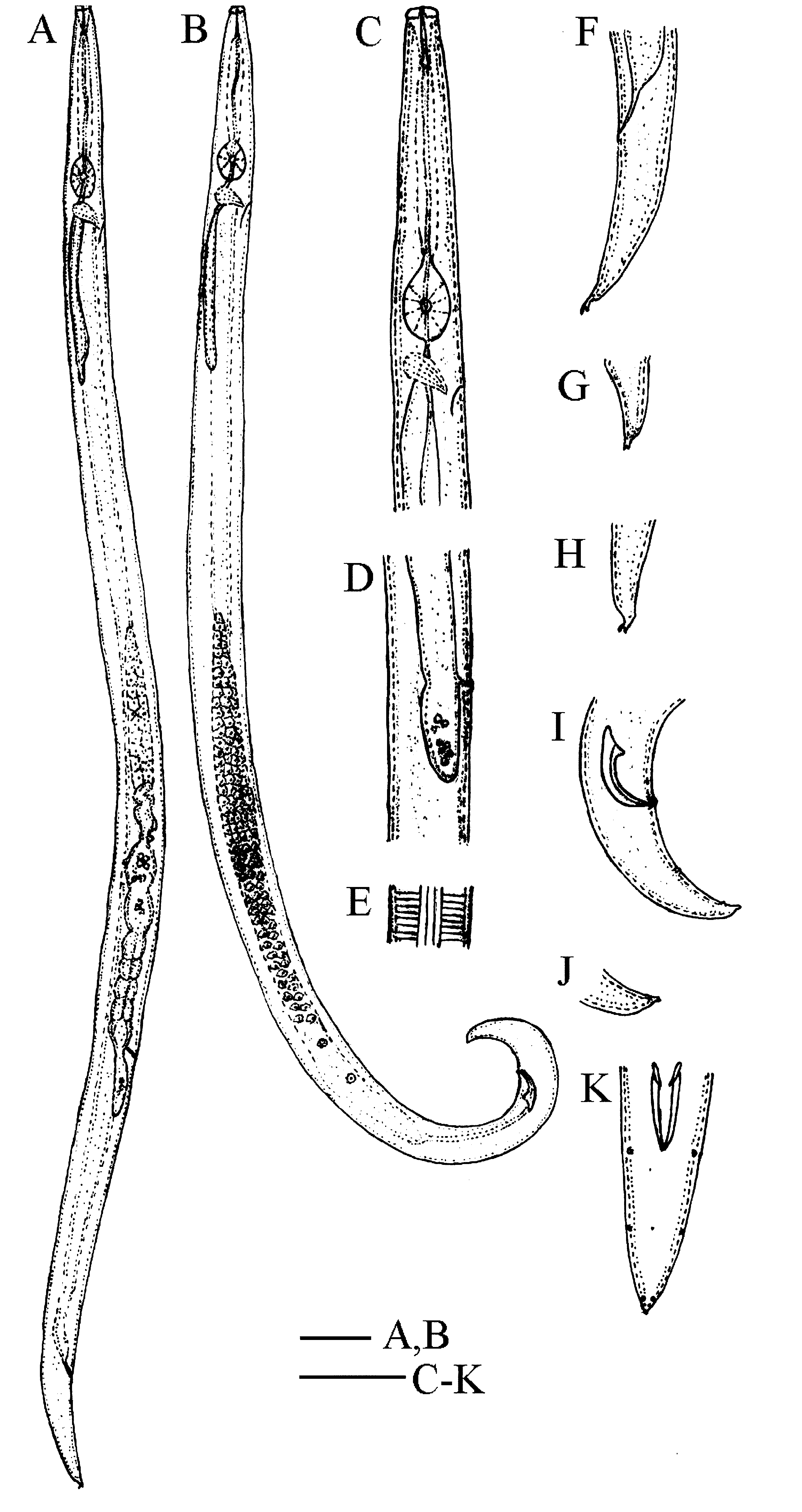
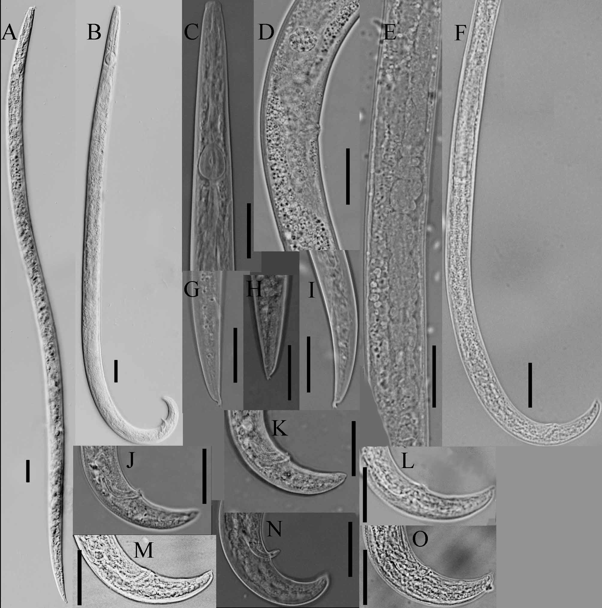
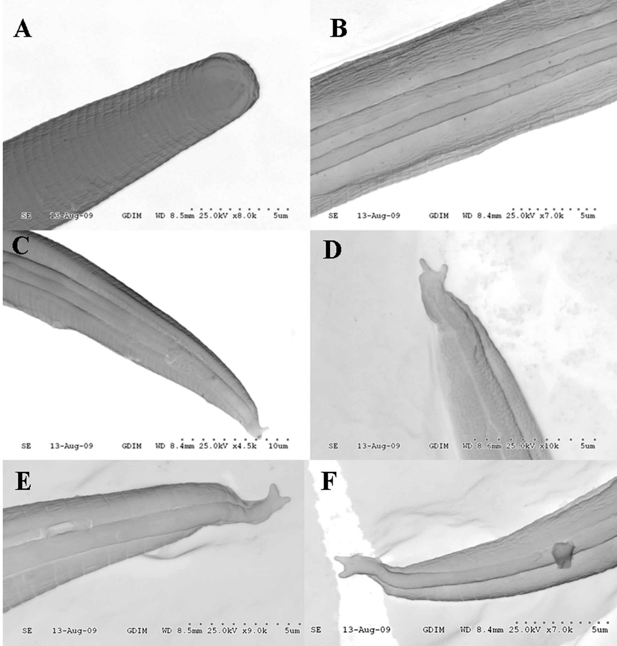
TABLE 1. Morphometrics of Aphelenchoides paradalianensis n. sp. [Measurements are in μm and in the format: mean ± SD (range)]
| Holotype Paratypes | Paratypes | |
|---|---|---|
| n | - 15 | 15 |
| L | 590 570±51.2 (485–683) | 452±31.2 (393–514) |
| a | 36.3 37.7±4.3 (31.1–46.7) | 27.8±1.3 (25.1–29.3) |
| b | 7.2 7.0±0.5 (6.2–7.9) | 6.3±0.4 (5.7–6.8) |
| c | 16.9 16.6±1.0 (14.6–17.7) | 15.4±1.1 (12.8–16.5) |
| cˏ V or T | 4.0 3.5±0.3 (3.3–4.0) 72.0 70.5±2.2 (65–73.3) | 2.7±0.3 (2.4–3.6) 48.1±4.7 (41.6–55.4) |
| Stylet | 10 10.8±1.0 (10–12.5) | 10.3±0.5 (9.2–10.5) |
| Vulva-anus distance Distance anterior end to distal end of median bulb | 130.0 134±20.3 (113–173) 72.5 70±3.0 (67.5–77.5) | - 50.6±3.7 (45–57.5) |
| Tail length Postuterine sac length | 35.0 34.4±2.9 (30–40) 18.8 22±2.6 (17.5–27.5) | 29.5±2.8 (26.1–32.3) - |
| Spicule | - - | 14.8±1.2 (14.1–18.6) |
| Postuterine sac length /vulva-anus distance(%) | 15.4 16.6±2.1 (12.9–19.3) | - |
No known copyright restrictions apply. See Agosti, D., Egloff, W., 2009. Taxonomic information exchange and copyright: the Plazi approach. BMC Research Notes 2009, 2:53 for further explanation.
|
Kingdom |
|
|
Phylum |
|
|
Class |
|
|
Order |
|
|
Family |
|
|
Genus |