
Phobeticomyia digitiformis, Shi, Li, Li, Wenliang & Yang, Ding, 2009
|
publication ID |
https://doi.org/ 10.5281/zenodo.187466 |
|
DOI |
https://doi.org/10.5281/zenodo.6218735 |
|
persistent identifier |
https://treatment.plazi.org/id/A738878A-FFFF-AD10-FF7D-9430FE73FA61 |
|
treatment provided by |
Plazi |
|
scientific name |
Phobeticomyia digitiformis |
| status |
sp. nov. |
Phobeticomyia digitiformis View in CoL sp. nov.
( Figs. 1–2 View FIGURES 1 – 8 , 9 View FIGURES 9 – 12 , 13–17 View FIGURES 13 – 17 )
Diagnosis. Antenna yellow, 1st flagellomere brownish yellow on apical 1/2. Wing with four hyaline bracket–shaped median spots between R2+3 and R4+5; two hyaline spots below r-m and a hyaline stripe on dmcu; a small hyaline triangular spot beyond dm-cu along lower edge of m1 cell separated from hyaline stripe on dm-cu, and a hyaline subapical stripe in m1 cell.
Description. MALE. Body length 3.7–4.0 mm, wing length 3.0– 3.6 mm. FEMALE. Body length 3.4–3.9 mm, wing length 3.0– 3.4 mm.
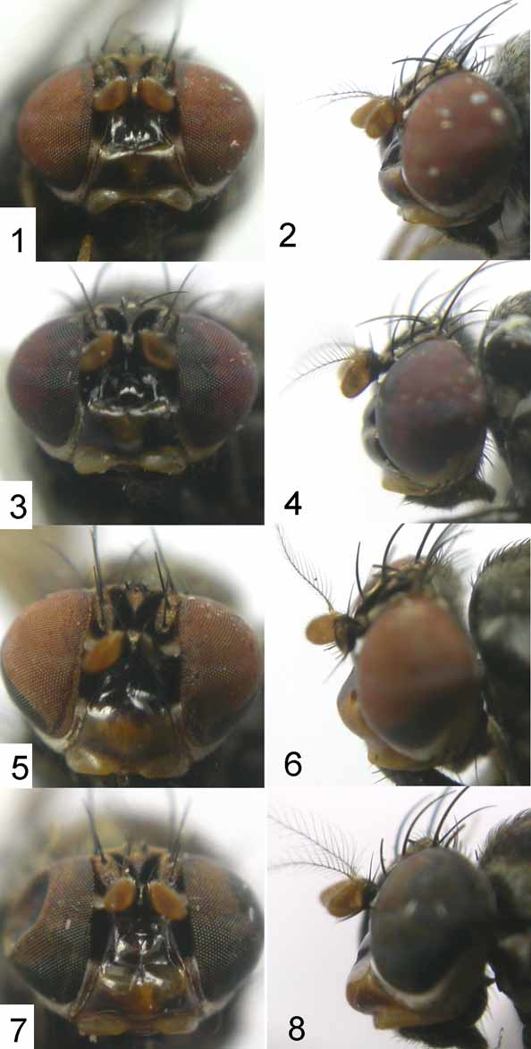
Head ( Figs. 1–2 View FIGURES 1 – 8 ) brownish yellow. Frons yellow, wider than long and parallel–sided, with two velvety blackish brown stripes extending to ocellar triangle, basally confluent with a W–shaped black or brown transverse band. Ocellar triangle black, oc strong and longer than anterior or; anterior or inclinate, shorter than posterior one. Face shining, black or brown on dorsal 1/2 and yellow on ventral 1/2 with a pair of brown triangular spots. Parafacia blackish brown on dorsal 1/2 and pale yellow with a thin reddish yellow stripe on ventral 1/2. Gena pale yellow with silver whitish pollen along eye margin, about 1/4 height of eye. Antenna yellow, 1st flagellomere brownish yellow on apical 1/2, nearly 1.6 times longer than high; arista long plumose, black, with longest hairs longer than height of 1st flagellomere. A silver white spot present between antenna and eye. Proboscis brown with yellow and blackish hairs; palpus black, with black hairs.
Thorax brown with grayish white pollen. Mesoscutum with a thin grayish white stripe between two wide brown median stripes and two brown lateral stripes behind suture, and a thin grayish white stripe along dc rows; 0+3 dc, with a brownish spot on base of each dc; acr in 8 rows; prsc shorter than 1st post–sutural dc. anepisternum brown, with a wide grayish white median stripe; 1 anepst, 2 kepst. Scutellum brown with yellowish brown pollen. Legs blackish brown, except all tarsi yellow (apical tarsomere pale brown in male); all tibiae with a pale yellow subapical ring. Fore femur with 5 pv and 6–7 pd, ctenidium with 15 short bristles; fore tibia with 1 preapical ad and 1 short apv. Mid femur with 6–7 a; mid tibia with 1 preapical ad and 3 apv (2 strong, 1 weak). Hind femur with 1–2 preapical ad and a row of av (only 5–6 apical bristles distinct); hind tibia with 1 preapical ad and 1 short apv. Wing ( Fig. 9 View FIGURES 9 – 12 ) brown, with a hyaline stripe at extreme tip; four hyaline bracket–shaped median spots between R2+3 and R4+5; a hyaline spot basal to r-m and two hyaline spots below r-m in the discal cell and a hyaline stripe on dm-cu; a small hyaline triangular spot beyond dm-cu along lower edge of m1 cell and a hyaline subapical stripe constricted at middle in m1 cell; a hyaline round median spot near CuA1 and an undulating hyaline stripe along hind margin of cua1 cell; subcostal cell with a hyaline spot; costa with 2nd (between R1 and R2+3), 3rd (between R2+3 and R4+5) and 4th (between R4+5 and M1) sections in proportion of 1.6 mm: 0.6 mm: 0.3 mm; r-m at middle of discal cell; ultimate and penultimate sections of M 1 in proportion of 1.0 mm: 1.6 mm; ultimate section of CuA1 about 1/8 of penultimate. Halter yellow.
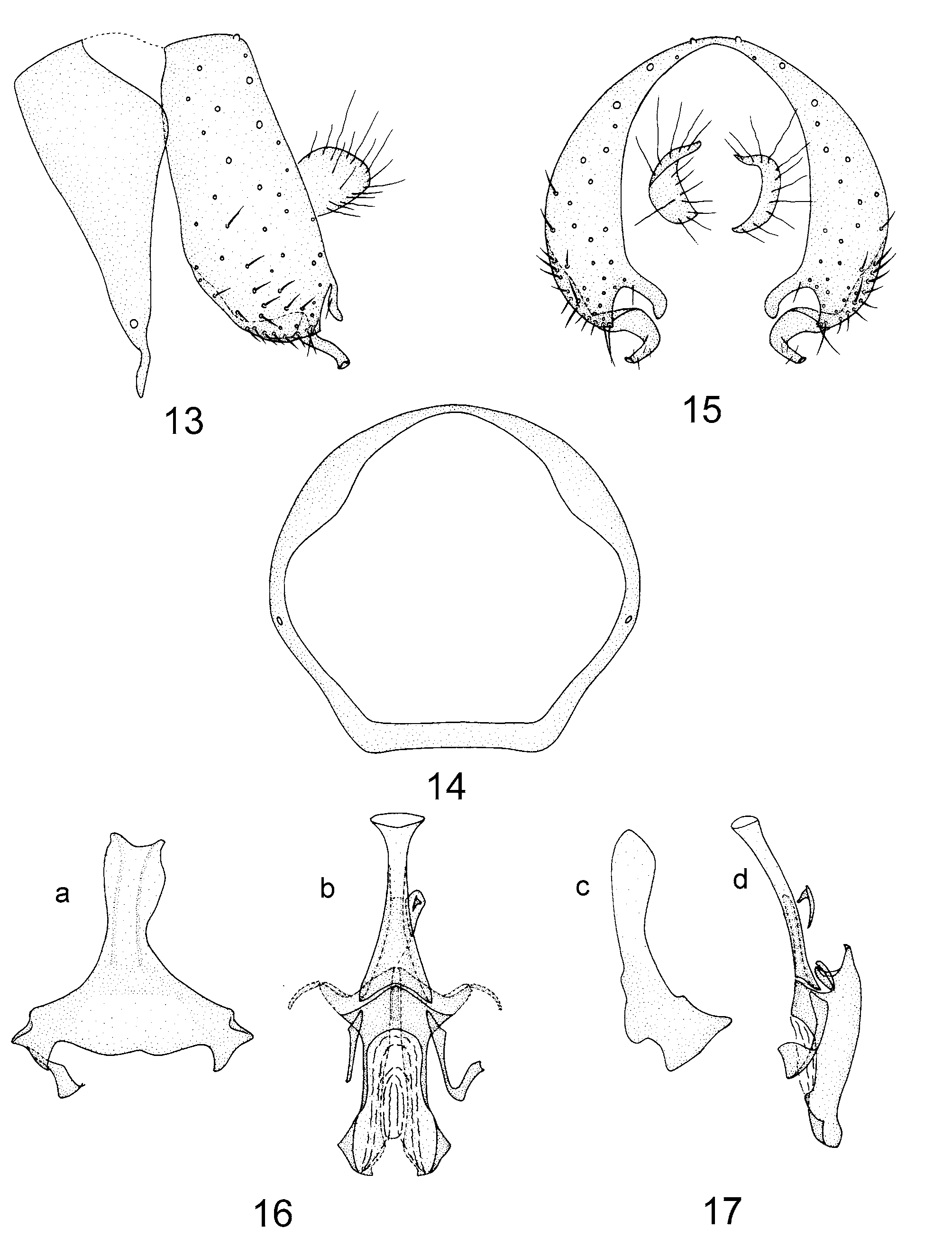
Abdomen black with sparse grayish white pollen. Male genitalia ( Figs. 13–17 View FIGURES 13 – 17 ): protandrium circular with a median incision; epandrium nearly rectangular with short hard hairs; surstylus with a small digitiform subapical process and a curved claviform inner process in posterior view; hypandrium nearly Y–shaped, gonopod very narrow and hook–shaped; aedeagus with a pair of triangular basal processes and subapical lateral processes, and a hooked apical process in ventral view.
Type material. Holotype male, Yunnan: Menglun No. fifty-five zone (920 m), 24. IV. 2007, Wenliang Li. Paratypes: 1 male, 2 females, Yunnan: data same as holotype; 4 males, Yunnan: Menglun No. fifty-five zone (920 m), 24. IV. 2007, Hui Dong.
Distribution: China (Yunnan).
Remarks. The new species is very similar to Phobeticomyia lunifera (de Meijere) in the wing with a hyaline subapical stripe in the m1 cell, a hyaline round median spot near CuA1 and an undulating hyaline stripe along the hind margin of the cua1 cell. But it can be separated from the latter by the antennal scape and pedicel yellow, the wing with four hyaline bracket–shaped median spots between R2+3 and R4+5, a small hyaline triangular spot beyond dm-cu along lower edge of m1 cell. In lunifera , the antennal scape and pedicel are black; four hyaline median spots between R2+3 and R4+5 are separated entirely on the wing, a large hyaline triangular spot is confluent with a hyaline spot on dm-cu in the m1 cell ( Sasakawa 1987).
Etymology. Latin, ‘ digitiformis ’ = digitiform, referring to the surstylus with a small digitiform subapical process; a feminine adjective.

No known copyright restrictions apply. See Agosti, D., Egloff, W., 2009. Taxonomic information exchange and copyright: the Plazi approach. BMC Research Notes 2009, 2:53 for further explanation.