
Myconus florae Bahder & Bartlett, 2024
|
publication ID |
https://doi.org/10.11646/zootaxa.5443.4.6 |
|
publication LSID |
lsid:zoobank.org:pub:36D4DFA2-9F33-41C3-8FB8-82824E7F221C |
|
DOI |
https://doi.org/10.5281/zenodo.11064624 |
|
persistent identifier |
https://treatment.plazi.org/id/A91B8633-FFEF-6A2F-3B8B-C8957828FE5C |
|
treatment provided by |
Plazi |
|
scientific name |
Myconus florae Bahder & Bartlett |
| status |
sp. nov. |
Myconus florae Bahder & Bartlett sp. n.
Type locality. La Tarde Ecolodge, Puntarenas Province, Costa Rica.
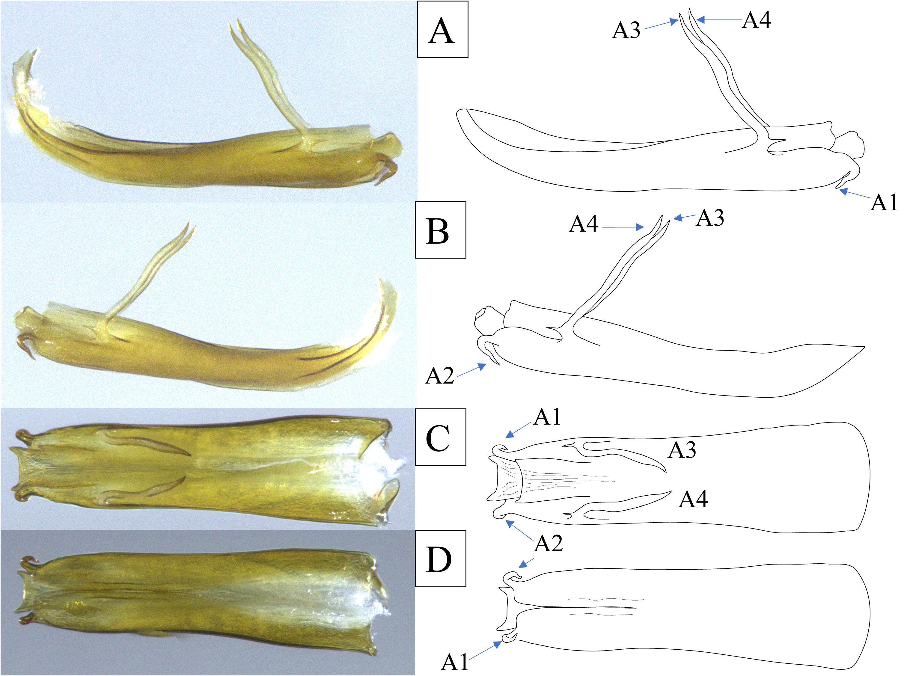
Diagnosis. Medium sized(~5.0– 6.5 mm), pale, light mottling over body, clavus with series (~10) of irregular fuscous marks between A1 and wing margin. Gonostyli with large, bifurcated capitulum with crescent-shaped basal projection and beak-shaped distal projection. Aedeagus slender with two pairs of processes, first pair at apex short, curved ventrally and second pair subapical, long and slender on dorsal surface, extending dorsally.
Description. Color. General body color pallid tan with irregular light fuscous wash, mottled. Pronotum from dorsal view with fore pale spots in transverse row each side of midline. Anterior portion of mesonotum bearing serpentine fuscous line laterad of lateral carina extending from leading margin curved laterad toward tegulae. Legs more uniformly pale. Forewings generally transparent, with most veins bearing a series of irregular, shallowly “V” shaped fuscous markings and fuscous mottling.
Structure. Males ( n =6) 4.99–5.01 mm long with wings, 3.33–3.34 mm without wings, females ( n =10) 6.41–6.45 mm long with wings, 5.11–5.13 mm without wings ( Table 2 View TABLE 2 ).
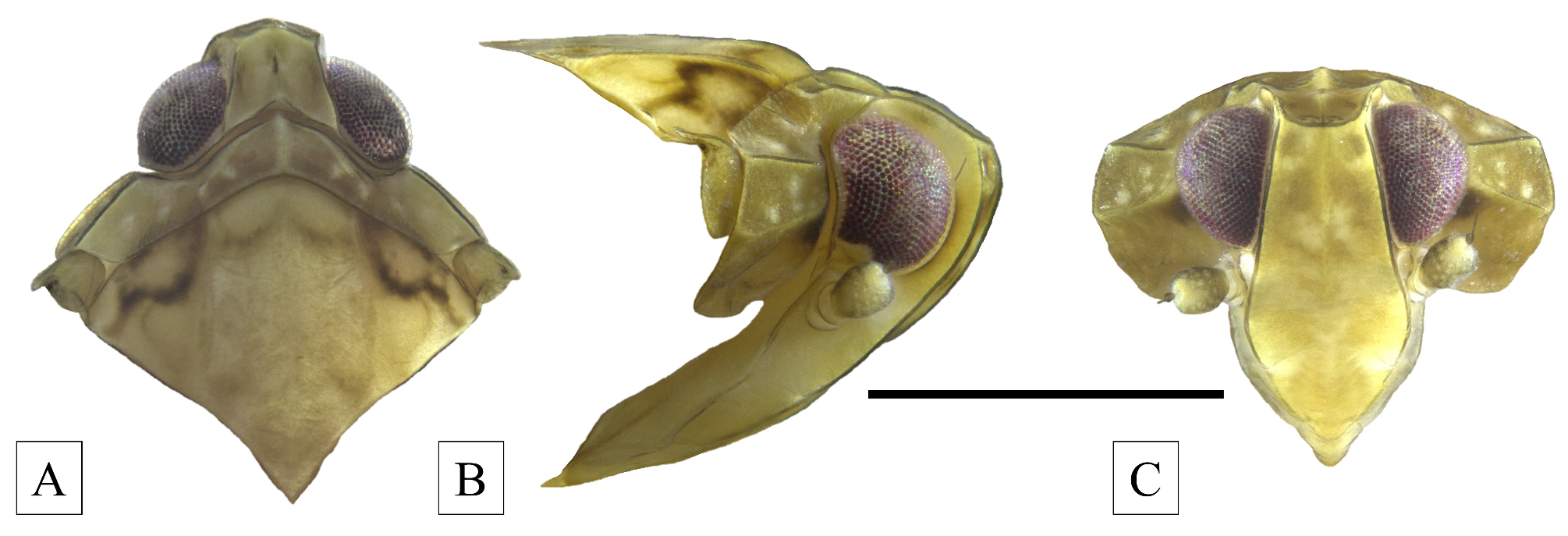
Head. In dorsal view, vertex approximately 1.45x wider than long at midline, disc slightly depressed, median carina present, weakly narrowed anteriorly; anterior margin convex and carinate, posterior margin irregularly sinuate, concave, lateral margins sinuate ( Fig. 3A View FIGURE 3 ). In lateral view, head projected slightly beyond eyes, angulate at carina of fastigium, slightly rounded ventrad of fastigium ( Fig. 3B View FIGURE 3 ). In frontal view, face elongated, roughly rhomboid (widest near level of antennae), dorsal margin broadly convave (with transverse carina), lateral margins of frons expanding from fastigium, constricting immediately below antennae, median carina present ( Fig. 3C View FIGURE 3 ); frontoclypeal suture convex. Lateral ocelli below eyes (hidden by antenna in lateral view). Compound eyes irregular in shape; anterior margin broadly rounded, posterior margin following contours of head.
Thorax. In dorsal view, pronotum relatively short (about ¾ length of vertex at midline), with anterior margin irregularly sinuate (tracing posterior margin of head), convex, posterior margin concave, tricarinate, median carina distinct, lateral carinae arched, obsolete just before posterior margin ( Fig. 3A View FIGURE 3 ); in lateral view, paradiscal region broad, with two distinct carinae, from anterior margin behind eye to posterior margin near tegula (carinae diverging such that they nearly embrace tegula at posterior margin, Fig. 3B View FIGURE 3 ). Mesonotum in lateral view raised above level of pronotum, in dorsal view tricarinate, lateral carinae sinuate, curving distad from anterior margin ( Fig. 3A View FIGURE 3 ).
Forewing with early fork of MP from SCP+R near basal cell, fork of RP from ScP+RA in proximal third of wing; CuA forked well before apex of clavus; ScP appears to have 2 branches before wing margin, each joining RA; branching pattern RA 2-branched, RP 3-branched, MP 5-branched, and CuA 2-branched but CuA 2 near wing apex linked to the marginal vein of uncertain affinity.
Hindlegs with three lateral spines spaced evenly along tibiae, progressively larger toward apex. Spinulation at tibial apex and tarsomeres 5-4-6.
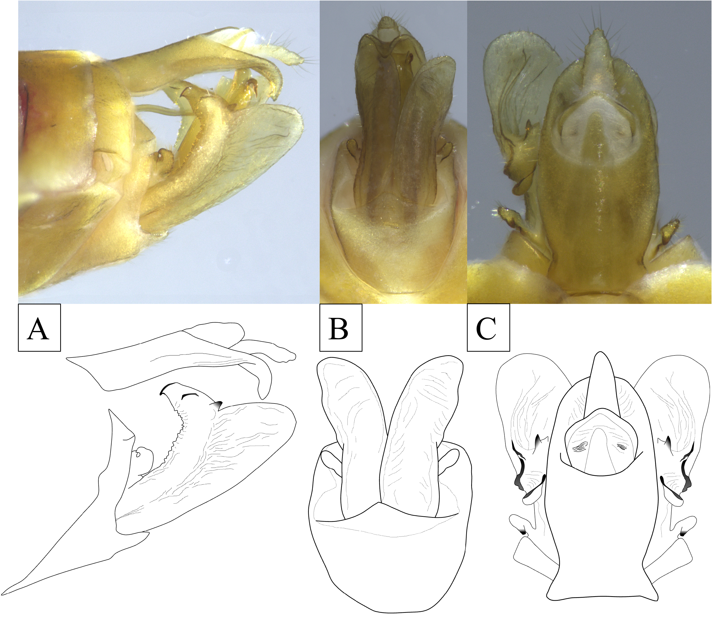
Terminalia. Pygofer in lateral view narrow (abruptly broadened at ventral margin), narrowed dorsally and appearing not to meet medially (in dorsal view) above base of anal tube; in lateral view, irregularly sinuate on anterior and posterior margin, narrowest dorsally, widest at ventral margin, strongly angled on lateral margin just below dorsal margin ( Fig. 5A View FIGURE 5 ). In ventral view, medioventral process present, short and broadly triangular (approximately 2X wider than tall ( Fig. 5B View FIGURE 5 ). Gonostyli in lateral view club-like, narrow at base, ventral margin gently curving dorsad to rounded apex, dorsal margin bearing two processes, a strongly bifurcated, basal process in form of robust crescent, with caudal side more strongly hooked than anterior side; distal projection in form of large avicephaliform hook, anterior face irregularly serrulate, apex nearly truncate, lateral margin bearing small triangular process at base (angled caudad and on oblique ridge near midlength Fig. 5A View FIGURE 5 ); gonostyli from ventral view of uniform width, inner margins smoothly arched laterad, lateral margins irregularly concave. Aedeagus cylindrical, simple, curved near pase (to incline upward), nearly bilaterally symmetrical, bearing two pairs of processes: first pair (A1 & A2) elongated, arising subapically on lateral margins, long, slender, slightly sinuate, extending dorso-cephalad, posterior margin of each process near base bearing single spine pointing caudad ( Fig. 6 View FIGURE 6 ): second pair (A3 & A4) arising at apex, short, strongly curved ventrad, margins sinuate ( Fig. 6 View FIGURE 6 ). Anal segment (in lateral view) subequal in length to gonostyli, slightly sinuate on dorsal and ventral margins, apex hooked downward to blunt apex ( Fig. 5A View FIGURE 5 ); in dorsal view broad, ovate (broadened at base).
Etymology. The specific name is given as an honorific to the senior author’s late grandmother, Flora Bahder.
Material examined. Holotype male “ Costa Rica, Puntarenas Province / Ecolodge La Tarde / 16.VII.2021 / Coll.: B.W. Bahder, light trap / Holotype Myconus florae ♂ ” ( FLREC) ; Paratype same as holotype ( 2 males, 2 females FSCA, 3 males, 8 females FLREC).
Sequence data. For the COI locus, a 757 bp sequence was generated (GenBank Accession No. OR838818); for 18S, a 1,429 bp sequence was generated (GenBank Accession No. OR840548); and for H3, a 346 bp sequence was generated (GenBank Accession No. OR902459).
Remarks. The novel taxon belongs to the genus Myconus based on morphological characters and limited molecular data. Myconus florae n. sp. differs from the type species, M. conspersinervis , most conspicuously by its small size, hind leg spinulation (5-4-6 vs. apparently (6(1+5)- 7-11 in M. conspersinervis ), fewer branches on some longitudinal veins of the forewing, and the lora adjacent to the clypeus being smaller and more strongly inflected so that they are less apparent from the frontal view. Despite these differences, the new species is more similar to Myconus than to other New World genera in Myconini (vis. Myrophenges Fennah, 1965 and Myconellus Fennah, 1950 ), but not sufficiently different to warrant the description of a new genus at this time.
Myconus florae n. sp. is most similar to M. jacquelinae than to other species in the genus. Superficially, Myconus florae n. sp. is a much smaller species (~ 5–6 mm) than M. jacquelinae (~ 12 mm). The general form of the genitalia of the two species is similar The gonostyli differ in that both processes on the capitulum are much more robust in M. jacquelinae compared to M. florae sp. n. The differences between the aedeagus between these two species are much more subtle, but consistent, with the dorsal processes in M. florae (A3 & A4) being much less sinuate than those in M. jacquelinae . Additionally, these processes in M. jacquelinae are angled mesad and cross at the midpoint whereas they do not cross in M. florae sp. n.
The close relationship of M. florae sp. n. and M. jacquelinae is made more evident based on the 18S locus, which has 100% similarity between the two species. However, for the COI barcoding region M. florae sp. n. differs from M. jacquelinae by 11.5%, a level normal with interspecific variability within a genus based on observations in cixiids ( Humphries et al. 2021), derbids ( Bahder et al. 2023) and nogodinids ( Bahder et al. 2024). Robust analyses of achilid species within genera for the region of COI analyed here has not been performed. While it is possible achilids might display lower intraspecific (or higher) variability for the 5’ region of COI than other families, based on levels observed in these other families, it is reasonable to assume those percentages are consistent in this genus of achilids, especially given the morphological support for generic placement and the low levels of variance observed in the other loci.
| FSCA |
Florida State Collection of Arthropods, The Museum of Entomology |
No known copyright restrictions apply. See Agosti, D., Egloff, W., 2009. Taxonomic information exchange and copyright: the Plazi approach. BMC Research Notes 2009, 2:53 for further explanation.