
Spratelloides atrofasciatus Schultz, 1943
|
publication ID |
https://doi.org/ 10.11646/zootaxa.4028.4.4 |
|
publication LSID |
lsid:zoobank.org:pub:CC57BC2C-7484-4644-AD7A-A875D8410373 |
|
DOI |
https://doi.org/10.5281/zenodo.5662816 |
|
persistent identifier |
https://treatment.plazi.org/id/AC2A87FC-882D-BA47-FF64-FA8543635420 |
|
treatment provided by |
Plazi |
|
scientific name |
Spratelloides atrofasciatus Schultz, 1943 |
| status |
|
Spratelloides atrofasciatus Schultz, 1943 View in CoL
(English name: Small-banded Round Herring) (Japanese name: Ryukyu-kibinago)
(Okinawan local name: Baka-jyako, Sururu, Sururugwa) ( Fig. 1 View FIGURE 1 A, B, Table 1 View TABLE 1 )
Spratelloides atrofasciatus Schulz 1943: 8 View in CoL , fig. 1 (type locality: Fagasa Bay, Tutuila I., American Samoa); Schulz & Welander 1953: 27, Table 1 View TABLE 1 , fig. 7 (Bikini Atoll & Rongelap Atoll, Marshall Is.); Nishijimamoto 1963: 69, Table II, fig. 4 (Yonaguni I., Miyako I., Motobu, Okinawa I., Ryukyu Is.); Uyeno & Sato 1984, 1988: 19, pl. 337-G (Okinawa I.); Aonuma 2002: 244, (Okinawa I.).
Diagnosis. A species of Spratelloides with the following combination of characters: Dorsal fin rays 10–14; anal fin rays 10–13; pectoral fin rays 10–14 ( Fig. 3 View FIGURE 3 ); vertebrae 43–47 ( Fig. 4 View FIGURE 4 ) and lower gill rakers 20–24 ( Fig. 5 View FIGURE 5 ); head length short 19.3–24.1% SL (mean 22.2%); upper jaw length short 7.0–9.3% SL (8.3%); snout length equal or slightly shorter than eye diameter, its length 5.9–7.4% SL (6.7%); lateral band usually wider than eye diameter, its width 5.7–7.9% SL (6.7%) ( Fig. 2 View FIGURE 2 ); numerous black pigment spots on inner side of gill opening visible from the outside in preserved specimens ( Fig. 6 View FIGURE 6 ).
Description. Counts and measurements of the holotype, paratype, and non-type specimens of Spratelloides atrofasciatus are given in Table 1 View TABLE 1 . Frequency distributions of selected counts (dorsal fin rays, pectoral fin rays, anal fin rays, total vertebrae, and lower gill rakers) are shown in Figs. 3–5 View FIGURE 3 View FIGURE 4 View FIGURE 5 . Characters stated in the diagnosis are not repeated. Body elongated, subcylindrical, slightly compressed; body depth less than head length; snout pointed; eye diameter greater than interorbital width; upper jaw longer than snout; maxillary extending posteriorly to a vertical through front of eye but not through front of pupil; nasal openings separated by a dermal flap; two supramaxillae, the posterior with the lower part of the expanded portion larger than the upper part; lower jaw slightly projecting; gill rakers long and slender; pseudobranch with basal part of filaments covered by membrane; frontalparietal region smooth; posterior frontal fontanelles broadly divided anteriorly by a wedge of bone; rear of isthmus with usual notch and fleshy lobe on shoulder girdle; scale in lateral series from rear of head to base of caudal fin usually 42–45; vertical striae on scales discontinuous and posterior margin of scales not toothed; upper and lower lobes of caudal fin each with a pair of elongate scales on both sides; belly without scutes except a single W-shaped abdominal scute at the pelvic fin base; origin of dorsal fin equidistant between snout and caudal base or just nearer to snout than to caudal base; pelvic insertion under middle of dorsal fin base; anus immediately in front of anal fin origin; origin of anal fin a little closer to caudal fin base than to pelvic insertions.
Spratelloides atrofasciatus Spratelloides gracilis Color of preserved specimens: Head and upper and lower surfaces of body light brown; midlateral band blackish brown (silver outlined above by a thin blackish line when fresh); tip of snout, tip of lower jaw, dorsal surface of brain with black pigment, numerous black pigment spots on inner side of operculum visible from outside in preserved specimens ( Figs. 1 View FIGURE 1 , 5 View FIGURE 5 ); dorsal margin of iris with a black blotch in some specimens (not visible in the holotype and paratype); mid-dorsal line with a double row of black pigment cells from occiput to caudal fin base, the line irregular along base of dorsal fin; dorsolateral scale pockets fringed with black; ventral edge of abdomen pigmented; scales on caudal not pigmented.
Distribution and ecological notes. Schulz (1943) stated that the holotype of Spratelloides atrofasciatus was collected from Fagasa Bay, Tutuila Island, American Samoa. Schulz and Welander (1953) also reported S. atrofasciatus from Bikini, Rongelap and Rongerik Atolls, Marshall Islands. We collected specimens of S. atrofasciatus from the Ryukyu Islands: from south to north, Hatoma Island, Ishigaki Island, Miyako Island, Ohjima, Okinawa Island, and Takara-jima. A series of 24 specimens, NSMT-P 106605, 16.3–20.1 mm SL, from Palikoulo bay, Espiritu Santo Island, Vanuatu, 9 November 1964, collected by RV Shunyo, were identified as S. atrofasciatus in this study. Despite our two decades of search efforts, S. atrofasciatus is still not known from Kyushu (including Nagasaki, the type locality of S. gracilis ), Shikoku, nor Honshu, Japan. Thus the present known distribution of S. atrofasciatus is restricted to the Samoa, Marshall, Vanuatu, and Ryukyu Islands, and Takara Island in the Tokara Islands is the most northern distribution. Direct microscopic examinations of one specimen of URM-P 44245, 36.2 mm SL, 16 Aug. 2004; three stained and cleared specimens of URM-P 20001 32.7–37.9 mm SL, 1st June 1961; and one specimen of URM-P 44248, 45.2 mm SL, 17 July 2003, revealed they were gravid with 0.9–1.0 mm diameter ripe eggs. Maximum size of these specimens was 45.2mm SL and minimum mature size was 32.7 mm SL.
Comparisons. Spratelloides delicatulus and S. robustus are omitted from this comparison by lacking a lateral stripe and having continues striae on body scales. Striped round herrings, S. atrofasciatus , S. gracilis and S. lewisi , are similar to one another in general body appearance, especially in having a midlateral band and discontinuous vertical striae on their scales. Spratelloides lewisi is distinguishable from S. atrofasciatus and S. gracilis by having an incomplete midlateral band that fades anteriorly near the tips of the pectoral fins and in the number of lower gill rakers ( S. atrofasciatus : 20–24, S. lewisi : 28–32, S. gracilis : 30–39) ( Wongratana 1983; this study).
Spratelloides atrofasciatus is distinguished from S. gracilis in having a wider lateral band (5.7–7.9 % vs. 3.8– 7.5%SL), and shorter head length (19.3–24.1% vs. 20.8–26.9%), upper jaw length (7.0–9.3% vs. 7.7–10.5%) and snout length (5.9–7.4% vs. 6.6–9.0%) ( Fig.2 View FIGURE 2 ), and fewer dorsal fin rays (10–14 vs. 12–14), anal fin rays (10–13 vs. 12–15), pectoral fin rays (10–14 vs. 12–14), total vertebrae (43–47 vs. 47–50), and lower gill rakers (20–24 vs. 30– 39) ( Figs. 3–5 View FIGURE 3 View FIGURE 4 View FIGURE 5 ). Numerous black pigment spots on the inner side of the operculum are visible from the outside on preserved specimens of S. atrofasciatus that are absent in S. gracilis ( Figs. 1 View FIGURE 1 AB and 6A).
Spratelloides atrofasciatus View in CoL is a small species characterized by its minimum mature size of 32.7 mm SL and maximum size of 45.2 mm SL. By contrast, Shirafuji et al. (2007) reported mature S. gracilis View in CoL from 54.9 mm SL and growing to 100.9 mm SL in Wakayama, Japan and Weng et al. (2005) reported mature S. gracilis View in CoL from 54 mm FL (about 50 mm SL) and growing to 84mm FL (about 80mm SL) in Penghu, central Taiwan Strait. In this study, five stained and cleared specimens of S. gracilis View in CoL (URM-P 17548, 39.1–49.5 mm SL) examined were not yet mature.
Discussion. Non-types of Spratelloides gracilis examined here share the same morphological characters with the lectotype of S. gracilis (RMNH 3260a, 94.0mm SL) ( Table 1 View TABLE 1 ).
Spratelloides atrofasciatus View in CoL was originally described by Schultz (1943) based on two specimens. Although in his original description Schultz (1943) did not give detailed comparisons with congeners, Schultz and Welander (1953) mentioned the following diagnostic characters: fewer anal fin rays (10–11 vs. S. japonicus View in CoL = S. gracilis View in CoL : 12– 14), scales (41–42 vs. 44–49) and lower gill rakers (19–23 vs. 26–36), which distinguished it from Spratelloides japonicus View in CoL (= S. gracilis View in CoL ). Nishijimamoto (1963) reported the occurrence of S. atrofasciatus View in CoL and compared it with the other two species, S. japonicus View in CoL (= S. gracilis View in CoL ) and S. delicatulus View in CoL , found in Japan. Almost all specimens that Nishijimamoto studied are damaged or lost, but fortunately the samples of S. atrofasciatus View in CoL collected from Miyako Island were salvaged. Those specimens were registered in our collection as URM-P 20000 and URM-P 20001 and used here.
Sprateloides gracilis was originally described by Temmick and Schlegel (1846) from specimens collected at Nagasaki, Japan. Boeseman (1947) designated the specimen RMNH 3060a as the lectotype of S. gracilis . Whitehead (1963) treated S. atrofasciatus View in CoL as a subspecies of S. gracilis based on fewer counts of lower gillrakers (19–23 vs. 26–36) mentioned by Schultz and Welander (1953) and assumed the fewer counts of lower gillrakers representing an ecophenotypic variation of S. gracilis . Although Wongratana (1980) examined the holotype of S. atrofasciatus View in CoL , he treated it as a junior synonym of S. gracilis because he found two specimens of S. gracilis with counts of fewer gill rakers from different areas of the Indo-West Pacific. Whitehead (1985) designated S. atrofasciatus View in CoL as a junior synonym of S. gracilis by following Wongratana (1980). Whitehead’s (1963) study lacked direct examination of specimens of S. atrofasciatus View in CoL . Two specimens showing counts of fewer gill rakers in Wongratana (1980) are here considered to be misidentifications of S. atrofasciatus View in CoL , because those data all fall within our counts of S. atrofasciatus View in CoL (see Fig. 42 in Wongratana, 1980).
We regard Clupea argyrotaeniata Bleeker, 1849 View in CoL as a junior synonym of Spratelloides gracilis View in CoL in having a larger number of lower gill rakers (31) and its larger body size (62.4mm SL), following Whitehead et al. (1966). Arambourg (1927) described a fossil species Spratelloides lemoinei from the Miocene, from Oran, northwestern coast of Algeria. In this study, we found that S. lemoinei is easily distinguishable from S. atrofasciatus View in CoL by its larger size (82 mm SL) and larger number of anal fin rays (14). Gaudant (1979) and Grande (1985) treated S. lemoinei as a valid species, while Sorbini (1987) treated S. lemoinei as a junior synonym of S. gracilis View in CoL . The relationships between S. lemoinei and S. gracilis View in CoL are beyond the scope of this study.
Little is known about the biology and ecology of Spratelloides atrofasciatus View in CoL . Schultz and Welander (1953) reported juveniles and male specimens with enlarged testes from Bikini Lagoon. In this study, minimum female maturation of S. atrofasciatus View in CoL is 32.7 mm and maximum size is 45.2 mm SL. By contrast, Shirafuji et al. (2007) reported mature female S. gracilis View in CoL from 54.9 mm SL and maximum size of 100 mm SL from Wakayama, Japan, and Weng et al. (2005) reported the first maturation size of female S. gracilis View in CoL at 54 mm FL (about 50 mm SL) and maximum size about 90 mm SL from Penghu, central Taiwan Strait. These reports indicate that S. atrofasciatus View in CoL matures at a smaller size and does not grow to the minimum mature size of S. gracilis View in CoL . Milton et al. (1991) reported on the age and growth of S. gracilis View in CoL from the Solomon Islands, near Samoa (the type locality of S. atrofasciatus View in CoL ), and according to their study, S. gracilis View in CoL showed two growth modes (slow and fast). From its estimated maximum size of 43mm SL, so-called ‘slow S. gracilis View in CoL ’ were probably S. atrofasciatus View in CoL and from its estimated maximum size of 60mm, so-called ‘fast S. gracilis View in CoL ’ were probably S. gracilis View in CoL . Thus, it appears that these two species are distributed sympatrically not only in the Ryukyu Islands, but also in the Solomon Islands.
Mohan and Kunhikoya (1985) reported S. japonicus (Houttuyn) View in CoL (= S. gracilis View in CoL ) matured from 45mm TL (about 39 mm SL) in Minicoy (formerly part of the Maldive Islands). Their study also seems to include S. atrofasciatus View in CoL because of its small size at maturity.
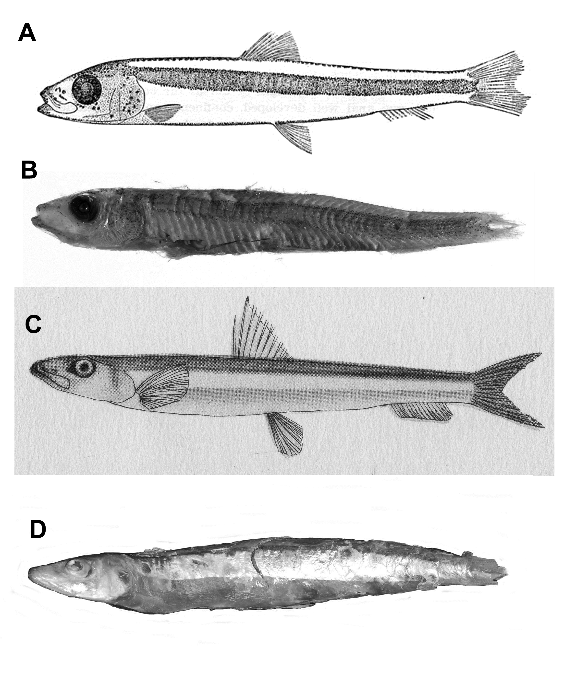
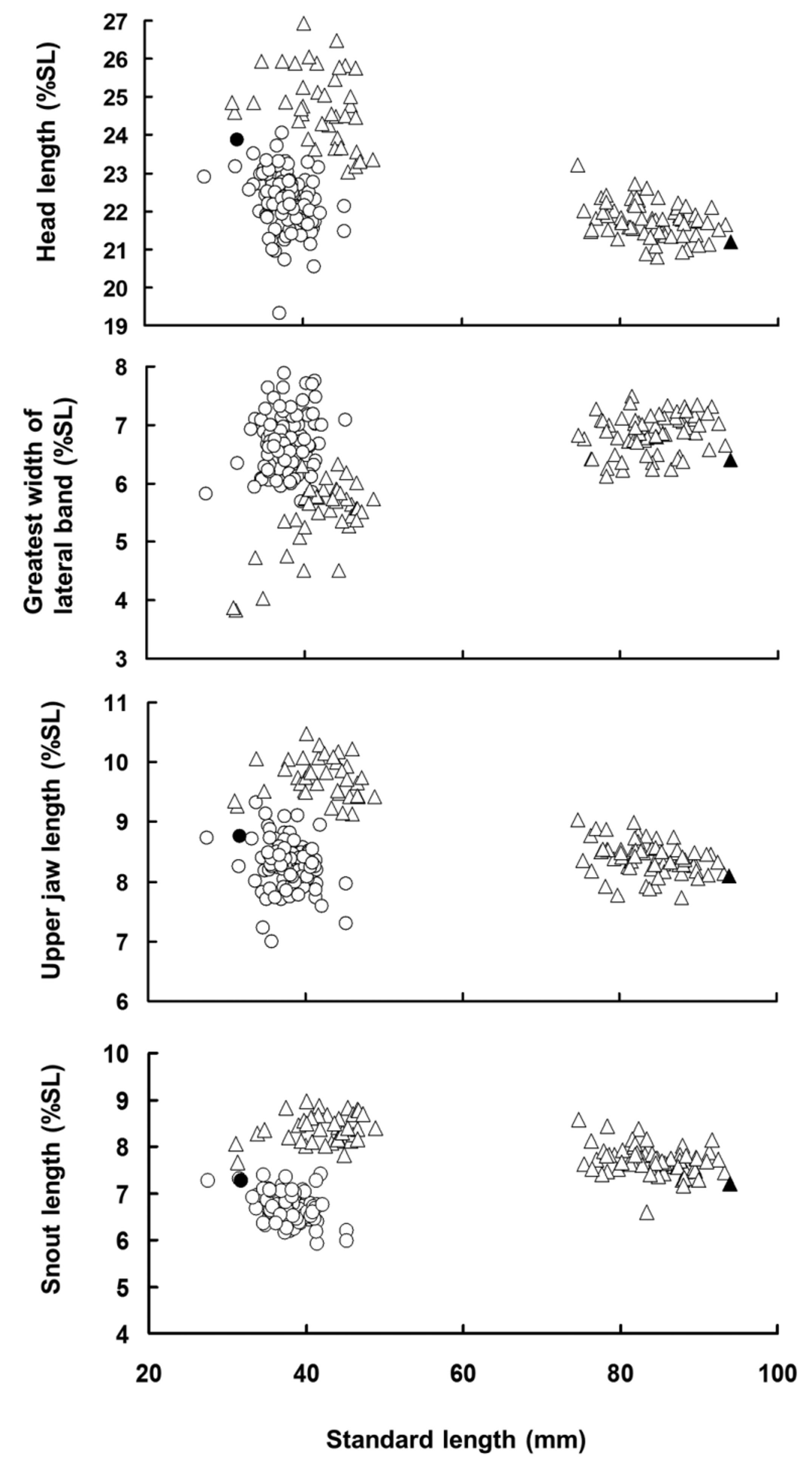
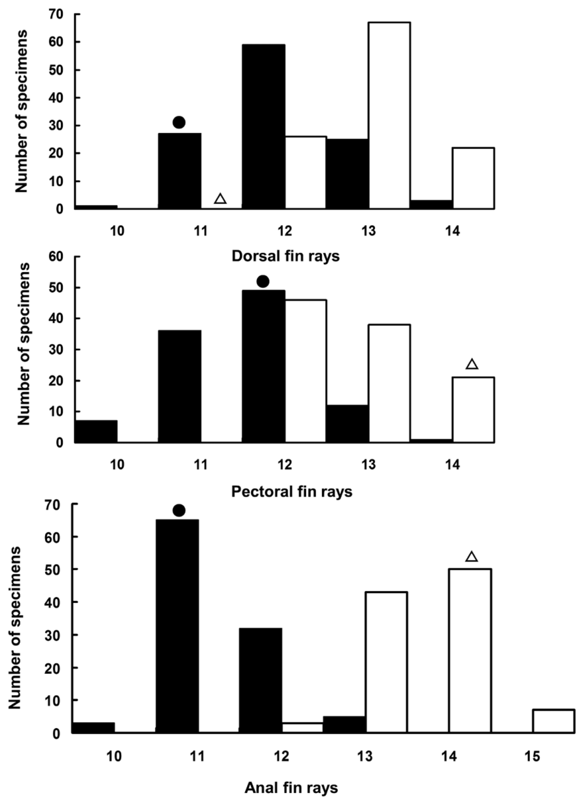
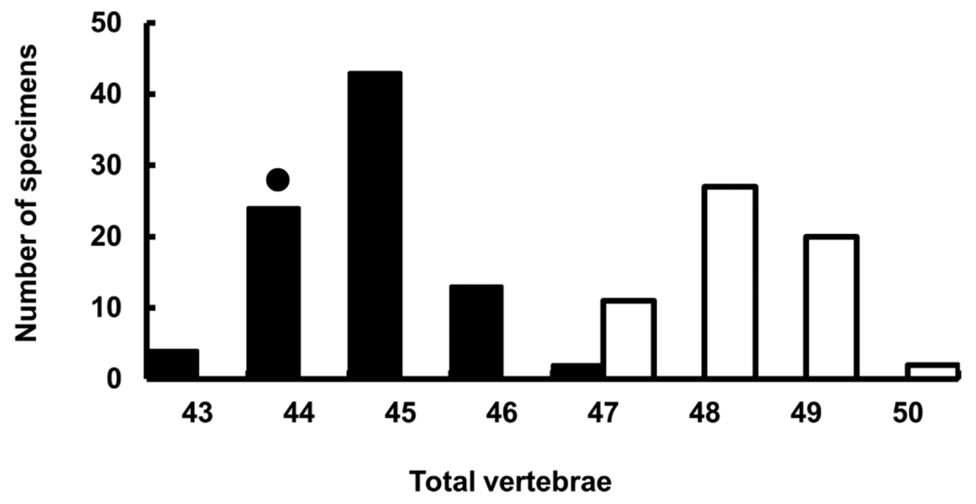
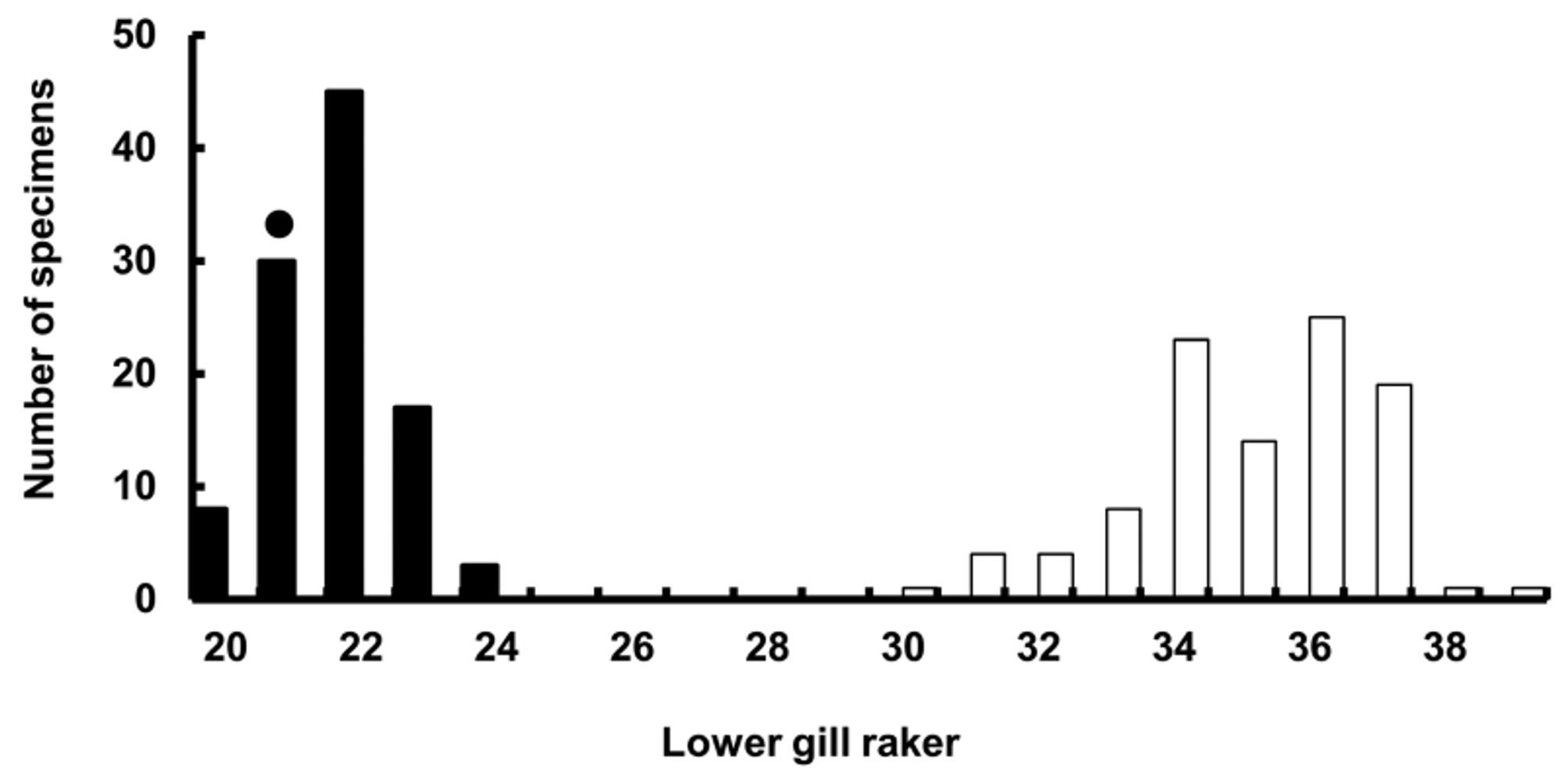
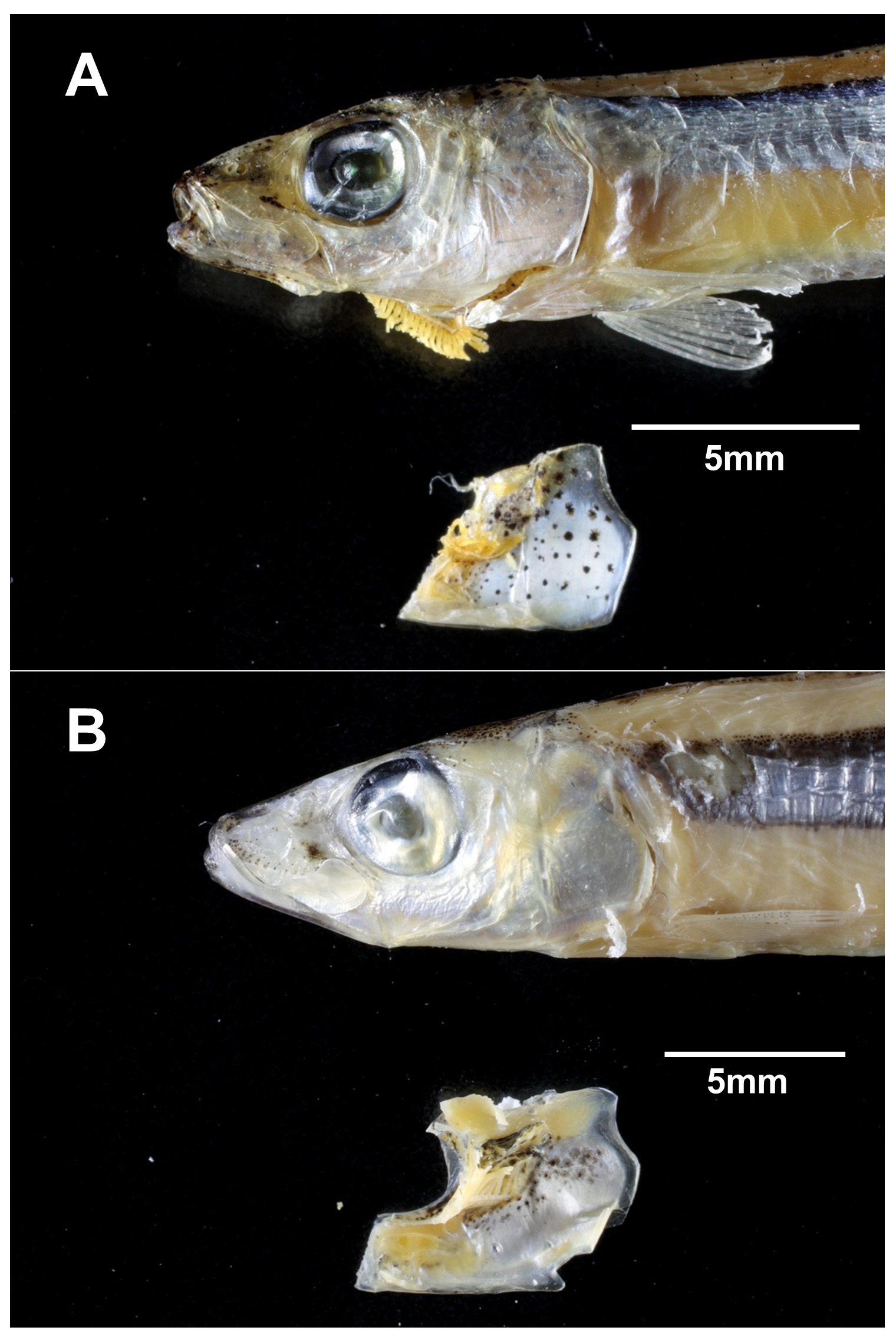
TABLE 1. Meristics and proportional measurements expressed as percentage of standard length (SL), of the type and non-type specimens of Spratelloides atrofasciatus and S. gracilis. (Figures in parentheses indicate mean values; a = data from Schultz 1943; b = data from lectotype description of Boeseman 1947).
| Holotype | Paratype | non-types | Lectotype | non-types |
|---|---|---|---|---|
| USNM 115099 | USNM 115100 | n = 110 | RMNH 3260a | n = 105 |
| Standard length (mm) 31.7 a Counts - dorsal-fin rays 11 a - pectoral-fin rays 12 a | 30.5 a 12 a – | 27.0–45.2 10–14 10–13 | 94.0 11 b 14 b | 31.0–93.3 12–15 12–14 |
| - pelvic-fin rays 8 a - anal-fin rays 11 a - lower gill rakers 21 | 8 a 10 a 19 a | 8–9 10–14 20–24 | 8 b 14 b – | 8 12–14 30–39 |
| - scales 44 a - vertebrae 44 Measurements as % SL - head length 24 a - body depth 14.5 a - body width – - eye diameter 7.3 a | 42 a – 24.3 a 14.7 a – 7.2 a | – 43–47 19.3–24.1 (22.2) 12.2–16.3 (14.0) 4.8–7.9 (6.2) 5.4–7.5 (6.3) | – – 21.2 – – – | – 47–50 20.8–26.9 (22.9) 11.6–18.2 (15.4) 4.8–7.3 (6.1) 4.4–8.6 (6.2) |
| - snout length 7.3 a - interorbital width 2.8 a - upper jaw length 8.8 a | 7.2 a 2.9 a 9.8 a | 5.9–7.4 (6.7) 3.1–5.2 (4.0) 7.0–9.3 (8.3) | 7.2 – 8.1 | 6.6–9.0 (8.0) 3.2–4.9 (3.9) 7.7–10.5 (8.9) |
| - greatest width of lateral band – - predorsal length 45.7 a - prepectoral length – | – 47.5 a – | 5.7–7.9 (6.7) 41.1–51.2 (47.0) 21.5–25.1 (23.3) | 6.4 – – | 3.8–7.5 (6.3) 46.4–50.9 (48.3) 21.3–28.2 (23.9) |
| - prepelvic length 52.1 a - preanal length 79 a - dorsal-fin base 9.5 a | 53 a 81.3 a 9.8 a | 49.7–58.4 (53.7) 76.2–83.5 (80.3) 7.9–12.6 (10.5) | – – – | 53.6–58.9 (56.5) 77.5–84.6 (82.5) 8.4–12.3 (10.3) |
| - anal-fin base 9.5 a | 9.8 a | 8.3–13.6 (10.3) | – | 7.9–11.8 (10.0) |
| - length of depressed dorsal fin 16.4 a | 16.4 a | 13.3–18.2 (15.9) | – | 13.1–18.2 (15.5) |
| - length of depressed anal fin – - length from pectoral-fin insertion to 30 a pelvic-fin insertion - length from pelvic insertion to anal-fin 24.3 a origin | – 30.2 a 26.2 a | 10.9–16.5 (13.9) 13.3–34.6 (30.2) 10.9–29.8 (26.5) | – – – | 11.3–16.0 (13.7) 29.8–39.8 (33.5) 23.9–29.8 (27.0) |
No known copyright restrictions apply. See Agosti, D., Egloff, W., 2009. Taxonomic information exchange and copyright: the Plazi approach. BMC Research Notes 2009, 2:53 for further explanation.
|
Kingdom |
|
|
Phylum |
|
|
Class |
|
|
Order |
|
|
Family |
|
|
Genus |
Spratelloides atrofasciatus Schultz, 1943
| Hidaka, Kouichi, Yamamuro, Taihei & Yoshino, Tetsuo 2015 |
Spratelloides atrofasciatus
| Aonuma 2002: 244 |
| Nishijimamoto 1963: 69 |
| Schulz 1953: 27 |
| Schulz 1943: 8 |