
Bifaxaria submucronata Busk, 1884
|
publication ID |
https://doi.org/10.11646/zootaxa.3856.1.4 |
|
publication LSID |
lsid:zoobank.org:pub:8A50EFE8-83AE-4298-B1EB-13805EB93AFF |
|
DOI |
https://doi.org/10.5281/zenodo.6141664 |
|
persistent identifier |
https://treatment.plazi.org/id/AC5C8787-FFC8-FFE7-42B0-FD7ECB7CFCB6 |
|
treatment provided by |
Plazi |
|
scientific name |
Bifaxaria submucronata Busk, 1884 |
| status |
|
Bifaxaria submucronata Busk, 1884 View in CoL
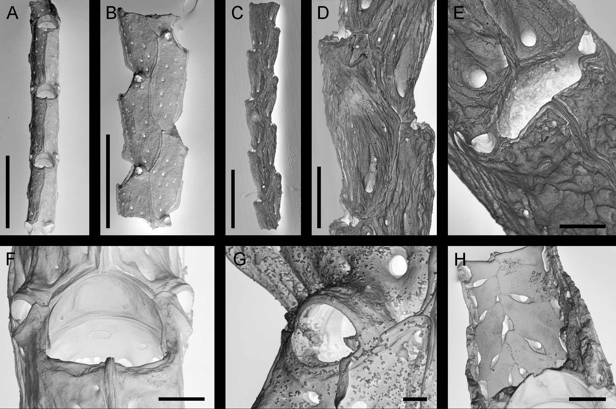
( Fig. 5 View FIGURE 5. A – B, F – H , A–B, F–H)
Bifaxaria submucronata Busk, 1884: 80 View in CoL , pl. 13, figs 1, 1a; Gordon 1993: 309, figs 3a–d, 18a–e (cum syn.).
Material examined. SMF 15003, SO 205 Stn 61, 13.174° N – 3.194° N, 118.106° W – 118.0953° W, Equatorial East Pacific, 3996–4007 m, collected 16 May 2010. Bleached.
Description. Colony candelabriform, jointed, with two lateral, slightly curved branches emanating from a branching point; mode of attachment to substratum not seen. Branches formed by laterally compressed zooids arranged in two alternating rows ( Fig. 5 View FIGURE 5. A – B, F – H , A–B). Lateral suture prominent, carinate, forming a sinuate line curving from avicularium to avicularium ( Fig. 5 View FIGURE 5. A – B, F – H , B). Outer shield layer umbonuloid, with mostly 3 rows of pores along main axis and additional smaller pores along sutures; frontal suture with distinct carina, forming a tip in middle of proximal orificial rim ( Fig. 5 View FIGURE 5. A – B, F – H , F). Inner orifice subcircular, proximal boundary rather straight; outer secondary orifice transversely D-shaped, with straight proximal rim. Avicularia distolateral, one on each side of orifice in distal third ( Fig. 5 View FIGURE 5. A – B, F – H , F), small and circular with tiny mandibular pivots that do not touch ( Fig. 5 View FIGURE 5. A – B, F – H , G). Spinocystal costae sinuous, touching each other thus leaving intercostal spaces ( Fig. 5 View FIGURE 5. A – B, F – H , H). Fertile zooids not observed.
Measurements.
Zooid length 785–1023 µm, σ = 82 µm, N = 6; width 327–403 µm, σ = 24 µm, N = 6
Branch width 451–552 µm, σ = 41 µm, N = 6
Orifice diameter 193–227 µm, σ = 12 µm, N = 6.
Remarks. Distinguishing B. submucronata from other similar species, mainly B. compacta Gordon, 1993 , B. gracilis Gordon, 1993 and B. modesta Gordon, 1993 , can be achieved by comparing zooidal dimensions and the shape and dimensions of the spinocystal costae. In B. modesta the costae are simple and do not touch each other, whereas in all other species they are somewhat sinuous and adjacent costae touch each other. Bifaxaria submucronata can be distinguished from the other two species by both its greater zooidal length and width compared with B. gracilis and greater zooidal length compared to B. compacta .
| SMF |
Forschungsinstitut und Natur-Museum Senckenberg |
No known copyright restrictions apply. See Agosti, D., Egloff, W., 2009. Taxonomic information exchange and copyright: the Plazi approach. BMC Research Notes 2009, 2:53 for further explanation.
|
Kingdom |
|
|
Phylum |
|
|
Class |
|
|
Order |
|
|
SubOrder |
Neocheilostomina |
|
Family |
|
|
Genus |
Bifaxaria submucronata Busk, 1884
| Matsuyama, Kei, Janssen, Annika, Arbizu, Pedro Martínez, Martha, Silviu O. & Freiwald, André 2014 |
Bifaxaria submucronata
| Gordon 1993: 309 |
| Busk 1884: 80 |