
Taeniacanthus mcgroutheri, Tang, Danny, Uyeno, Daisuke & Nagasawa, Kazuya, 2011
|
publication ID |
https://doi.org/ 10.5281/zenodo.201761 |
|
DOI |
https://doi.org/10.5281/zenodo.6189828 |
|
persistent identifier |
https://treatment.plazi.org/id/AC718795-212D-4C45-FF64-FD0FFBAFF9A7 |
|
treatment provided by |
Plazi |
|
scientific name |
Taeniacanthus mcgroutheri |
| status |
sp. nov. |
Taeniacanthus mcgroutheri n. sp.
( Figs 9–11 View FIGURE 9 View FIGURE 10 View FIGURE 11 )
Type material. Ƥ holotype ( WAM C38683) and 24 Ƥ paratypes (1 damaged) ( WAM C38555, C38605, C38610, C38684, C38773–C38774), ex 4 Monacanthus chinensis (Osbeck) ( WAM P.25095.025), Exmouth Gulf, Western Australia, Australia, October 1974.
Other material examined. 27 Ƥ (AM P69677), ex 1 M. chinensis (AM I20230-004), Cockburn Sound, Western Australia, Australia, 27 March 1978; 12 Ƥ ( WAM C38677–C38678, C38762), ex 2 M. chinensis ( WAM P.24985.001), Cockburn Sound, Western Australia, Australia, August 1974; 1 Ƥ ( WAM C38590), ex M. chinensis ( WAM P.31985.001), Cockburn Sound, Western Australia, Australia, 1975; 1 Ƥ ( MAGNT Cr014946), ex M. chinensis ( MAGNT S.12894-001), Broome, Western Australia, Australia, 20 May 1987; 2 Ƥ ( MAGNT Cr014948), ex 1 M. chinensis ( MAGNT S.13917-006), Darwin Harbour, Northern Territory, Australia, 5 April 1994; 35 Ƥ (1 damaged) ( MAGNT Cr014950–Cr014954), ex 1 M. chinensis ( MAGNT S.10352-003), Shoal Bay, Northern Territory, Australia, 5 April 1977; 1 Ƥ ( MAGNT Cr015005), ex Paramonacanthus choirocephalus (Bleeker) ( MAGNT S.12768-001), Darwin Harbour, Northern Territory, Australia, 30 May 1989; 5 Ƥ ( MAGNT Cr015006), ex 1 P. choirocephalus ( MAGNT S.13735-009), Darwin Harbour, Northern Territory, Australia, 9 September 1993; 4 Ƥ ( MAGNT Cr015007), ex 1 P. choirocephalus ( MAGNT S.13736-011), Darwin Harbour, Northern Territory, Australia, 9 September 1993.
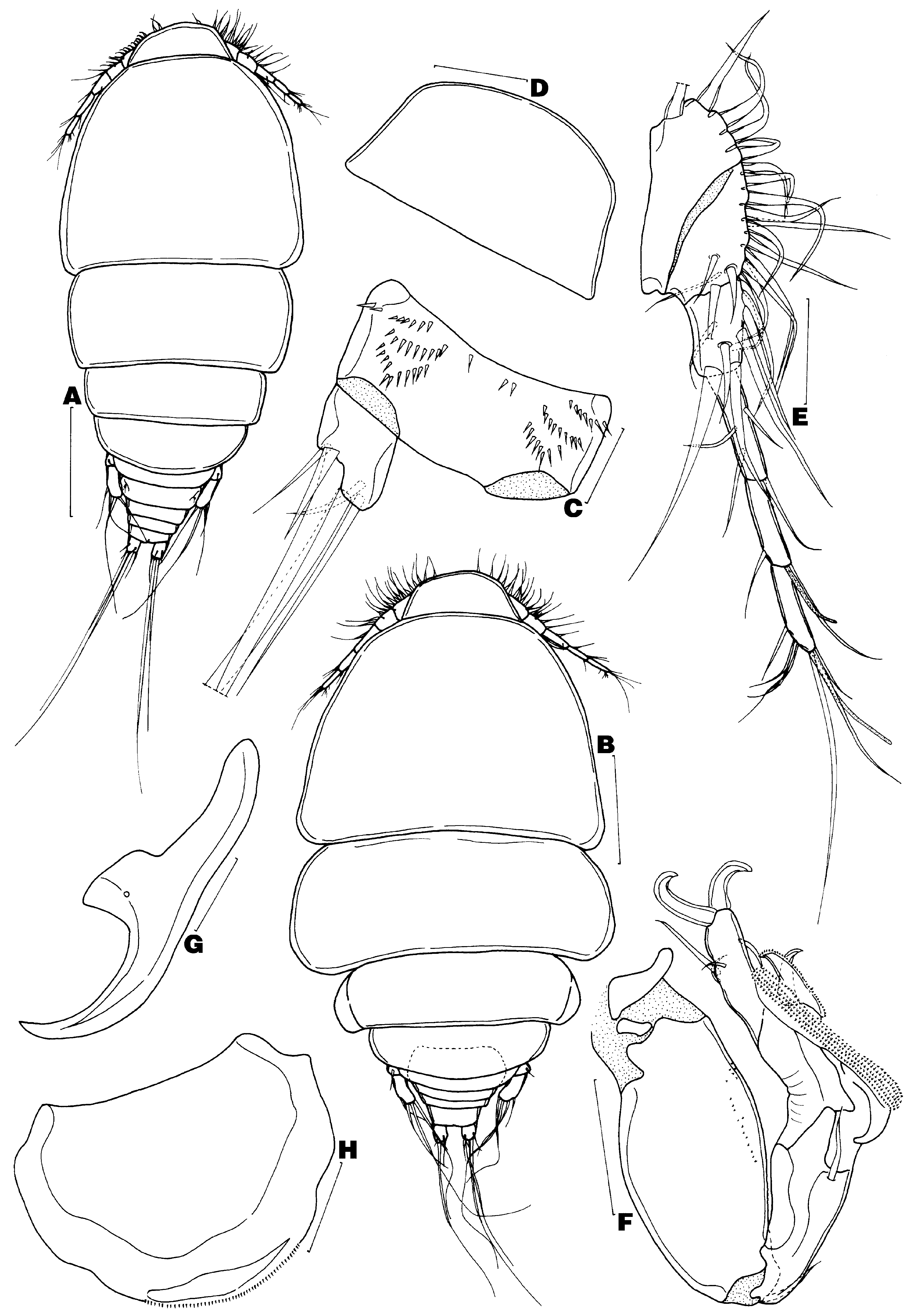
Description of adult female. Body 0.60 mm long (excluding caudal setae) and 254 µm wide (n = 11) ( Fig. 9 View FIGURE 9 A). Several specimens with second pedigerous somite broader (290 µm) than cephalothorax (280 µm) ( Fig. 9 View FIGURE 9 B). Prosome 487 µm long, composed of cephalothorax (cephalosome plus first pedigerous somite) and 3 free pedigerous somites. Second pedigerous somite 244 µm wide; 3rd and 4th pedigerous somites decreasing in width posteriorly. Urosome comprised of 5th pedigerous somite, genital double-somite and 3 free abdominal somites. Genital double-somite wider (89 µm) than long (52 µm). Abdomen 54 µm long and 67 µm wide; first 2 abdominal somites unornamented; ventral surface of anal somite ( Fig. 9 View FIGURE 9 C) with 3 interrupted rows of spinules. Caudal ramus ( Fig. 9 View FIGURE 9 C) longer (17 µm) than wide (14 µm), bearing 6 naked setae (seta I not observed).
Rostral area ( Fig. 9 View FIGURE 9 A, D) highly protuberant, lacking ventromedian sclerotised structure. Antennule ( Fig. 9 View FIGURE 9 E) 6-segmented (articulation between ancestral segments XIV–XVII and XVIII–XX not expressed); armature formula: 5, 14, 8, 4, 2 + 1 aesthetasc, and 7 + 1 aesthetasc. Antenna ( Fig. 9 View FIGURE 9 F) indistinctly 4-segmented. Coxobasis robust, bearing minute spinules on outer border and distal seta. First endopodal segment with long inner seta. Second endopodal segment bearing 2 unequal pectinate processes and claw-like spine; large pectinate process with multiple rows of spinules (usual seta not observed, possibly broken off); small pectinate process with row of spinules and minute seta. Third endopodal segment elongate, extending well-beyond pectinate processes, armed with 4 setae and 2 claw-like spines. Postantennal process ( Fig. 9 View FIGURE 9 G) with wide base and sharply curved tine.
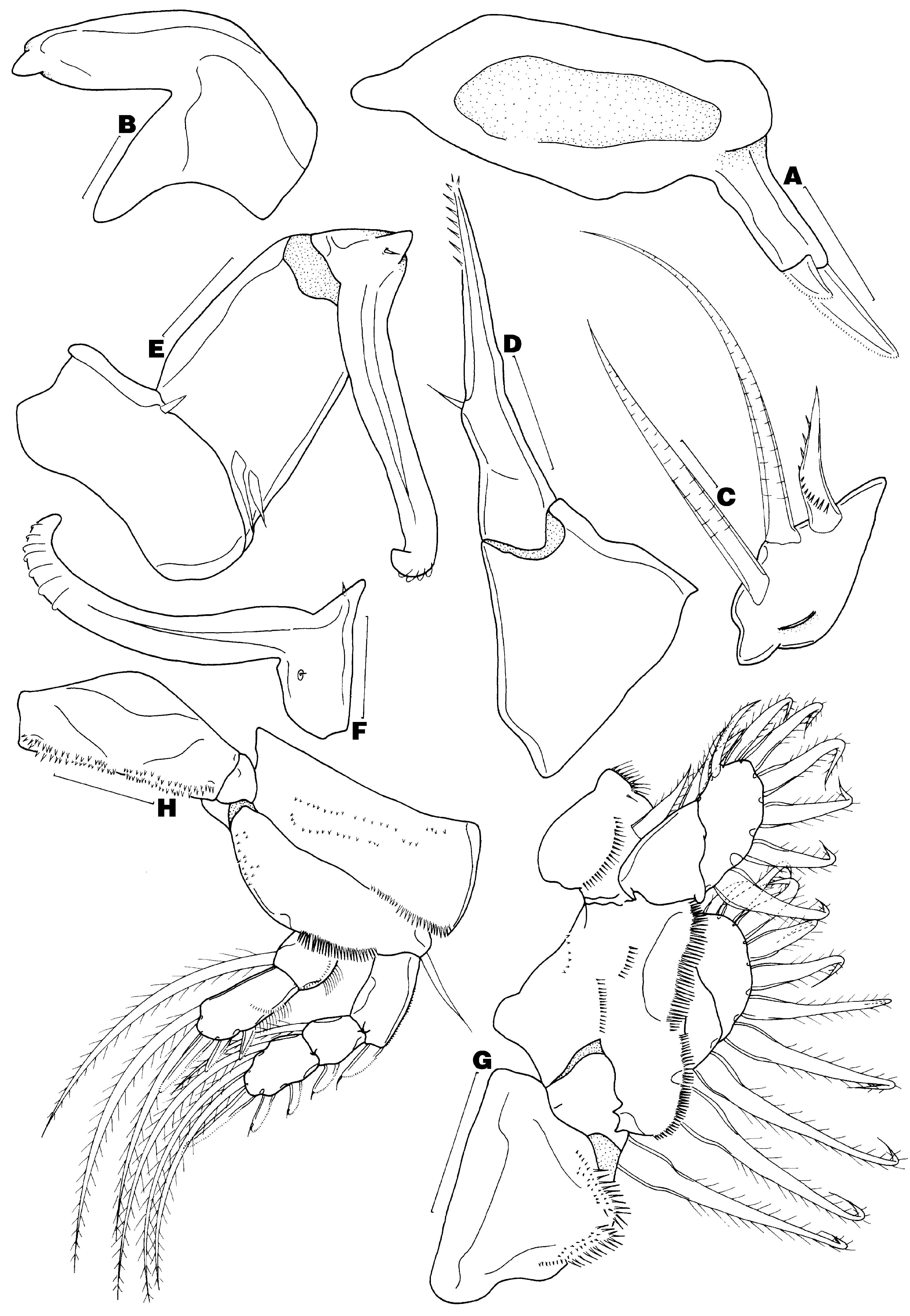
Labrum ( Fig. 9 View FIGURE 9 H) spinulate along posterior margin. Mandible ( Fig. 10 View FIGURE 10 A) with 2 unequal spinulate blades. Paragnath ( Fig. 10 View FIGURE 10 B) with nipple-like protuberance at tip. Maxillule ( Fig. 10 View FIGURE 10 C) lobate, bearing 2 long naked setae, spinulate seta, lateral protrusion and small anterior ridge. Maxilla ( Fig. 10 View FIGURE 10 D) 2-segmented; syncoxa naked; basis armed with spinulate terminal process and naked seta. Maxilliped ( Fig. 10 View FIGURE 10 E–F) 3-segmented; syncoxa with naked seta; basis armed with 2 proximal naked setae; endopod segment a curved claw, bearing 2 minute setae, basal conical protrusion and transverse flanges along inner distal margin.
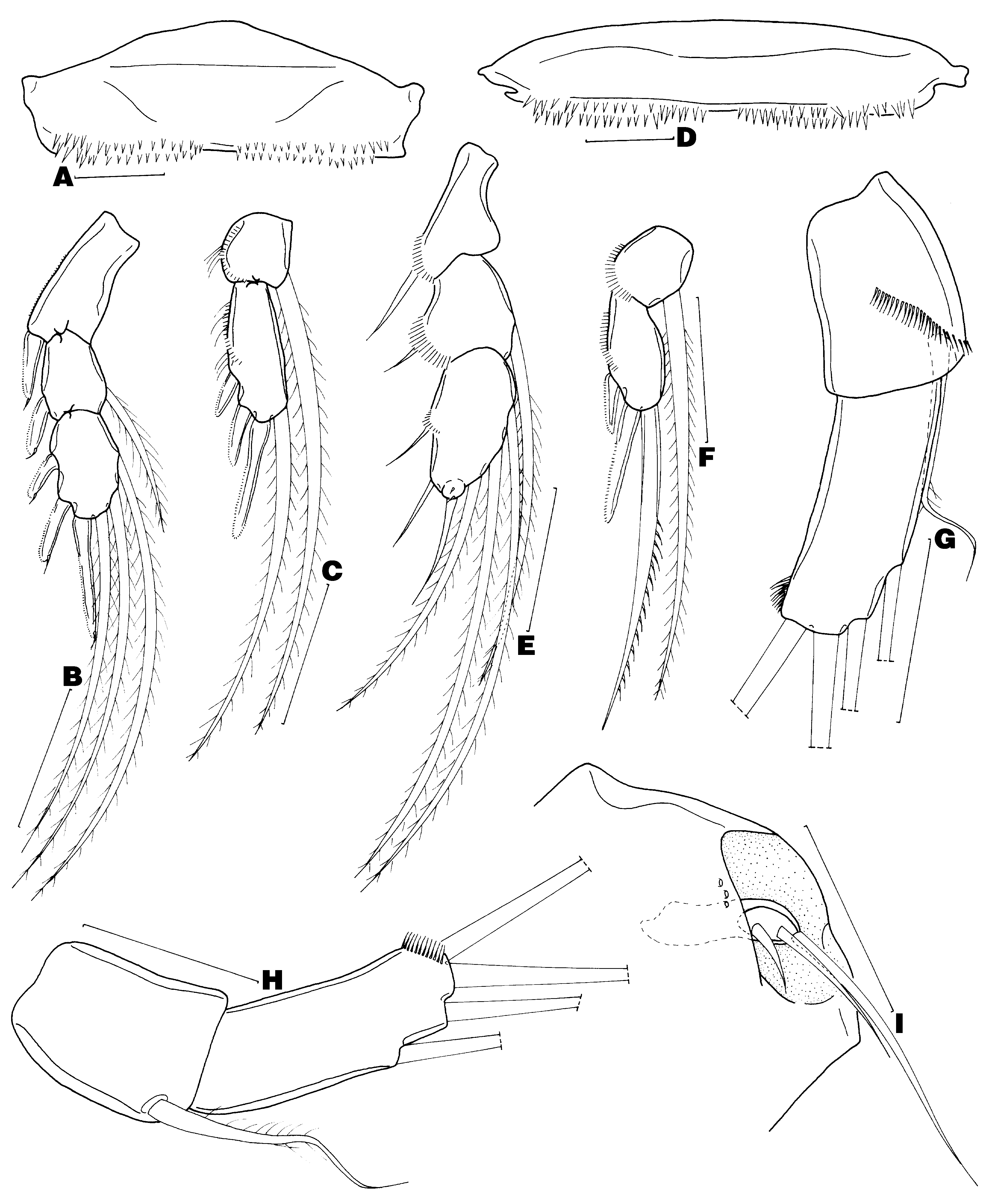
Legs 1–4 biramous ( Figs 10 View FIGURE 10 G–H, 11A–F), with 2-segmented rami on leg 1 and 3-segmented exopod and 2 - segmented endopod on legs 2–4. Armature on rami of legs 1 to 4 as follows (Roman numerals = spines; Arabic numerals = setae; int. = intermediate spine).
Leg 1 ( Fig. 10 View FIGURE 10 G) flattened and expanded. Intercoxal sclerite with short and long spinules posteriorly. Coxa and basis with several rows of spinules. Intercoxal sclerite of legs 2–4 ( Figs 10 View FIGURE 10 H, 11A, D) with several rows of spinules on posterior edge; leg 4 intercoxal sclerite widest. Leg 2 coxa ( Fig. 10 View FIGURE 10 H) with row of long spinules on posterolateral margin and several rows of spinules on ventral surface; basis with row of large spinules on posterior border and patch of small spinules along inner margin. Leg 2 exopod ( Fig. 10 View FIGURE 10 H) with outer row of spinules on first segment; exopodal spines spinulate along lateral margin and armed with terminal flagellum; inner seta on second segment relatively short. Leg 2 endopod ( Fig. 10 View FIGURE 10 H) with outer row of setules and spinules; terminal spines spinulate. Coxa and basis of legs 3 and 4 similar to those of leg 2, except spinules along inner margin of basis absent. Leg 3 exopod ( Fig. 11 View FIGURE 11 B) identical to leg 2 exopod. Leg 3 endopod ( Fig. 11 View FIGURE 11 C) similar to that of leg 2, except with 1 fewer seta on terminal segment. Leg 4 exopodal segments ( Fig. 11 View FIGURE 11 E) each with outer row of spinules; spines weakly sclerotised; inner seta on second segment longer than that of legs 2 and 3; third segment with distal protrusion bearing 2 minute spinules. Leg 4 endopod ( Fig. 11 View FIGURE 11 F) with outer row of spinules; first 2 spines spinulate along outer border; intermediate spine with row of large spinules along inner margin. Leg 5 ( Fig. 11 View FIGURE 11 G–H) uniramous, 2-segmented. Protopodal segment armed with dorsolateral, weakly pinnate seta and row of long spinules. Free exopodal segment with distomedial row of long spinules and 2 subterminal and 2 terminal naked setae (outer terminal seta longest of 4). Leg 6 ( Fig. 11 View FIGURE 11 I) vestigial, represented by 3 unequal naked setae at egg sac attachment area.
Adult male. Unknown.
Attachment site. Orbit.
Etymology. This species is named in honour of Mark McGrouther, Collection Manager (Ichthyology) at the Australian Museum.
Remarks. Taeniacanthus mcgroutheri n. sp. resembles T. brayae n. sp. in the protuberant rostrum without a ventromedian plate, the 6-segmented antennule, structure of the third endopodal segment of the antenna, spinulate seta on the maxillule, maxilla with a spinulated terminal process and one seta, armature formula of II, I, 3 on the third exopodal segment of legs 2–4 and leg 5 armed with four long setae on the free exopodal segment. Both species are also parasitic on the external surface of the eyes of several monacanthid hosts. Taeniacanthus mcgroutheri n. sp. can be differentiated from T. brayae n. sp. by having 14 setae (rather than 15) on the second antennulary segment, numerous rows of spinules (rather than one) on the large pectinate process of the antenna, three setae (rather than four) on the maxillule and a bimerous (rather than trimerous) endopod on legs 2–4. Comparisons of the ornamentation and setation patterns of the endopod of legs 2–4 between these two species indicates that the 2-segmented condition exhibited by T. mcgroutheri n. sp. is most likely derived from the fusion of the middle and terminal segments (or failure of the middle and terminal segments to separate) rather than the amalgamation of the first and middle segments (or failure of the first and second segments to divide).
No known copyright restrictions apply. See Agosti, D., Egloff, W., 2009. Taxonomic information exchange and copyright: the Plazi approach. BMC Research Notes 2009, 2:53 for further explanation.