
Eremophylus nakagawai, Yasunaga, 2022
|
publication ID |
https://doi.org/10.11646/zootaxa.5094.1.1 |
|
publication LSID |
lsid:zoobank.org:pub:72F6E1D9-E9E4-41F6-9AC0-97A249F94E68 |
|
DOI |
https://doi.org/10.5281/zenodo.6301166 |
|
persistent identifier |
https://treatment.plazi.org/id/AC7FEA3B-080A-FFE0-FF78-FC09FD2C70D8 |
|
treatment provided by |
Plazi |
|
scientific name |
Eremophylus nakagawai |
| status |
sp. nov. |
Eremophylus nakagawai n. sp.
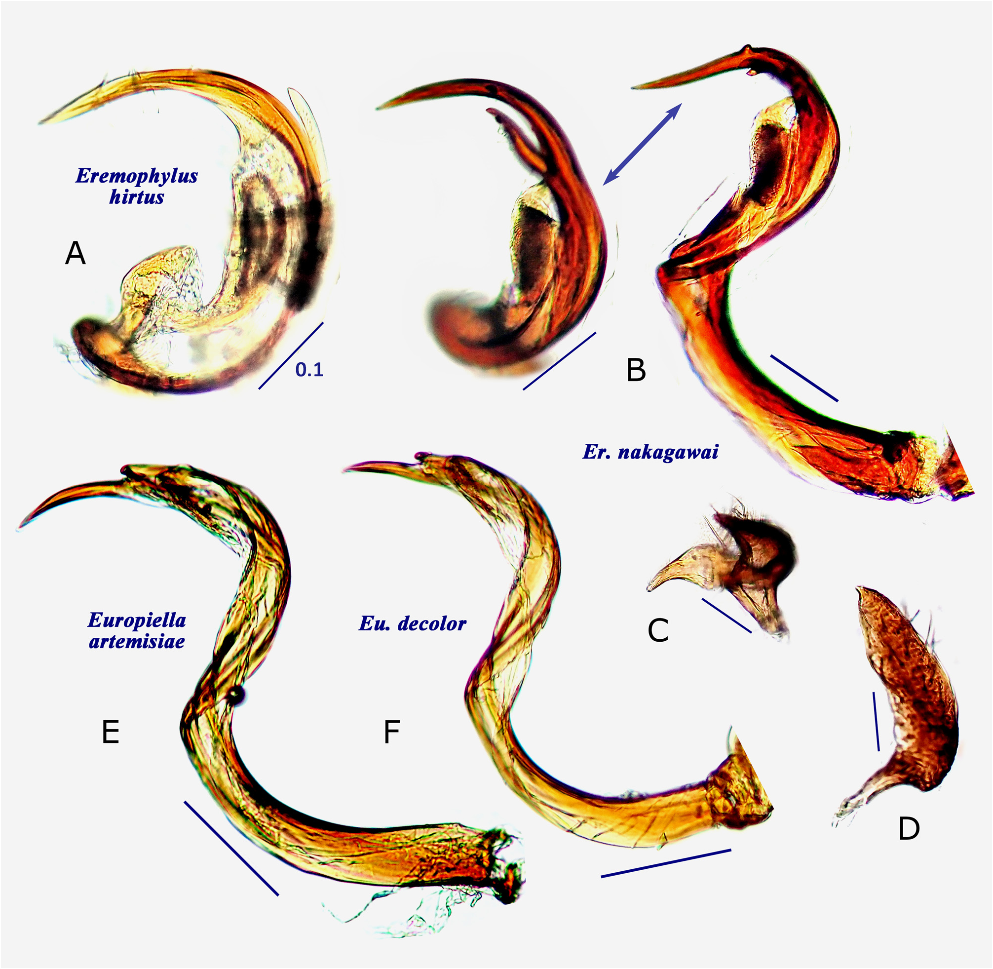
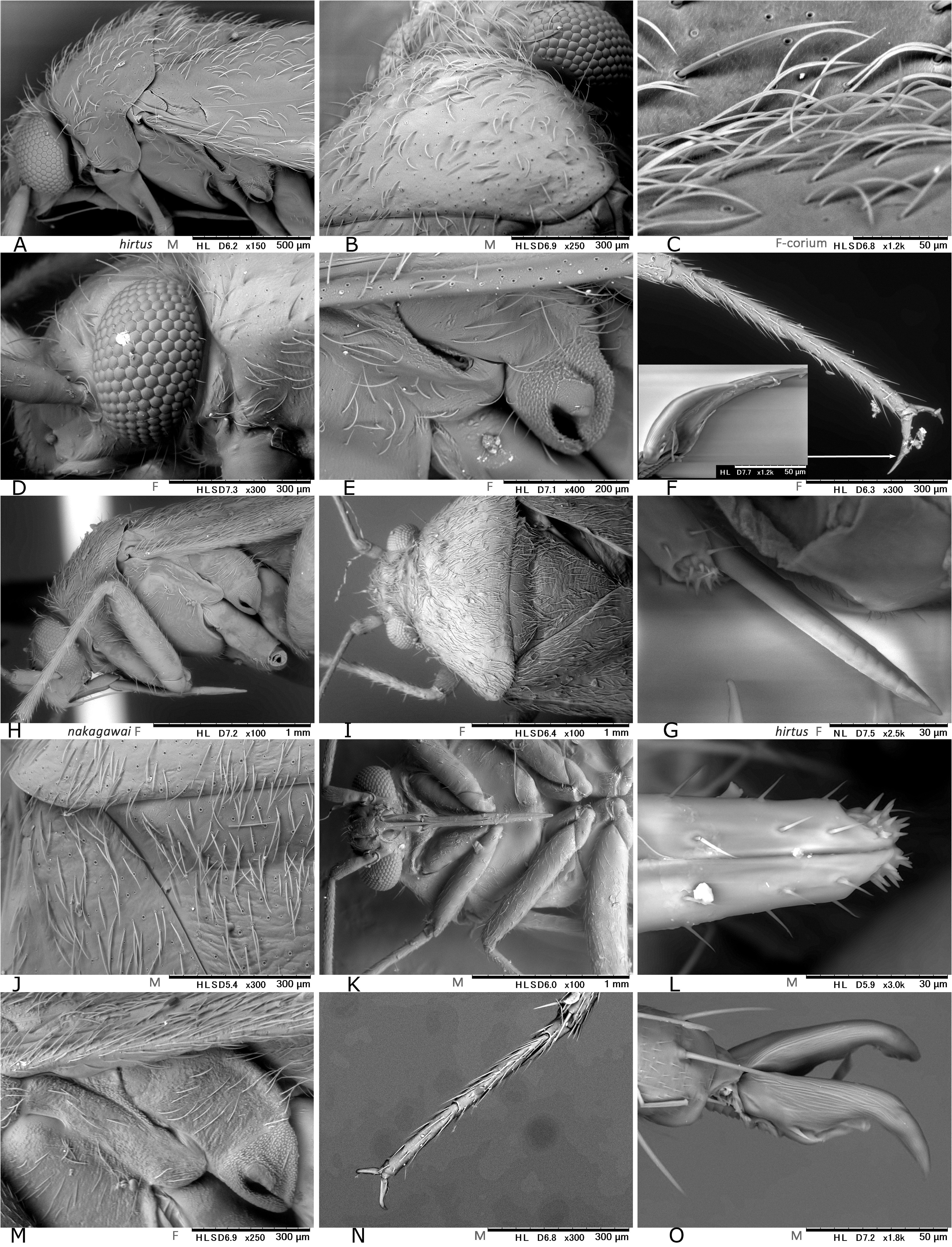
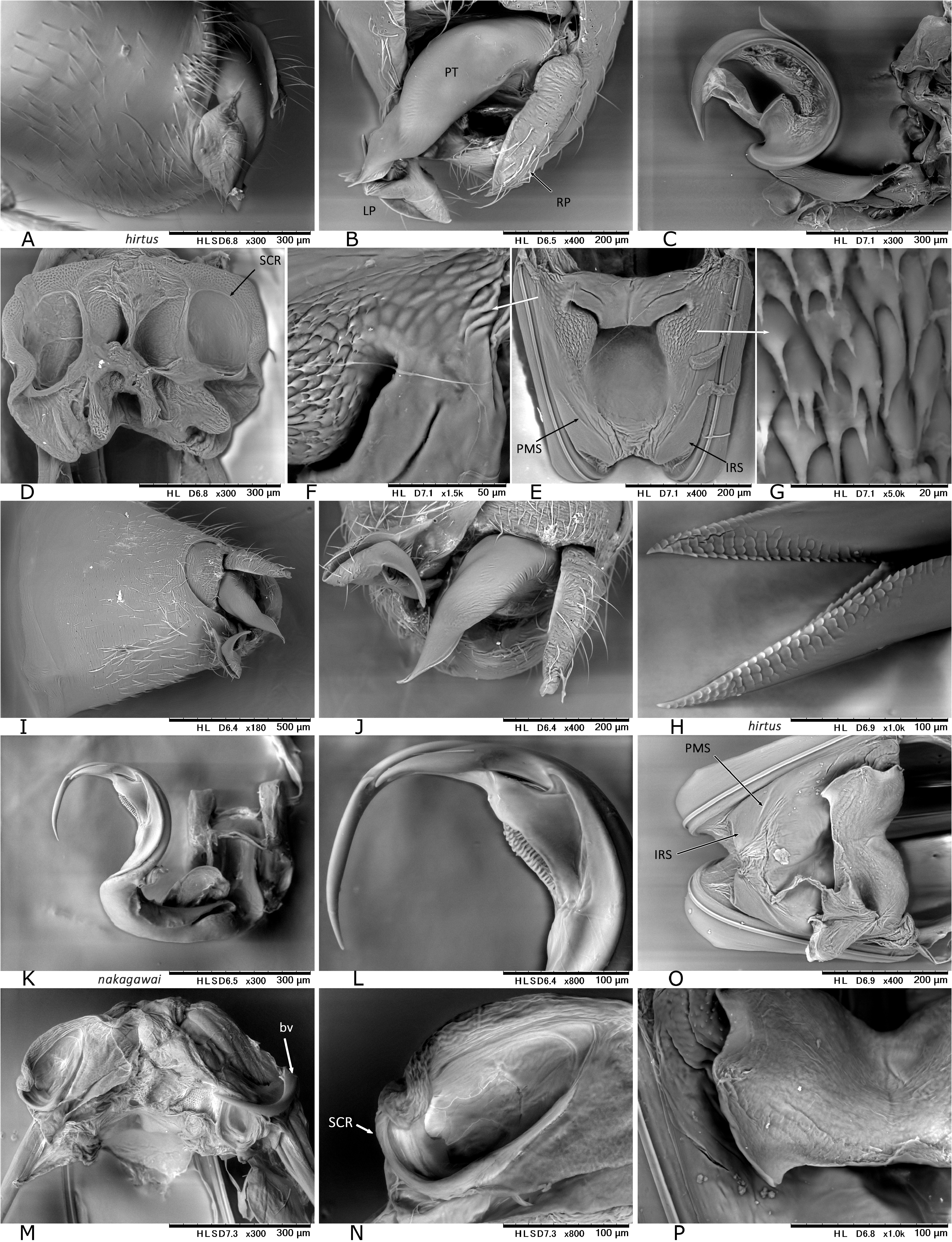
( Figs. 1 View FIGURE 1 A−E; 2C−I; 3B, D; 4B−D; 16; 18H−O; 19I−P)
Material examined. Holotype ( ♂). JAPAN: Honshu, Akita Pref., Yurihonjo City, Mt. Chokai , Chokai Town , Sarukura , 39.17 140.14, 610 m alt., on Phellodendron amurense Rupr. , 13 Jun 2019, Y. Nakagawa ( AMNH) ( AMNH _ PBI 00380697 About AMNH ) . Paratypes: JAPAN: Same data as for holotype 5♂ ( TYCN); same data, except for date, 27 Jun 2017, 3♂ 4♀ & 26 Jun 2018, 1♀ ( CNC, TYCN) .
Diagnosis. Easily distinguished from the other congener, E. hirtus , by the following characters: Antennal segment II almost entirely pale; metafemur as long as or shorter than antennal segment II; and metatibia yellowish brown. The final instar nymph is recognized by its pale green, elongate-ovoid body; uniformly distributed, dark spines on dorsum; and small dark spots on head, antennae, thorax, and legs ( Fig. 1E View FIGURE 1 ).
Description. General coloration not sexually dimorphic, but female has slightly larger size, wider vertex, and shorter antennal segment II ( Table 1 View TABLE 1 ) as in many other mirids. Body elongate, medium-sized; basic coloration variable, usually dark brown, but widely smoky yellow in pale variant (cf. Fig. 2H–I View FIGURE 2 ); dorsal surface usually dark brown except for inner margin of corium and cuneal margin ( Fig. 1C–D View FIGURE 1 ), matte, with densely distributed, brown, simple setae and silvery, lanceolate setae ( Fig. 18I–J View FIGURE 18 ). Head dark brown, partly or widely smoky yellow; eyes small; vertex wide, about twice as wide as an eye in dorsal view. Antenna yellowish brown; each segment generally linear; segment I dark brown; extreme bases of segments II and III infuscate; combined length of segments III+IV slightly longer than II. Labium shiny yellowish brown; its length shorter than antennal segment II; its apex not exceeding middle of mesocoxa ( Fig. 18H, K View FIGURE 18 ); apical 1/3 of segment IV darkened; sensilla on labial apex as in Fig. 18L View FIGURE 18 . Pronotum weakly shining, somewhat shagreened, varying from smoky yellow ( Fig. 2H View FIGURE 2 ) to dark brown ( Fig. 2C, F View FIGURE 2 ); calli weakly elevated; mesoscutum, scutellum and thoracic pleura including scent efferent system varying from yellowish brown to fuscous; scutellum (if darkened) with a mesal pale stripe ( Fig. 1C–D View FIGURE 1 ); peritreme of scent efferent system small ( Fig. 18M View FIGURE 18 ). Hemelytron variable in color; inner margin of corium and margins of cuneus always pale; membrane smoky brown, with pale median part and veins. Coxae varying from smoky yellow ( Fig. 2I View FIGURE 2 ) to dark brown ( Fig. 2G View FIGURE 2 ); legs yellowish brown, variable in color; profemur spotted apically, sometimes with darkened base; metafemur widely darkened ( Fig. 2D, G View FIGURE 2 ) or with 3 rows of dark spots ventrally ( Fig. 2E, I View FIGURE 2 ), about as long as or shorter than antennal segment II; each tibia with dark spots at bases of spines; apical part of each tarsomere III dark brown; meta-tarsomere II slightly longer than III ( Fig. 18N View FIGURE 18 ); claw thick, with relatively developed pulvilli ( Fig. 18O View FIGURE 18 ). Abdomen varying from pale to fuscous. Male genitalia ( Figs. 4 View FIGURE 4 B−D; 19I−L): Pygophore with a ventral mesal keel at apical 1/3; phallotheca stout ( Fig. 19J View FIGURE 19 ); right paramere about twice as large as left; ( Fig. 4C–D View FIGURE 4 ); vesica not inflated at middle ( Fig. 19I View FIGURE 19 ), with sharp subapical branch ( Fig. 19L View FIGURE 19 ). Female genitalia ( Figs. 3B, D View FIGURE 3 ; 19 View FIGURE 19 M−P): Sclerotized rings thick-rimmed, elongate-oval ( Figs. 3D View FIGURE 3 , 19N View FIGURE 19 ); posterior wall relatively smooth ( Fig. 19O View FIGURE 19 ), with interramal sclerite sparsely spinulate ( Fig. 19P View FIGURE 19 ); ovipositor (gonapophysis) I expanded subapically ( Fig. 3D View FIGURE 3 ).
Measurements: See Table 1 View TABLE 1 .
Etymology. Named for Mr. Y. Nakagawa ( Akita, Japan) who enthusiastically collected the specimens of this new species, observed its ecology, and shared the images.
Biology. Nakagawa (pers. obs.) verified the host association of Eremophylus nakagawai with inflorescence of Phellodendron amurense Rupr. (Rutaceae) ( Fig. 1A–B View FIGURE 1 ), on which both the adults and late instar immature forms were found together ( Fig. 1E View FIGURE 1 ). A univoltine life cycle is assumed for this new species. The adults appear in mid to late June.
Remarks. A ‘horn-like sclerite’ on the female seminal depository (‘bv’ in Figs. 3B View FIGURE 3 , 19M View FIGURE 19 ) was not regarded as a unique character but simply a fragment of apical part of the vesica (endosoma) after dissecting two additional female specimens. It was presumed to be ‘accidentally broken off’ during copulation and left in the female genital chamber, which may represent an unprecedented case.


No known copyright restrictions apply. See Agosti, D., Egloff, W., 2009. Taxonomic information exchange and copyright: the Plazi approach. BMC Research Notes 2009, 2:53 for further explanation.
|
Kingdom |
|
|
Phylum |
|
|
Class |
|
|
Order |
|
|
Family |
|
|
Tribe |
Phylini |
|
SubTribe |
Oncotylina |
|
Genus |