
Sarax yayukae, Rahmadi, Cahyo, Harvey, Mark S. & Kojima, Jun-Ichi, 2010
|
publication ID |
https://doi.org/10.5281/zenodo.197810 |
|
DOI |
https://doi.org/10.5281/zenodo.6207611 |
|
persistent identifier |
https://treatment.plazi.org/id/AD1687F3-FF82-FF95-0CEB-48172A95FE13 |
|
treatment provided by |
Plazi |
|
scientific name |
Sarax yayukae |
| status |
sp. nov. |
Sarax yayukae View in CoL sp. nov.
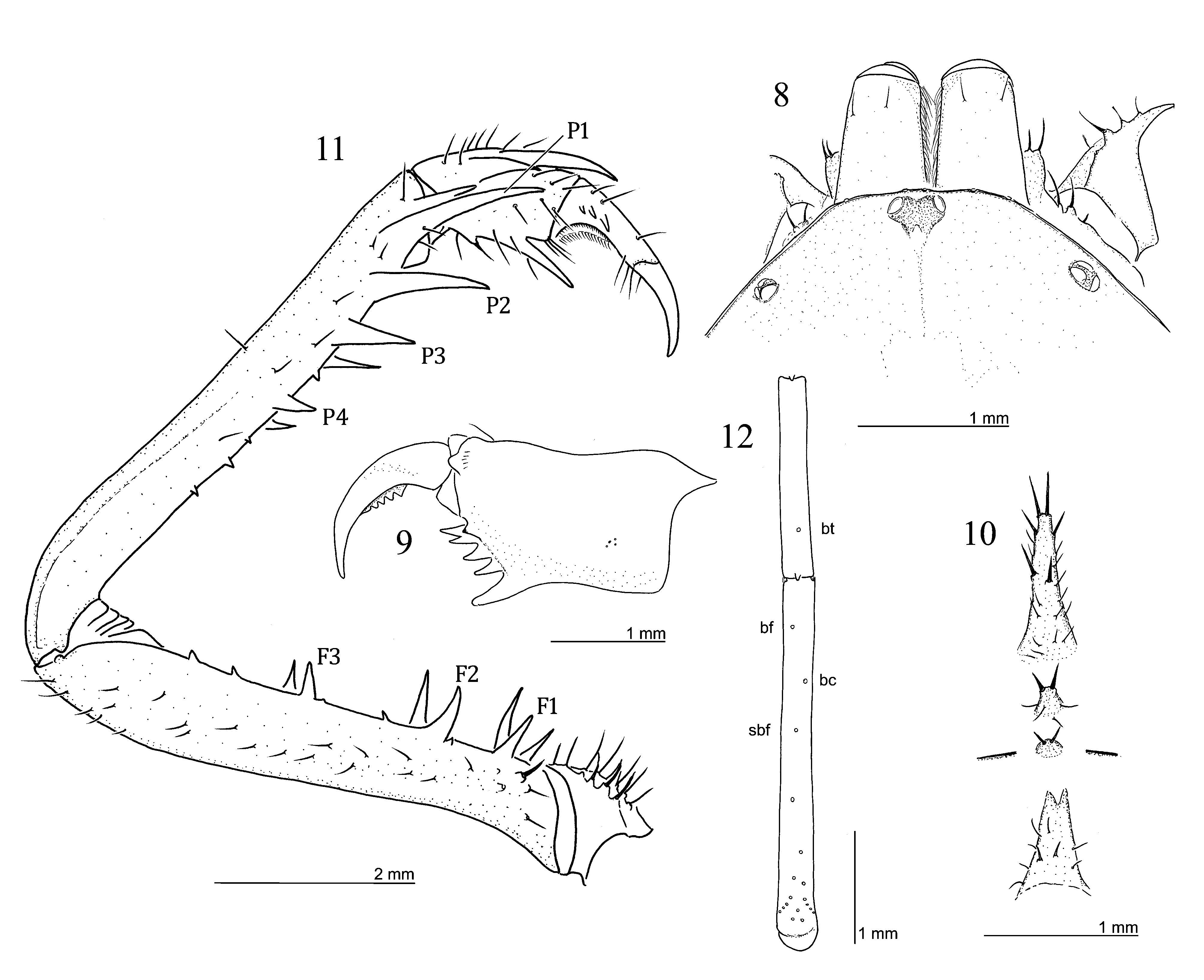
( Figs. 8 – 15 View FIGURES 8 – 12 View FIGURES 13 – 15 , 35 – 36 View FIGURES 33 – 40 )
Material examined: Male holotype ( MZB.Ambl.056), INDONESIA: Central Kalimantan: Murung Raya Regency, Tumbang Topus, Liang Puruk, GPS coordinates 0o27’45.0”N, 115o00’55.5”E, 9 June 2004, C. Rahmadi, Y.R. Suhardjono & D. Silam. Paratypes: 2 females and 1 juvenile ( MZB.Ambl. 057-059), 1 female (MNHN-Am.09), Liang Puruk, GPS coordinates 0o27’45.0”N, 115o00’55.5”E, 9 June 2004, C. Rahmadi, Y.R. Suhardjono & D. Silam; 1 male ( MZB.Ambl.0 93), Liang Hajuq, GPS coordinates 0o27’37.9”N, 115o00’47.6”E, 11 June 2004, C. Rahmadi; West Kalimantan: 1 male ( MZB.Ambl.148), Gua Kelasi in buffer zone, Bukit Raya-Bukit Baka National Park, Menukung, Melawi, 7 August 2008, K.P.G. Himakova. 1 male, 3 females ( CAS, CASENT 9036127), MALAYSIA: Sabah: Kota Kinabalu, Manukan Island, under logs in forest, 6 May 2006, T. Briggs; 1 juvenile ( CAS, CASENT 9036126), Kota Kinabalu, Manukan Island, jogging track on leaf litter, under logs, 6 May 2006, H. Tu & T. Briggs.
Diagnosis: Sarax yayukae has distinct sexual dimorphism, the male with strong and slender pedipalp whereas the female shorter and stouter. The species is medium-sized with an adult body length of about 8.8- 11.8 mm. Pedipalpal femur with three major spines on antero-dorsal and antero-ventral margins; patella with four major spines on antero-dorsal margin; tarsus with three spines on antero-dorsal margin in adult specimens; the juveniles of S. yayukae have two spines on the pedipalpal tarsus. Tibia leg IV with 19 trichobothria with bc much closer to sbf than to bf; bt on fourth basitibial segment of leg IV close to the distal margin of the segment.
Description: Male: Color in alcohol: Carapace brown, centrally with darker marks; pedipalp brown except for tarsus reddish-brown; legs yellowish-brown, but patella dark brown; abdomen yellowish-brown. The paratype male from Sabah is similar to the holotype. The paratype male from West Kalimantan much darker than other specimens: carapace, pedipalps and legs dark brown.
Carapace ( Fig. 8 View FIGURES 8 – 12 ): Width about 1.4 – 1.5 times its length; surface finely granulate, without setiferous tubercles, with several short setae in frontal area; median sulcus deep in posterior one-third of the carapace; paired lateral sulci present. Flange wide and bent upward. Anterior margin of carapace rounded, with 5 (in holotype) or 6 (in paratypes) frontal setae and 11 fine setae. Median eye tubercle black, without apical setae, slightly emarginate antero-medially to form heart-shape; eyes facing antero-laterally. Lateral eyes close to lateral margin of carapace. Frontal process not visible from above.
Chelicera ( Fig. 9 View FIGURES 8 – 12 ): Dorsal surface smooth, with 2 fine frontal setae and 2 fine setae. Basal segment with 4 teeth: lower-most tooth largest, upper-most tooth bicuspid, with upper cusp larger than lower cusp; inner surface with several setae arranged in vertical row; outer surface with small tooth opposite bicuspid tooth, ventrally with several setae near proximal margin. Movable article with 5 ( holotype) or 3 ( paratype (MZB.Ambl.148)) teeth; basal-most tooth largest, subsequent teeth decreasing in size distally.
Sternum ( Fig. 10 View FIGURES 8 – 12 ): First sternite (= tritosternum) elongate, with paired apical and 17 other setae. Second and third sternites rounded and slightly elongate, with 5 and 2 setae, respectively, in addition to paired apical setae. Fourth sternite (= metasternum) with 5 setae.
Pedipalp ( Fig. 11 View FIGURES 8 – 12 ): Strong and slender. Trochanter: antero-dorsal margin with several setiferous tubercles and 2 setae, antero-ventral margin with one spine medially and seven setiferous tubercles; distal margin with ventro-anterior apophysis equipped with several setiferous tubercles. Femur: antero-dorsal margin with 3 major spines (length F1>F2>F3), several setiferous tubercles and small tubercles ( Fig. 11 View FIGURES 8 – 12 ); area without setiferous tubercles or small tubercles forming narrow band running length-wise; antero-ventral margin with 3 major spines (length FI>FII>FIII), several minor spines and small tubercles; 1 spine present dorsally of FI and as long as half length of FI, minor spine present between FI and FII and between FII and FIII, 3 minor spines between FIII and distal margin of femur. Patella: antero-dorsal margin with 4 major spines (length P1>P2>P3>P4) clumped distally, several minor spines, several setiferous tubercles and small tubercles; major spines located on distal half of patella, 1 minor spine between P1 and distal margin of patella, and as long as half of P1 length, 1 spine between P3-P4, three minor spines between P4 and proximal margin ( Fig. 11 View FIGURES 8 – 12 ); antero-ventral margin with 3 major spines (length PI>PII>PIII), several setiferous tubercles and small tubercles present, 2 minor spines between PIII and proximal margin. Tibia with outer surface smooth and with spines as follows: 2 major spines on antero-dorsal margin, proximal spine less than half as long as distal spine; 1 major spine on antero-ventral margin close to distal margin of tibia. Tarsus completely divided (claw clearly demarcated by articulation), with 3 spines on antero-dorsal margin: proximal spine shortest, medial spine about half as long as distal one, proximal and medial spines separated from each other by about one basal diameter of median one, medial spine separated proximal spine by about 3 basal diameters of medial one; cleaning organ ventrally with about 29 modified hairs; apotele present ( Fig. 11 View FIGURES 8 – 12 ).
Legs ( Fig. 12 View FIGURES 8 – 12 ): Femora of legs I–IV with small tubercles bearing setae. Tibia and tarsus of leg I with 23 and 41 segments, respectively; tibiae of legs II and III 2 -segmented; basitibia of leg IV 4 -segmented, fourth segment with 1 trichobothrium (value in parentheses: ratio of the distance from the trichobothrium to the proximal margin of the segment against the length of the segment), bt (0.74); distitibiae of legs II–IV each with 18 trichobothria ( Fig. 12 View FIGURES 8 – 12 ), bf (0.14), sbf (0.43), bc (0.29), bt close to distal margin of fourth basitibial segment, bc closer to sbf than to bf. Tarsi of legs II–IV 4 -segmented; first segment about as long as length of subsequent three segments combined; second segment with light-yellow transverse line; fourth segment without oblique slit; pulvilli present.
Genitalia ( Figs. 35–36 View FIGURES 33 – 40 ): Covered ventrally by genital operculum rounded apically and with several setae; paired with 2 small lobes apically, 2 brown sclerotized curves, the apical curve larger than the basal in the white weakly sclerotized area, brownish sclerotized area with two projected point medially ( Fig. 36 View FIGURES 33 – 40 ). In dorsal view, paired brown bands extend posteriorly, apical sclerotized area and small ventral lobes present ( Fig. 35 View FIGURES 33 – 40 ).
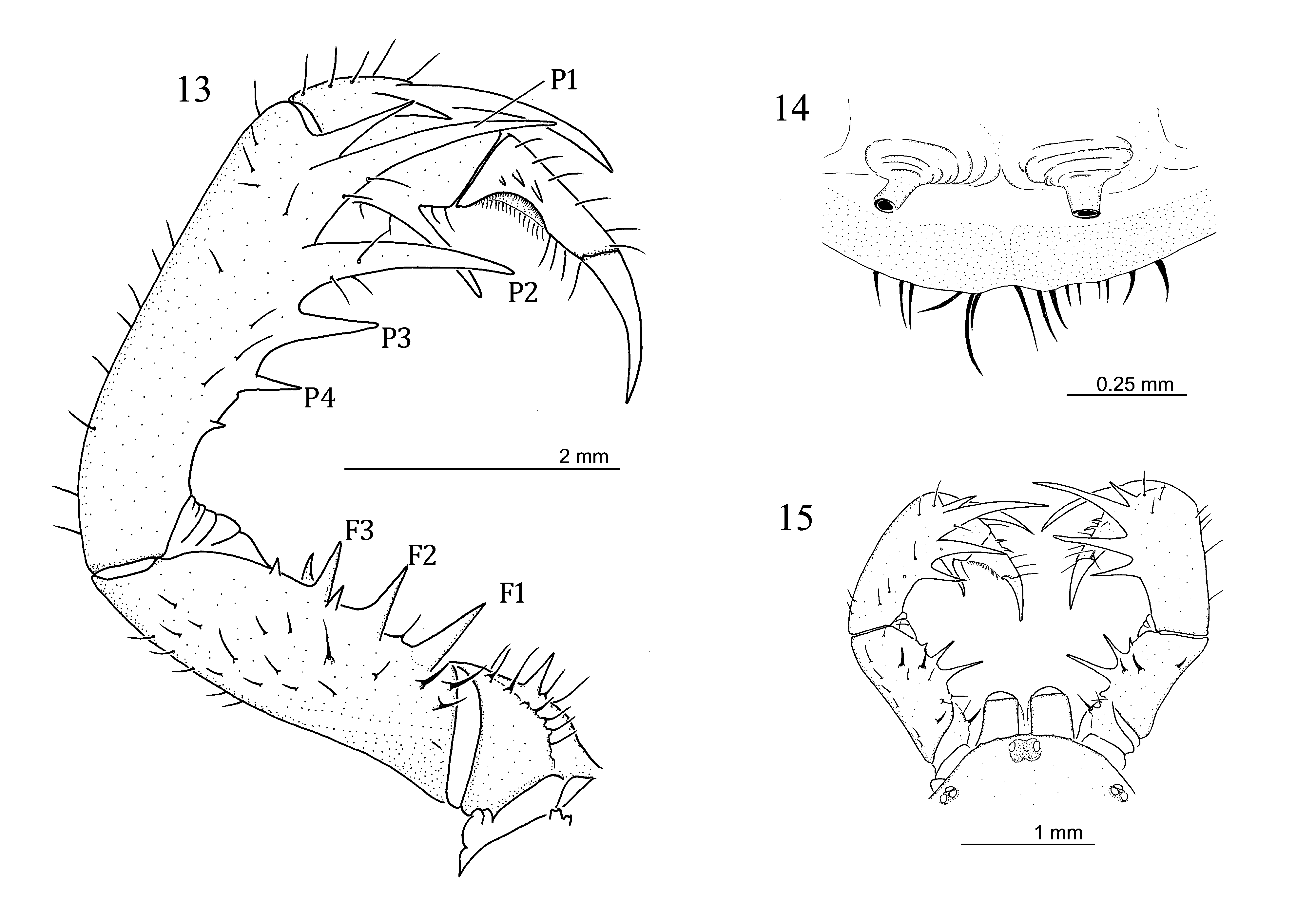
Female ( Figs. 13–14 View FIGURES 13 – 15 ): Color in alcohol much darker and distinctly different from male as follows: pedipalp distinctly shorter and stouter ( Fig. 13 View FIGURES 13 – 15 ) (see Fig. 11 View FIGURES 8 – 12 for male); trochanter with 4 setiferous tubercles, 3 of them arranged in row along antero-dorsal margin; femur with single minor spine each between F2-F3 and between F3 and distal margin; antero-ventral margin with 4 minor spines. Patella with four major spines distributed across most of patella. Tarsus with 3 spines: proximal spine the shortest, distal spine the longest, and medial spine about half as long as distal spine, medial and distal spines separated from each other by about three basal diameters of medial spine. Gonopods with paired tube-like, apically-pointed projections ( Fig. 14 View FIGURES 13 – 15 ).
Juvenile ( Fig. 15 View FIGURES 13 – 15 ): Similar to adult, but color in alcohol much paler, and pedipalpal tarsus with two instead of three spines ( Fig. 15 View FIGURES 13 – 15 ).
Measurements (in mm): male (n =3) [female (n =5)]; values for segments of the appendages are their lengths. Body length (excluding chelicera) 9.80–10.60 [8.80–11.80]. Carapace: median length 3.19–4.20 [3.40–3.80]; width 5.40–5.80 [5.00–5.48]; median eyes to anterior margin of carapace 0.04 [0.04]; distance between lateral eyes 2.40–2.52 [2.40–2.60]; lateral eye to anterior margin of carapace 0.60 [0.52–0.60]; lateral eye to lateral margin of carapace 0.20 [0.12–0.20]. Pedipalps: trochanter 1.44–1.72 [1.40]; femur 5.60–6.80 [2.96–3.40]; patella 6.20–7.40 [3.60–3.80]; tibia 1.92–2.20 [1.40–2.00]; tarsus 1.60–2.00 [2.00–2.40]. Leg I: femur 14.80 [10.00–12.20]; patella 0.80 [0.80]. Leg II: femur 8.80 [6.20–7.20], patella 1.20 [1.20]; basitibia 6.40–8.00 [4.80–6.40]; distitibia 3.56–4.20 [3.04–3.40]; metatarsus + tarsus 3.00–3.60 [2.40–2.80]. Leg III: femur 8.00–11.20 [6.80–8.00]; patella 1.20–1.40 [1.20]; basitibia 8.80–10.00 [6.40–7.80]; distitibia 3.60–3.80 [3.20–3.80]; metatarsus+ tarsus 3.60 [2.00–2.80]. Leg IV: femur 8.00–9.60 [7.00–8.00]; patella 1.20 [1.00]; basitibia 9.20–10.80 [7.00–8.00]; distitibia 3.40–3.60 [3.00–3.40]; metatarsus + tarsus 3.20 [2.00–3.00].
Etymology: This species is dedicated to Yayuk R. Suhardjono who collected the type material from caves in Central Kalimantan, and has contributed much to the knowledge on cave biology in Indonesia.
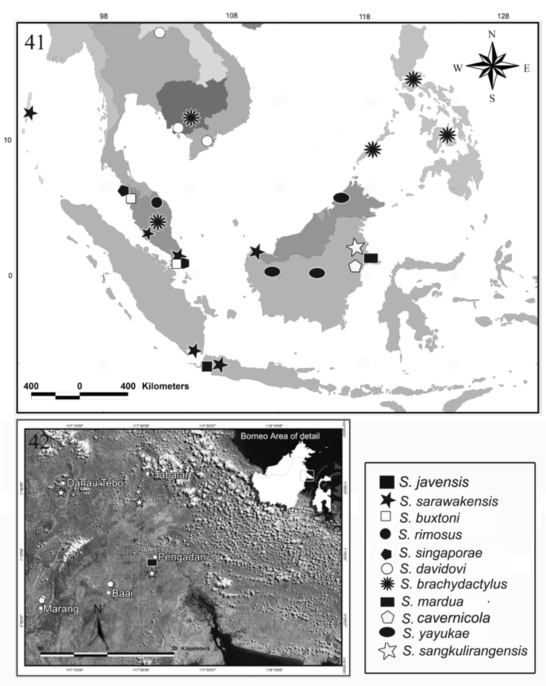
Distribution: Sarax yayukae is found in Central and West Kalimantan ( Indonesia), and Manukan Island, Sabah ( Malaysia) ( Fig. 41 View FIGURES 41 – 42 ).
Natural history: This species inhabits caves (Liang Puruk, Liang Hajuq and Gua Kelasi), as well as occurring under logs in forest (Manukan Island, Sabah). Detailed descriptions of the caves in Central Kalimantan were given in Rahmadi & Suhardjono (2006). The species was observed preying on cave crickets, Diestramenna sp., in Liang Puruk, Central Kalimantan.
Remarks: Sarax yayukae can be distinguished from other congeners by its distinct sexual dimorphism, the presence of three spines on the pedipalpal tarsus in adults ( Fig. 11 View FIGURES 8 – 12 ) and two in juveniles ( Fig. 15 View FIGURES 13 – 15 ), the tibia of leg IV having 19 trichobothria arranged with bc much closer to sbf than to bf, and bt on the fourth basitibial segment of leg IV being close to the distal margin of the segment ( Fig. 12 View FIGURES 8 – 12 ). The juvenile of S. yayukae is similar to S. sarawakensis in having two spines on the pedipalpal tarsus, but the spines in S. yayukae (both juvenile and adult) are long while those in S. sarawakensis are short and minute. Sarax sarawakensis has four spines on the antero-dorsal margin of the pedipalpal femur and three spines in the juvenile of S. yayukae . The pedipalpal patella of S. sarawakensis has four major spines on the antero-dorsal margin instead of three in juveniles of S. yayukae . The number and arrangement of the trichobothria on the tibia of leg IV also differ between both species: S. yayukae (both juvenile and adult) has 19 trichobothria while S. sarawakensis has 17 trichobothria ( Weygoldt 1996, 2000).
Sarax davidovi Fage 1946 View in CoL from Indochina shows distinct sexual dimorphism and has three spines on the antero-dorsal margin of the pedipalpal tarsus as in S. yayukae View in CoL . They differ in the number of spines on the antero-dorsal margin of the male pedipalpal patella (4 spines decreasing in size from distal to proximal in S. yayukae View in CoL ; 2 long spines in S. davidovi View in CoL (see Fage 1946, fig. 4)) and the shape of the carapace (anterior margin rounded in S. yayukae View in CoL , with broad flange on the antero-lateral corners in S. davidovi View in CoL see Fage 1946, fig. 2). The number and arrangement of the trichobothria on tibia leg IV of S. davidovi View in CoL is unknown.

No known copyright restrictions apply. See Agosti, D., Egloff, W., 2009. Taxonomic information exchange and copyright: the Plazi approach. BMC Research Notes 2009, 2:53 for further explanation.
|
Kingdom |
|
|
Phylum |
|
|
Class |
|
|
Order |
|
|
Family |
|
|
Genus |
Sarax yayukae
| Rahmadi, Cahyo, Harvey, Mark S. & Kojima, Jun-Ichi 2010 |
Sarax davidovi
| Fage 1946 |