
Leptolaimus cupulatus Lorenzen, 1972
|
publication ID |
https://doi.org/10.11646/zootaxa.3739.1.1 |
|
publication LSID |
lsid:zoobank.org:pub:79473E74-F230-40D5-8C15-55220DD6CA92 |
|
DOI |
https://doi.org/10.5281/zenodo.5271816 |
|
persistent identifier |
https://treatment.plazi.org/id/AD26453D-FFAD-FFB4-FF4E-0B73F872DA1D |
|
treatment provided by |
Felipe |
|
scientific name |
Leptolaimus cupulatus Lorenzen, 1972 |
| status |
|
Leptolaimus cupulatus Lorenzen, 1972
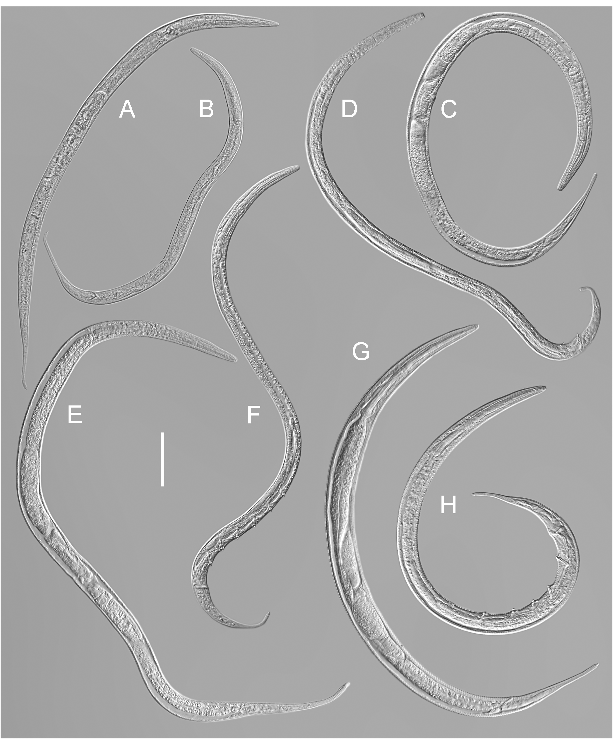
( Figs 2 E–F View FIGURE 2 , 4 View FIGURE 4 ; Table 3)
Material examined. 3 males and 4 females (slides # 129911, 129921–129923) deposited in the general invertebrate collection of the Department of Zoology , Swedish Museum of Natural History, Stockholm, Sweden .
Habitat and locality. Sediment (top layer), Baltic of the east coast of Sweden ( N 58° 46' 00'', E 17° 49' 52''), 26 May 2011, legit C. Raymond ( 3 males and 4 females) GoogleMaps .
Description. Adult. Body tapering anteriorly in pharyngeal region and posteriorly on tail; ventrally curved upon fixation, more strongly curved ventrad in posterior part in males. Maximum body diameter at vulva in female; male body more cylindrical. Cuticle annulated; annuli 1.3–1.9 µm wide at mid-body, without ornamentation. Lateral field present, consists of single ala (two incisures), 2.0–3.0 µm wide at mid-body; originating at level of isthmus (posterior to first body pore) and extending posteriorly to middle of tail. Prominent body pores in sublateral position at both sides of lateral field present, arising from oval lateral epidermal gland cells; usually three pairs of body pores are present along pharyngeal region; several body pores scattered over rest of the body. Anteriormost body pore located at level of procorpus. Somatic sensilla absent in female, present in male (see below). Labial region rounded, continuous with body contour, lips fused. Inner and outer labial sensilla indistinct. Cephalic sensilla setiform, equal to 20–40% of labial region diameter. Subcephalic and cervical sensilla and ocelli absent. Amphidial fovea round, located at level of middle of stoma. Nerve ring surrounding pharynx at level of anterior part of isthmus. Hemizonid not seen. Secretory-excretory system present; excretory pore located posterior to nerve ring. Buccal cavity uniformly tubular: cheilostom and gymnostom short, undifferentiated; stegostom tubular, with uniformly thickened lumen. Pharynx muscular, cylindrical anteriorly, with distinct oval basal bulb; valvular apparatus absent. Anterior cylindrical part of pharynx subdivided by breaks in muscular pharyngeal tissue into cylindrical procorpus, cylindrical metacorpus and narrow isthmus. Pharyngeal glands and their orifices indistinct. Cardia cylindrical, its posterior part embedded in intestine. Tail similar in shape in both sexes (shorter and more curved ventrad in male), elongate-conoid, gradually narrowing distally. Three caudal glands present, their nuclei are incaudal. Spinneret functional.
Male. Three short setae emerging through first, second and third pairs of body pores. Reproductive system diorchic; anterior testis outstretched; posterior one reflexed. Spicules paired, symmetrical, with arcuate conoid calamus and ovoid manubrium. Gubernaculum plate-like, with paired dorsal apophysis. Accessory apparatus composed of one midventral precloacal papilliform sensillum located on anterior cloacal lip, six–seven evenly spaced midventral tubular supplements extending for 117–130 µm from cloaca towards anterior end; 25–26 alveolar supplements. Tubular supplements straight or weakly arcuate, with dentate tips. Posteriormost tubular supplement 27–32 µm anterior to cloaca, anterior to spicules. Alveolar supplements with broad inner sclerotized ring. Anteriormost alveolar supplement 89–97 µm from anterior end, at level of isthmus. Sublateral caudal sensilla present: usually eight (four pairs) caudal setae arranged in two subventral and two subdorsal pairs; precloacal setae absent.
Female. Reproductive system didelphic, amphidelphic; ovary branches reflexed antidromously. Anterior genital branch 78–117 µm long (equal to 12.1–16.8% of total body length), located on right-hand side of intestine (n=2), posterior genital branch 91–122 µm long (equal to 14.5–17.5% of total body length), located on left-hand side of intestine (n=3). Oviduct a narrow tube. One offset, oval, sac-like spermatheca located on right side of anterior gonoduct and one on left side of posterior gonoduct. Spermathecae often filled with oval spermatozoa. Uterus a wide and short tube. Vagina straight, 0.3 times vulval body diameters long; pars proximalis vaginae encircled by single sphincter muscle; pars refringens vaginae present, angular. Vulva midventral. Epiptygmata and sensitive structures around vulva (advulval sensilla) absent. Supplements absent. Rectum 1.4–1.7 anal body diameters long; surrounded by three gland-like cells at intestine-rectum junction.
Diagnosis (supplemented with literature data). Leptolaimus cupulatus is particularly characterised by the 543– 698 µm long body; rounded labial region continuous with body contour; cephalic setae 1.0–2.0 µm long; amphid located 8.5–11.0 µm from anterior end; first body pore located 27.0–33.0 µm from anterior end; lateral field originating at level of isthmus; female without supplements, vagina with angular pars refringens, vulva midventral; male with 6–8 tubular and 25–27 alveolar supplements, tubular supplements straight with dentate tips, alveolar supplements with sclerotized ring; spicules arcuate and 16.0–20.0 µm long.
Remarks. Current material agrees well with the type specimens in general morphology and measurements, shape of supplements, spicules and gubernaculum, with only minor differences in few body measurements.

No known copyright restrictions apply. See Agosti, D., Egloff, W., 2009. Taxonomic information exchange and copyright: the Plazi approach. BMC Research Notes 2009, 2:53 for further explanation.
|
Kingdom |
|
|
Phylum |
|
|
Class |
|
|
Order |
|
|
Family |
|
|
Genus |