
Hymedesmia, 1864
|
publication ID |
https://doi.org/10.1111/j.1096-3642.2008.00498.x |
|
DOI |
https://doi.org/10.5281/zenodo.5114893 |
|
persistent identifier |
https://treatment.plazi.org/id/AF2487CE-146C-FFF2-FF05-EA60364CFDD9 |
|
treatment provided by |
Carolina |
|
scientific name |
Hymedesmia |
| status |
sp. nov. |
HYMEDESMIA (HYMEDESMIA) STELLIFERA View in CoL SP. NOV. ( FIG. 8A, B View Figure 8 )
Type material: Holotype: specimen in IMS, section and spicule preparation from tissue sample ( Rathlin Island Sponge Biodiversity Project ; Damicornis Bay, 55°17.436 ′ N, 006°15.003 ′ W; water depth, 30–35 m; Mc 2606). Collected by B. Picton and A. Mahon, 6 July 2005. GoogleMaps
Paratypes: specimen 1, specimen in IMS, section and spicule preparation from tissue sample ( Rathlin Island Sponge Biodiversity Project ; Damicornis Bay, 55°17.459 ′ N, 006°15.233 ′ W; water depth, 27–32 m; Mc 2790). Collected by C. Goodwin and D.Goodwin, 15 August 2005 GoogleMaps . Specimens 2 and 3: specimens in ethanol, sections and spicule preparations from tissue sample ( Sublittoral Survey Northern Ireland Project ; Russells Rock, The Maidens, 54°57.291 ′ N, 005°45.008 ′ W; water depth, 25–30 m; Mc 3955 and Mc 3960). Collected by C. Goodwin and D. Goodwin, 24 August 2007 GoogleMaps . Specimen 4: specimen in IMS, section and spicule preparation from tissue sample (south-west of Lunga, Firth of Lorne, western Scotland; 56°12.53 ′ N, 005°43.40 ′ W; Mc 492). Collected by B. Picton, 31 December 1981 GoogleMaps .
Etymology: Named from the Latin stella, meaning star, and fero, meaning to bear, because of the starshaped pattern on its surface.
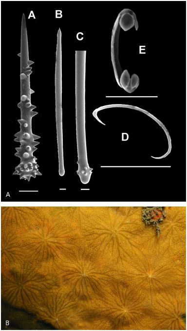
External morphology: Bright yellow/orange sponge forming large patches (over 20 cm in diameter), which are very conspicuous because of their bright colour and oscule form. Prominent oscules surrounded by numerous (10–20) oscular channels, some of which are branched. The oscules are regularly spread over the sponge surface, and are arranged in diamonds. The ends of the oscular channels touch those of neighbouring oscules, giving the surface a regular star-type pattern.
Skeleton: Basal layer of large and small acanthostyles, from which ectosomal spicules arise in columns that are 10–15 spicules thick. The smaller acanthostyles are more abundant. Sigmas are very abundant throughout the tissue. Chelae are present in certain regions of the sponge, but may not be apparent in some specimens. Where present, they are reasonably common and form a layer at the suface. The sponge is ~800-Mm thick.
Spicules:
1. Large acanthostyles: 265–440 Mm (358 Mm) by 8–12 Mm. These are fusiform acanthostyles with a tylote head. The shaft is smooth, and the head is sparsely spined with small, rounded spines. Occasional spicules are present, consisting of two of these acanthostyles fused at the head.
2. Small acanthostyles: 65–95 Mm (79 Mm) by 8–10 Mm. These acanthostyles are very characteristic in appearance; they taper evenly to a sharp point, with no development of the head. The majority of the shaft is spined with large spines, but the last eighth to one-quarter, towards the tip, is smooth, and there are smooth areas on the shaft above the head.
3. Ectosomal spicules: the styles are 210–290 Mm (247 Mm) in length, with the majority of being 8–10-Mm wide, but with some much thinner spicules of 3–5 Mm in width. These spicules are fusiform: one end is rounded and tylote, and the other end is pointed; some are mucronate at the tip.
3. Sigmata: 10–12 Mm in length.
4. Chelae: 15–18 Mm in length. These are small chelae, with leaf-like alae, and are only joined to the shaft by a short section at the top. The alae have a claw-like appearance when viewed under the light microscope.
Remarks: Based on its skeletal structure, this species is in the genus Hymedesmia . The smooth, large acanthostyles are unusual for this genus, and are very similar in form to Lissodendoryx (Ectyodoryx) atlanticus . However, in the subgenus Ectyodoryx , the choanosomal skeleton is composed of either styles or acanthostyles, with a separate category of ectosomal tornotes. The pronounced difference in the form of the acanthostyles is also unusual. The smaller acanthostyles are very similar in form to those of Hymedesmia zetlandica Bowerbank, 1864 , which is the type specimen of the genus Hymedesmia .
Chelae were not visible in tissue sections from either the Rathlin specimens or the Lunga specimen, despite numerous sections being taken from different tissue areas, although scarce chelae were visible in the spicule preparations. However, chelae were numerous in some areas of the surface of two specimens collected from the Maidens: this suggests that chelae are present only in certain regions of the sponge. It was not possible to determine which precise regions they were associated with.
Embryos were present in the Lunga specimen. These embryos have different spicules to the main tissue: thin, short styles (125 Mm by 2 Mm), and unguiferous anchorate chelae (20 Mm). The majority of the chelae have three claw-shaped alae on each end, but in some individuals there were up to five. In several cases, spines are present on the chelae shaft just below the alae.
SUBGENUS STYLOPUS FRISTEDT, 1885
No known copyright restrictions apply. See Agosti, D., Egloff, W., 2009. Taxonomic information exchange and copyright: the Plazi approach. BMC Research Notes 2009, 2:53 for further explanation.
|
Kingdom |
|
|
Phylum |
|
|
Class |
|
|
Order |
|
|
SubOrder |
Myxillina |
|
Family |