
Cletocamptus dominicanus Kiefer, 1934
|
publication ID |
https://doi.org/ 10.11646/zootaxa.4272.3.1 |
|
publication LSID |
lsid:zoobank.org:pub:0ECB3A74-2E13-4713-95FF-D5739035EE09 |
|
DOI |
https://doi.org/10.5281/zenodo.6002070 |
|
persistent identifier |
https://treatment.plazi.org/id/AF5D87B3-4A1B-FFDA-AF8C-1D85C617F8E4 |
|
treatment provided by |
Plazi |
|
scientific name |
Cletocamptus dominicanus Kiefer, 1934 |
| status |
|
Cletocamptus dominicanus Kiefer, 1934
( Figs. 15–26 View FIGURE 15 View FIGURE 16 View FIGURE 17 View FIGURE 18 View FIGURE 19 View FIGURE 20 View FIGURE 21 View FIGURE 22 View FIGURE 23 View FIGURE 24 View FIGURE 25 View FIGURE 26 )
Synonymy. Cletocamptus albuquerquensis: Lang 1948 (part.): 1277–1278, Abb. 508-1c; Wilson & Yeatman 1959: 838. Cletocamptus dominicanus: Kiefer 1934, 1936 ; Lang 1936.
Material examined. Type material. One female lectotype (slide 2076), and three paralectotypes (slides 2117, 2118, and 2119) deposited in The Kiefer Collection , State Museum of Natural History Karlsruhe ( Germany) .
Additional Material. The vials housed in the US National Museum (Smithsonian Institution) that were analyzed are labeled as follows (additional information about the collection site of vial USNM No. 306869 is given). The material housed in other collections is also provided.
Cletocamptus albuquerquensis , USNM No. 128929; Trinidad; Caroni ; Caroni Swamp; collector B. F. Bacon, 1966; identification by T. E. Bowman, 1969.
Cletocamptus albuquerquensis , USNM No . 278085, Accession No . 416030; Trinidad and Tobago; Chacachacare Island; surface water of salt pond; collector A. L. Kong, May 7, 1993.
Cletocamptus albuquerquensis , USNM No . 306869, Accession No . 2018756; British Virgin Islands; Anegada, Flaminco Pond; collected and identified by L. Jarecki, July 20, 2000.
Notes: Flaminco Pond is described as a typical Caribbean salt pond that seasonally varies widely in salinity (Jarecki, personal communication).
Cletocamptus dominicanus , USNM No. 1418187; 25 females and 9 males ; Laguna Fraternidad , Cabo Rojo, Puerto Rico; Salinity, 87.6 ppt; water temperature, 31.5°C ; pH, 8.56; dissolved oxygen, 8.5 mg L -1; collected by Ray Gerber, January, 2010.
Cletocamptus dominicanus , USNM No. 1418188; 14 females ; salt pond lagoon at Ensenada Dakity, Culebra Island, Puerto Rico; Salinity, 45.8 ppt; water temperature, 29.6°C ; pH, 9.23; dissolved oxygen,> 12 mg L -1; collected by Ray Gerber, December, 2008.
Cletocamptus dominicanus , USNM No. 1418189; 22 females and 8 males ; Laguna El Padre , Vieques Island, Puerto Rico; Salinity, 32.9 ppt; water temperature, 27.4°C ; pH, 8.14; dissolved oxygen, 9.18 mg L -1; collected by Ray Gerber, December, 2008.
Cletocamptus dominicanus , USNM No. 1418190 ; 29 females and 8 males; salt pond at Salt Pond Bay, St. John Island, US Virgin Islands; Salinity , 171.6 ppt; water temperature, 28.0°C ; pH, 9.2; dissolved oxygen, 7.34 mg L -1; collected by Ray Gerber, January, 2007.
Cletocamptus dominicanus , USNM No. 1418191; 15 females and 6 males ; Elliot Pond , San Salvador Island, Bahamas; Salinity, 41 ppt; collected by D. Barr, January, 1994.
Cletocamptus dominicanus , six males (ICML-EMUCOP- 010107 -03, and -07 to -11) and one female ( ICML – EMUCOP– 010107 –02), dissected; one undissected female mounted onto one slide ( ICML –EMUCOP– 010107 – 01); two females and one male preserved in alcohol ( ICML –EMUCOP– 010107 –04); St. John Island ( US Virgin Islands); collected by Ray Gerber, January, 2007.
Cletocamptus dominicanus , 15 females ( UARC 282 M) and 15 males ( UARC 283 M) preserved in alcohol; Pozos Colorado, Santa Marta , Colombia; 11° 14′ 10″ N, 74° 12′ 6″ W; found in a small temporal pond (0.3–0.6 m deep); temperature from 28 to 31°C GoogleMaps ; pH from 8.5 and 8.9; observed in both the limnetic region and in the mangrove zone, being more abundant in the latter during the rainy season when salinity was lowest (5 PSU); collected by Juan Manuel Fuentes-Reinés, August –November, 2015.
Cletocamptus dominicanus , 46 females and 33 males preserved in alcohol ( ICML –EMUCOP– 010815 –01); Pozos Colorado, Santa Marta , Magdalena, Colombia; 1° 14′ 10″ N, 74° 12′ 6″ W. All other information as above. GoogleMaps
Type locality. Enriquillo Lake , Dominican Republic, not far from the border with Haiti ( Kiefer 1934).
Distribution. Bahama Islands: San Salvador (present study). British Virgin Islands: Flaminco Pond, Anegada (present study). Colombia: Pozos Colorados, Santa Marta (present study). Dominican Republic: Enriquillo Lake ( Kiefer 1934, 1936). Trinidad and Tobago: Caroni Swamp, Caroni (present study), Chacachacare Island (present study). US: St. John Island ( US Virgin Islands) (present study), Puerto Rico: Laguna Candelaria (present study), Culebra (present study) and Vieques (present study). Based on our extensive samples from the Caribbean, C. dominicanus is the most abundant harpacticoid copepod in the coastal saline lagoons.
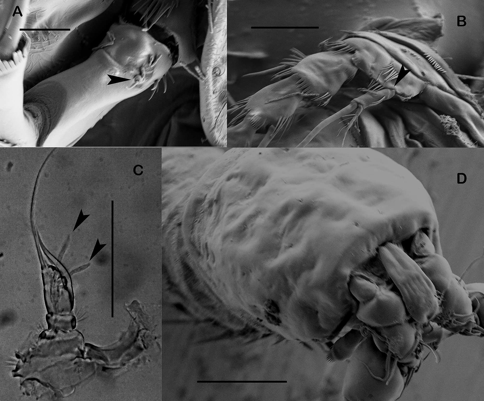
Redescription (based on material from the US Virgin Islands). Female. Habitus ( Fig. 15 View FIGURE 15 A, B) tapering posteriorly; total body length measured from tip of rostrum to posterior margin of caudal rami ranging from 520 µm to 644 µm (mean= 540 µm; n= 3). Rostrum set off, triangular, with pair of setules subapically and with row of spinules distally as in C. albuquerquensis (see Fig. 4 View FIGURE 4 A, B). Posterior margin of cephalothorax, pro- and urosomites ornamented with denticles conferring a serrated appearance (see Fig. 15 View FIGURE 15 C); denticles of P2–P5 bearing-somites coarser and blunt, those of second to fourth urosomites progressively smaller and pointed, of fifth urosomite very small; first (P2 bearing-somite) and third (P4 bearing-somite) prosomites (Pro1, 1’, and Pro3, 3’, in Fig. 15 View FIGURE 15 C) with additional row of medial denticles; P5 bearing-somite with additional row of minute spinules (see Uro 1 in Fig. 15 View FIGURE 15 C). Second and third urosomites distinct dorsally and laterally ( Fig. 15 View FIGURE 15 A, B), completely fused ventrally ( Fig. 17 View FIGURE 17 A) forming genital double-somite. Anal somite ( Figs. 15 View FIGURE 15 A, B, 16A, 17A) as shown; anal operculum crescentic medially, with spinules ( Figs. 15 View FIGURE 15 A, 16A); ventral spinular ornamentation as shown ( Fig. 17 View FIGURE 17 A). Caudal rami ( Figs. 15 View FIGURE 15 A, B, 16A, B, 17A) about 2 times as long as wide; surface covered with slender spinules; with comparatively stronger ventrolateral spinules close to insertion site of setae IV and V; with seven elements in all; seta I small, situated laterally close to anterior margin of ramus, close to setae II and III, the latter setae longer; setae IV and V fused basally, the former 15%, the latter 60% of total body length; seta VI situated on distal outer corner; seta VII situated dorsally midway length of ramus on inner edge.
Antennule ( Fig. 18 View FIGURE 18 A) six-segmented; surface of segments smooth except for two rows of spinules on first segment. Armature formula, 1-(1), 2-(9), 3-(5), 4-(1+[1+ae]), 5-(1), 6-(9+[1+ae]).
Antenna ( Fig. 18 View FIGURE 18 B): small coxa with outer spinules. Allobasis with two abexopodal setae. Free endopodal segment with inner spinules proximally, with two lateral inner spines and a slender seta, and five distal elements, two of them geniculate. Exopod minute, one-segmented, with one seta ( Figs. 18 View FIGURE 18 B, 26A).
Mandible ( Fig. 19 View FIGURE 19 A): robust, with rows of spinules proximally; chewing edge with teeth as figured, with one pyriform element and one lateral pinnate seta. Palp one-segmented, with three setae unequal in length.
Maxillule ( Fig. 19 View FIGURE 19 B): robust; arthrite of praecoxa with few spinules, with one surface seta, with seven spines and two slender setae distally. Coxa with some spinules, with two setae. Basis with spinules as figured, with two apical setae. Exopod and endopod incorporated to basis, represented by two setae each.
Maxilla ( Fig. 19 View FIGURE 19 C): syncoxa with inner and outer spinules, and close to joint with allobasis; with two endites, each bearing three setae. Allobasis drawn into strong claw with one accompanying seta. Endopod represented by three setae.
Maxilliped ( Fig. 19 View FIGURE 19 D): subchelate. Syncoxa with row of spinules, with one tiny seta on inner distal corner.
Basis without armature, with spinules as shown. Endopod drawn into long and slender claw with one accompanying small seta.
P1 ( Fig. 20 View FIGURE 20 A): praecoxa with spinules close to joint with coxa. The latter with anterior and posterior transverse rows of spinules as shown. Basis with inner and outer spine; with median rows of spinules, and with spinules at base of exopod, between rami and at base of inner and outer spines. Exopod three-segmented; EXP1 without, EXP2 with inner seta; EXP3 with four elements. Endopod two-segmented, slightly longer than exopod, both segments subequal; inner seta of ENP 1 shorter than both endopodal segments combined, with brush tip; ENP 2 with three elements.
P2 ( Fig. 20 View FIGURE 20 B): praecoxa and coxa ornamented as figured. Basis with spinules between rami and at base of exopod; outer element spine-like. Exopod three-segmented, ornamented as shown; EXP1 without inner seta; inner seta of EXP2 relatively short, about 0.5 times as long as outer apical seta of EXP3, with brush tip; EXP3 with five elements, of which inner about 0.7 times as long as outer apical seta, without brush tip. Endopod two segmented, reaching about distal third of EXP2; ENP 1 small, slightly longer than wide, with outer and inner spinules, without armature; ENP 2 about 3 times as long as wide, with long spinules as shown, with one inner, one apical and one outer element; inner and outer elements subequal, apical seta longest.
P3 ( Fig. 21 View FIGURE 21 A): praecoxa and coxa as in P2; basis as in P2 except for setiform outer element. Exopod as in P2; EXP1 without inner armature; inner seta of EXP2 about 0.4 times as long as outer apical seta of EXP3, with brush tip; EXP3 with five elements, of which inner seta about 0.8 times as long as outer apical seta, without brush tip. Endopod two-segmented, reaching distal third of EXP2; ENP 1 nearly as long as wide, with long spinules as shown, without armature; ENP 2 about 3 times as long as wide, ornamented as shown, with two inner and two apical setae, and one outer element; inner and outer elements subequal; apical setae longest.
P4 ( Fig. 21 View FIGURE 21 B, 26B): praecoxa, coxa and basis as in P3. Exopod as in P3, except for armature formula of EXP3 (without inner seta); EXP1 without inner seta; inner seta of EXP2 visibly shorter than outer apical seta EXP3, with brush tip; EXP3 with four elements. Endopod one-segmented, about 1.5 times as long as wide, barely reaching middle of EXP1, with slender spinules, with two apical setae (innermost smaller).
P5 ( Fig. 19 View FIGURE 19 E): exopod and baseoendopod fused and barely separated by small notch. Baseoendopod with outer seta of basis; endopodal lobe longer than exopod, with spinules as figured, with one outer, two apical and three inner setae, relative length of setae as shown. Exopod with outer spinules, with five setae in all.
P6 ( Fig. 17 View FIGURE 17 A, B): represented by median plate in anterior half of first genital somite, each vestigial leg represented by one pinnate small setae. Copulatory pore in the middle of genital double-somite.
Male. Total body length measured from tip of rostrum to posterior margin of caudal rami, ranging from 345 µm to 430 µm (mean= 383 µm; n= 5). General shape of habitus as in female except for separate second and third urosomites ( Fig. 22 View FIGURE 22 A, B). Posterior margin of cephalothorax, pro- and urosomites not serrated (not shown); with spinules close to posterior margin of cephalothorax; P2 bearing-somite slenderer than in succeeding somites; dorsal surface covered with slender spinules. Anal somite covered with slender spinules; anal operculum and caudal rami as in female ( Fig. 22 View FIGURE 22 A, B). Caudal seta IV and V 14 % and 48% of total body length, respectively. Ventral ornamentation of third, fourth and fifth urosomites as shown ( Fig. 22 View FIGURE 22 B).
Rostrum ( Figs. 23 View FIGURE 23 B, 26D): sexually dimorphic, elongate.
Antennule ( Fig. 23 View FIGURE 23 A): six-segmented; subchirocer; smooth except for the presence of spinules on first segment; last segment with two acute teeth. Armature formula difficult to define; most probably as follows: 1-(1), 2-(9), 3-(7), 4-(8+[1+ae]), 5-(3), 6-(5+[1+ae]). The armature on the last segment arises from a plate-like swelling, it seems not to be a true segment. The nature of this structure as well as an in-depth analysis of the segmentation of the antennule deserves further investigation.
Antenna, mandible, maxillule, maxilla and maxilliped (not shown) as in female.
P1 ( Fig. 24 View FIGURE 24 A): as in female except for dimorphic projection on inner distal corner of basis.
P2 ( Fig. 24 View FIGURE 24 B): as in female except for relatively stouter outer spines of male exopod, and for inner seta of ENP 2 reduced.
P3 ( Fig. 25 View FIGURE 25 A, 26C): as in female except for relatively stouter outer spines of exopod. Endopod clearly twosegmented; ENP 1 slightly wider than long, with long inner spinules, unarmed; ENP 2 with inner medial apophysis, about as long as supporting segment, with two apical setae of which outer well-developed, inner element very small, with paired elongated asprothekes (indicated in figure 26C; for definition see Discussion), without the spinular ornamentation typically found in other congeners.
P4 ( Fig. 25 View FIGURE 25 B) as in female, except for stouter outer spines of all the exopodal segments, and relatively shorter setae of ENP.
P5 ( Fig. 23 View FIGURE 23 C): both legs fused medially; exopod and baseoendopod fused; division between rami indicated by slight notch. Exopod with spinules at base of setae of basis; with four elements. Baseoendopod with outer seta of basis; endopodal lobe with inner spinules as shown, with three elements in all.
P6 ( Fig. 22 View FIGURE 22 B) represented by plate; without armature.
Armature formula in Table 3.
Variability. US Virgin Islands: The male P4 ENP may be one or two-segmented (first segment being very small; see Fig. 25 View FIGURE 25 C), being the one-segmented condition more common and is considered here as the normal condition. Also, as noted in Fig. 26 View FIGURE 26 B, the small proximal element can be an artifact due to a slight narrowing at the base of the endopodal segment.
Leg P1 P2 P3 P4 P5 Female EXP I-0; I-1;I,I1,1 I-0; I-1;II,2,1 I-0; I-1;II,2,1 I-0; I-1;II,2,0 5 ENP 0-1;0,I1,1 0-0;I,1,1 0-0;I,2,2 0,2,0 6 Male EXP I-0; I-1;I,I1,1 I-0; I-1;II,2,1 I-0; I-1;II,2,1 I-0; I-1;II,2,0 4 ENP 0-1;0,I1,1 0-0;I,1,1 0-0;0,2,Apophysis 0,2,0 3 Santa Marta, Colombia: The right P1ENP1 of one female was observed to possess a distal spine. The left P3EXP3 of another female was observed to possess two inner distal setae instead of one lateral inner seta. The left P5 endopodal lobe of another female was observed to possess five instead of six setae.


No known copyright restrictions apply. See Agosti, D., Egloff, W., 2009. Taxonomic information exchange and copyright: the Plazi approach. BMC Research Notes 2009, 2:53 for further explanation.
|
Kingdom |
|
|
Phylum |
|
|
Class |
|
|
Order |
|
|
Family |
|
|
Genus |