
Neostagonosporella bambusicola L.S. Dissan. & J.C. Kang, 2022
|
publication ID |
https://doi.org/ 10.11646/phytotaxa.573.2.6 |
|
DOI |
https://doi.org/10.5281/zenodo.7362692 |
|
persistent identifier |
https://treatment.plazi.org/id/B00187D1-3D59-FFBD-F7B5-6A232403FD8E |
|
treatment provided by |
Plazi |
|
scientific name |
Neostagonosporella bambusicola L.S. Dissan. & J.C. Kang |
| status |
sp. nov. |
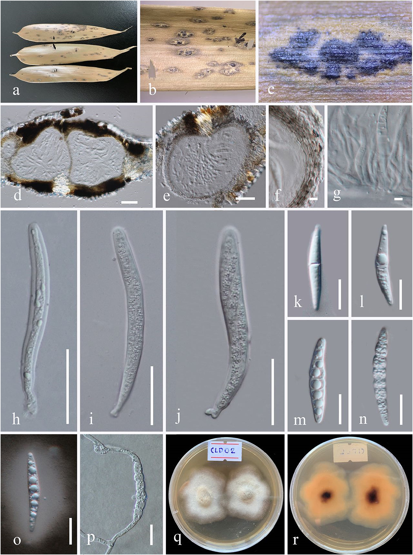
Neostagonosporella bambusicola L.S. Dissan. & J.C. Kang View in CoL sp. nov. ( FIGURE 2 View FIGURE 2 )
Index Fungorum number: IF559874; Facesoffungi number: FoF09712
Etymology— Referring to the host “bamboo” from which the holotype was isolated.
Holotype — HKAS115852 View Materials
Saprobic on dead leaves of bamboo ( Poaceae ). Sexual morph: Ascostromata 180–220 × 140–180 μm (x = 201 × 163 μm, n = 10), ellipsoidal, globose to subglobose or irregular in shape, semi-immersed in host epidermis, coriaceous, solitary to gregarious, multiloculate, erumpent through host tissue, with dark brown to black, glabrous, with a centrally located ostiole. Peridium 12–20 μm wide (x = 15 μm, n = 10), thin at base, thick at sides towards apex, upper part fused with host tissue, composed of two layers; outer layer: pale to brown pseudoparenchymatous cells of textura angularis, inner layer: hyaline, somewhat flattened cells of textura angularis. Hamathecium composed of 1–3 μm (x = 2 μm, n = 10) wide, cellular, septate pseudoparaphyses, embedded in a hyaline gelatinous matrix. Asci 70–130 × 13.5–15 μm (x = 92 × 13 μm, n = 20), 8-spored, bitunicate, fissitunicate, cylindrical to cylindric-clavate, short pedicellate (up to 10 μm), apically rounded with an ocular chamber. Ascospores 30–36 × 5.5–7.2 μm (x = 33 × 6 μm, n = 20), partially overlapping uni- to bi-seriate, hyaline, cylindrical to fusiform or cylindric-clavate, with acute ends, narrower towards end cells, straight or slightly curved, 5-7-septate, slightly constricted at septa, nearly equidistant between septa, guttulate, smooth-walled, surrounded by a 10–14 μm (x = 11 μm, n = 10) thick mucilaginous sheath. Asexual morph: Undetermined.
Culture characteristics— Colonies on PDA, reaching 2 cm diam., after 2 weeks at 20°C, medium dense, circular to slightly irregular with uneven margin, slightly raised and cottony surface, colony from above: white at the margin, pale-grey at the center. From below: yellowish margin, yellow to brown at the center; mycelium pale-grey.
Material examined— CHINA, Guizhou Province, Guiyang , Guizhou University , on dead leaves of bamboo, 28 May 2019, L. S . Dissanayake ( HKAS115852 View Materials , holotype), ex-type culture ( KUMCC 20-0031 ) .
Known distribution — Guizhou Province, China
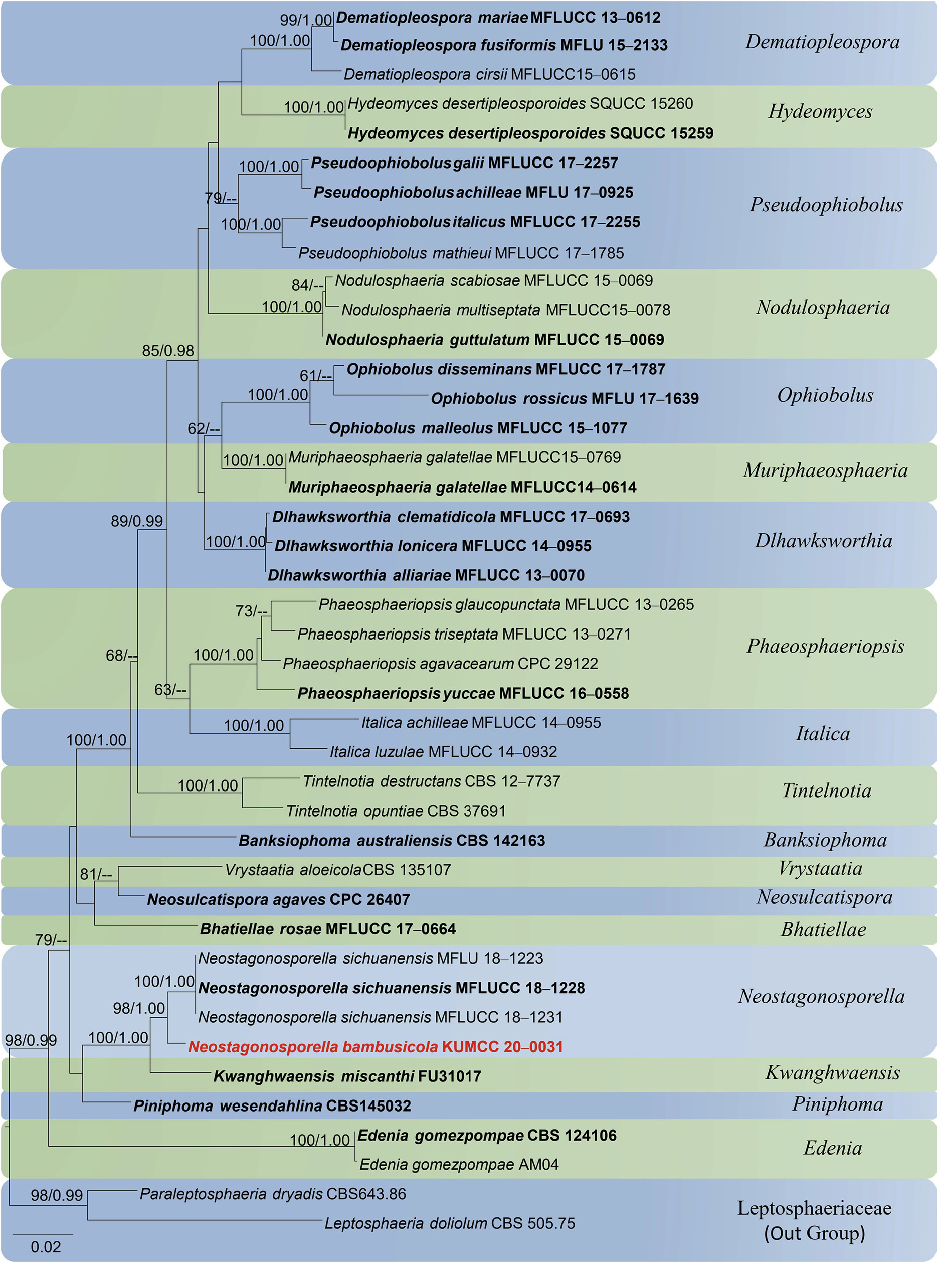
Notes: Neostagonosporella bambusicola shares similar morphology with N. sichuanensis showing multiloculate ascostromata and cylindrical to fusiform, transversely multi-septate, straight or curved ascospores, which are widest at the central cells. In the present phylogenetic analyses, N. bambusicola clusters sister to N. sichuanensis with relatively high statistical support ( Figure 1 View FIGURE 1 ). The two species are easily distinguishable since Neostagonosporella bambusicola has smaller ascomata (180–220 × 140–180 μm), cellular, septate pseudoparaphyses and ascospores with thick sheath (10–14 μm), whereas N. sichuanensis has large ascomata (1–2 mm × 230–340 μm), trabeculate pseudoparaphyses and ascospores with a thin sheath (5–9 μm).
| L |
Nationaal Herbarium Nederland, Leiden University branch |
| S |
Department of Botany, Swedish Museum of Natural History |
No known copyright restrictions apply. See Agosti, D., Egloff, W., 2009. Taxonomic information exchange and copyright: the Plazi approach. BMC Research Notes 2009, 2:53 for further explanation.
|
Kingdom |
|
|
Phylum |
|
|
Class |
|
|
Order |
|
|
Family |
|
|
Genus |