
Agapetus
|
publication ID |
https://doi.org/ 10.5281/zenodo.194445 |
|
DOI |
https://doi.org/10.5281/zenodo.6211865 |
|
persistent identifier |
https://treatment.plazi.org/id/B00E87B0-FF8A-FFB0-8595-FA7AFBC7F847 |
|
treatment provided by |
Plazi |
|
scientific name |
Agapetus |
| status |
|
KEY TO MALES OF AUSTRALIAN SPECIES OF Agapetus View in CoL
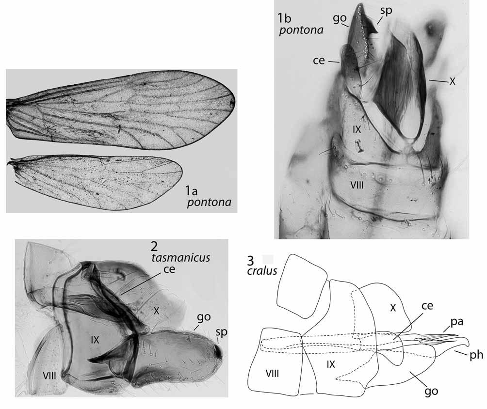
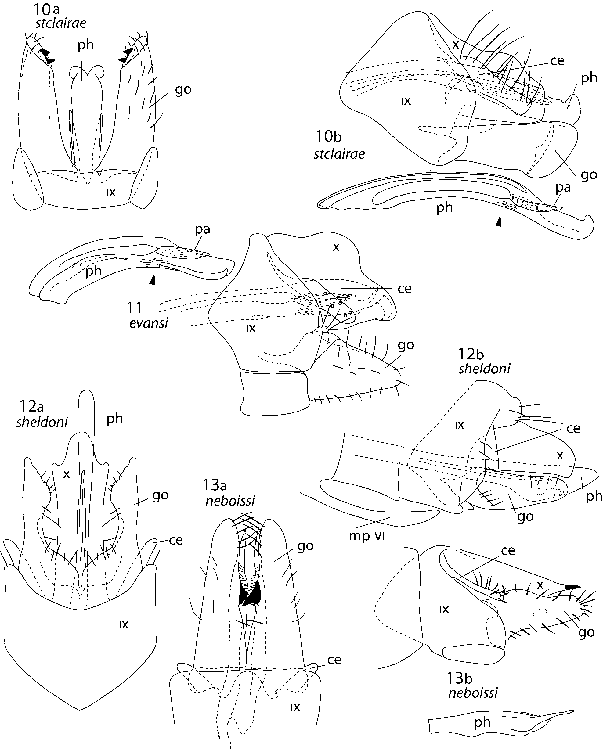
1. Cerci in lateral view slender, elongate, with length more than 6 times height ( Figs 2 View FIGURES 1 – 3 , 13 View FIGURES 10 – 13 b, 14b, 15b, 17b, 18) ... 2
-. Cerci in lateral view stout, generally club-shaped, length less than 5 times height ( Figs 3 View FIGURES 1 – 3 , 4 View FIGURES 4 – 6 b, 5b, 6b, 7b, 8b, 9b, 10b, 11, 12b, 16b, 20b) .......................................................................................................................................... 8
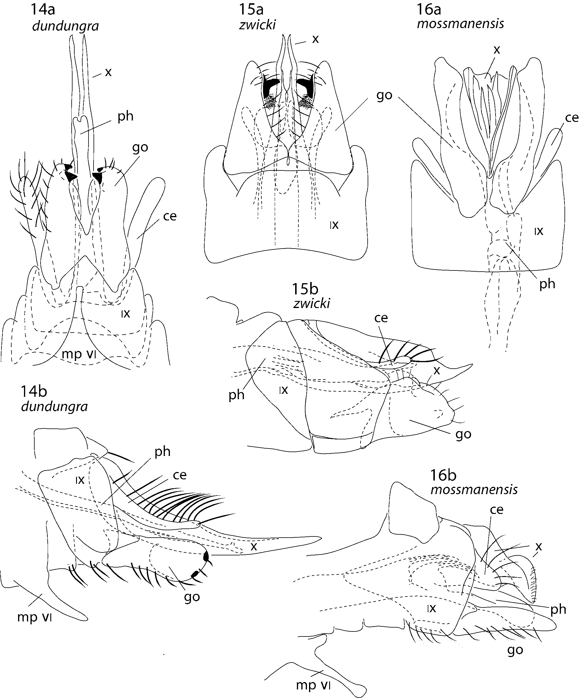
2(1). Gonopods in lateral view subquadrate ( Figs 15 View FIGURES 14 – 16 b, 18)........................................................................................... 3
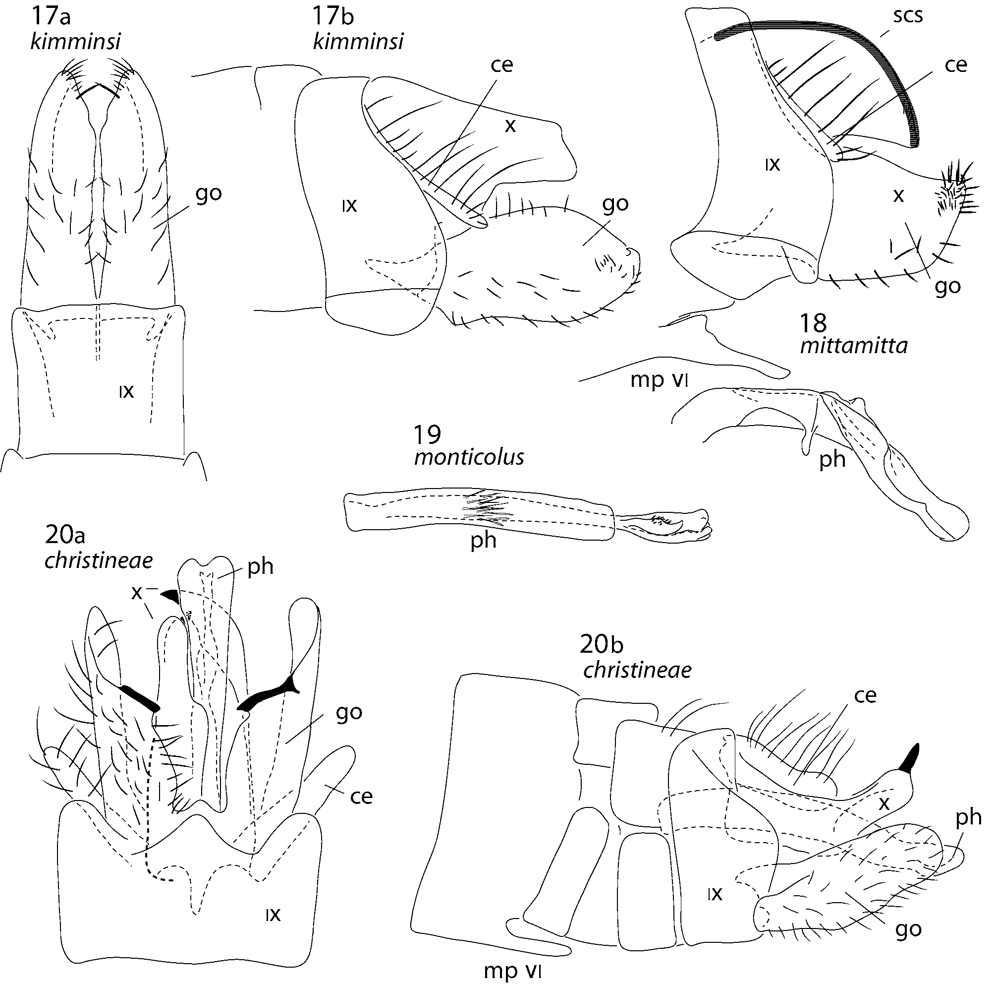
-. Gonopods in lateral view longer than tall ( Figs 2 View FIGURES 1 – 3 , 13 View FIGURES 10 – 13 b, 14b, 17b)......................................................................... 4
3(2). Segment X having dorsal and apical margins each with sclerotised band, in lateral view rounded apically ( Fig. 18 View FIGURES 17 – 20 ); gonopods each with single small spine or spur apically, visible in lateral view ( Fig. 18 View FIGURES 17 – 20 )................................ ............................................................................................................................................... A. mittamitta sp. nov.
-. Segment X entirely membranous, in lateral view projected distad, apically acute ( Fig. 15 View FIGURES 14 – 16 b); gonopods in ventral view each with pair of apicomesal spurs ( Fig. 15 View FIGURES 14 – 16 a) ..................................................................... A. zwicki sp. nov.
4(2). Segment X entirely membranous, in lateral view produced distad to about 2X length of gonopods, apically acute ( Fig. 14 View FIGURES 14 – 16 b); gonopods in ventral view each clearly bearing pair of sclerotised spurs apicomesally ( Fig. 14 View FIGURES 14 – 16 a) ........ ...................... ......................................................................................................................... A. dundungra sp. nov.
-. Segment X at least partly sclerotised, in lateral view shorter than, equal to, or only slightly longer than gonopods, irregularly rounded ( Figs 2 View FIGURES 1 – 3 , 17 View FIGURES 17 – 20 b), truncate (see Neboiss 1986: 56), or tapered and apically acute ( Fig. 13 View FIGURES 10 – 13 b); gonopods in ventral view each with only a single apical spur visible, or no apical spur visible ( Figs 13 View FIGURES 10 – 13 a, 17a) .. 5
5(4). Gonopods in ventral view each with pair of spurs on mesal margin, one at apex and second at about 2/3 from

No known copyright restrictions apply. See Agosti, D., Egloff, W., 2009. Taxonomic information exchange and copyright: the Plazi approach. BMC Research Notes 2009, 2:53 for further explanation.
|
Kingdom |
|
|
Phylum |
|
|
Class |
|
|
Order |
|
|
Family |