
Prendalona barbulata ( Megard, 1967 ) Sinev & Sousa & Elmoor-Loureiro, 2023
|
publication ID |
https://doi.org/ 10.11646/zootaxa.5293.1.4 |
|
publication LSID |
lsid:zoobank.org:pub:3D768D7C-FD9E-452A-B83A-9FE800F66402 |
|
DOI |
https://doi.org/10.5281/zenodo.7968430 |
|
persistent identifier |
https://treatment.plazi.org/id/B0355A27-FFA5-FFBA-FF33-3625FDAE5190 |
|
treatment provided by |
Plazi |
|
scientific name |
Prendalona barbulata ( Megard, 1967 ) |
| status |
comb. nov. |
Prendalona barbulata ( Megard, 1967) comb. nov.
Megard, 1967: 37–57, Figs. 9 View FIGURE 9 –19, pl. 2a–b ( Alona View in CoL ); Smirnov, 1971: 382, Fig. 458 ( Alona View in CoL ).
Type locality Island Lake in the Wind River Mountains, about 20 km northeast of Pinedale, Wyoming, U.S.A.
Type material. The holotype (parthenogenetic female) and several paratypes deposited in the British Museum (No. 1966. 3.21.4).
Material examined. Six parthenogenetic females, over 20 ephippial females, 7 adult males, 2 juvenile males of instar II from Salmon Lake , Missoula County, Montana, U.S.A., 15.11.1977, coll. K. Brakke, sample DGF 4458 in collection of Prof. D. G. Frey at National Museum of Natural History (Washington DC, USA), general access number 403774 .
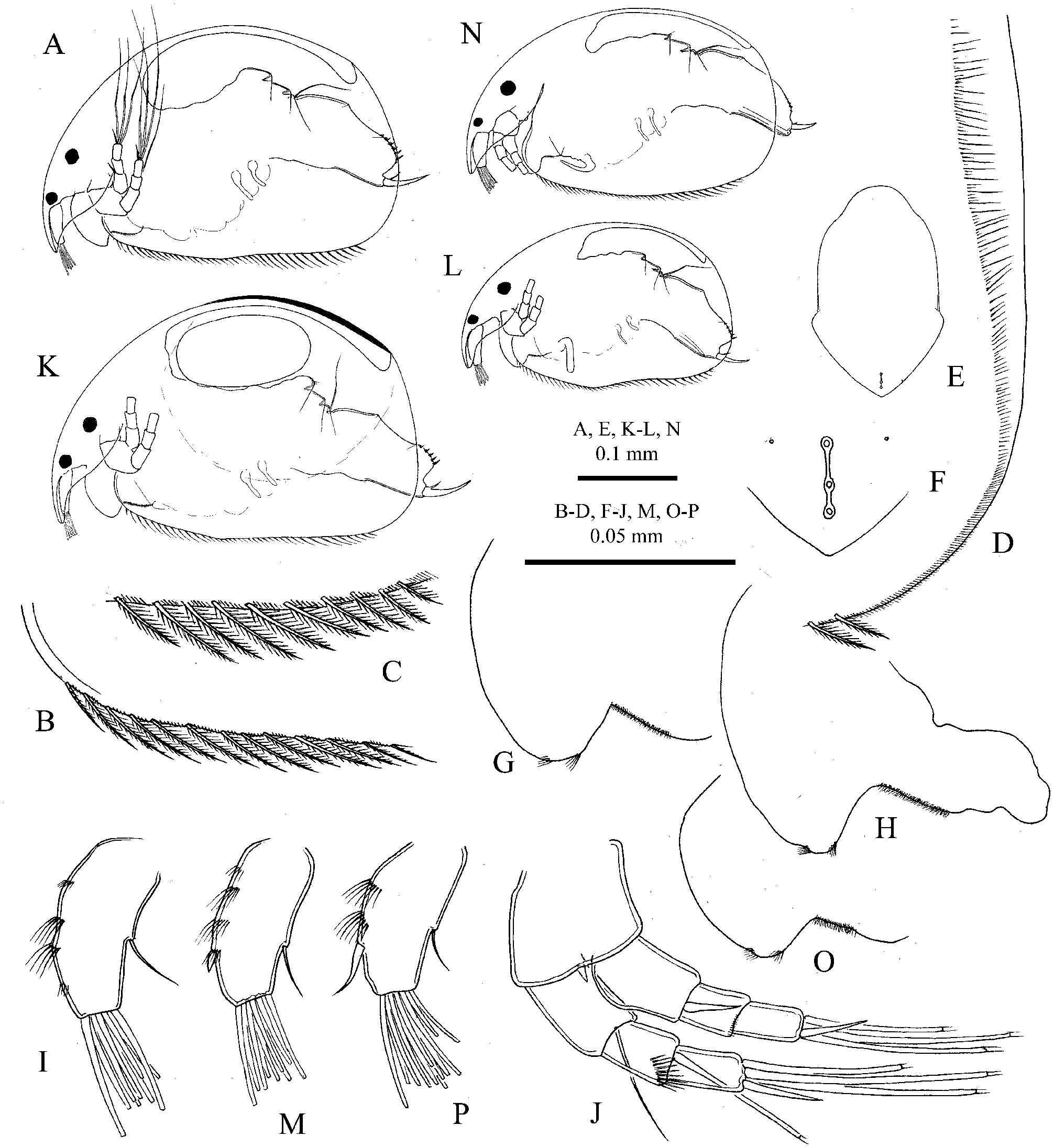
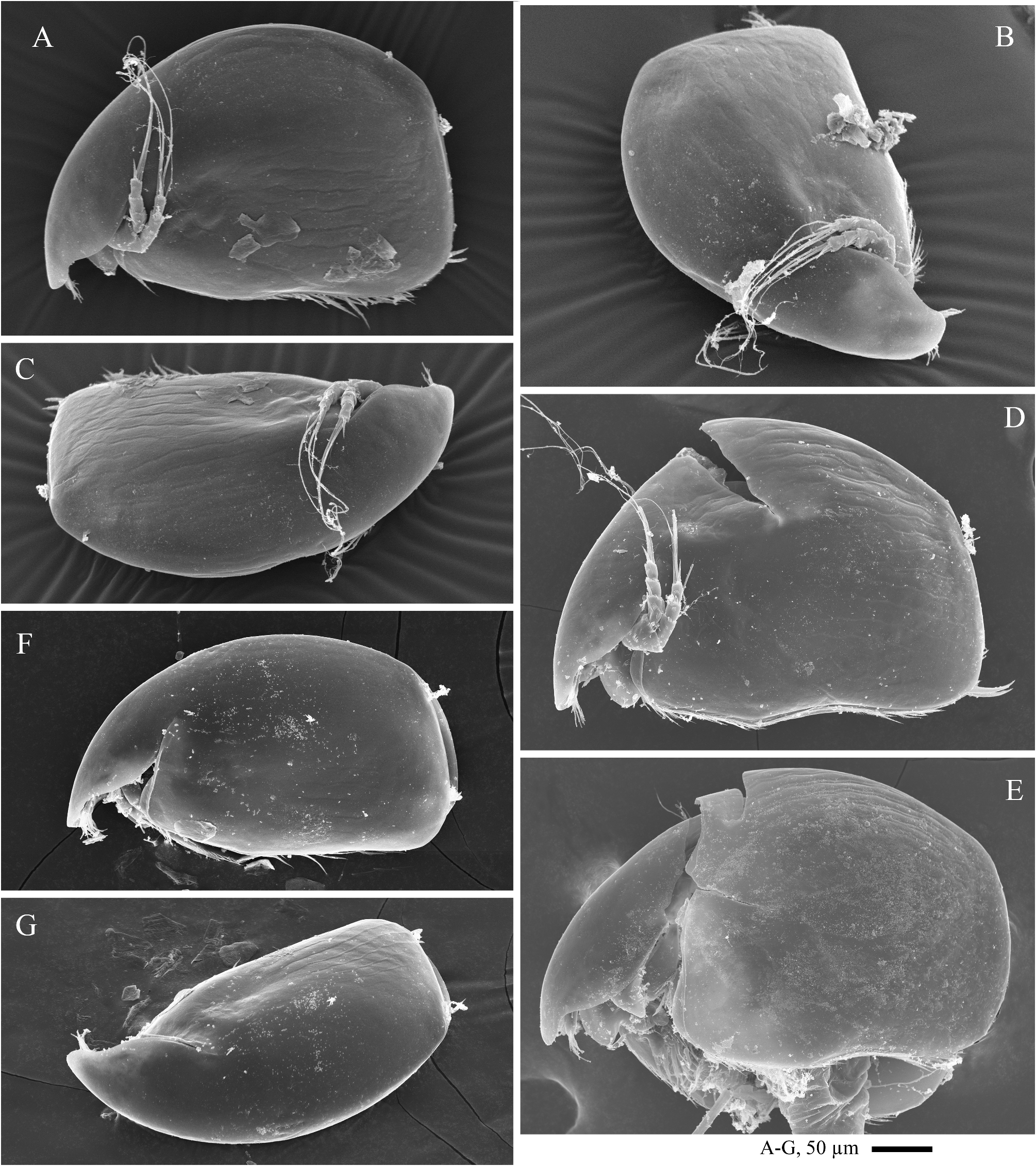
Description. Parthenogenetic female. General. In lateral view body ( Figs. 2A View FIGURE 2 , 3A–C View FIGURE 3 ) oval, of moderate height, maximum height at middle of body, in adults height/length ratio 0.65. Dorsal margin uniformly curved; postero-dorsal and postero-ventral angles broadly rounded; posterior margin uniformly curved; ventral margin almost straight; antero-ventral angle rounded. Body moderately compressed laterally.
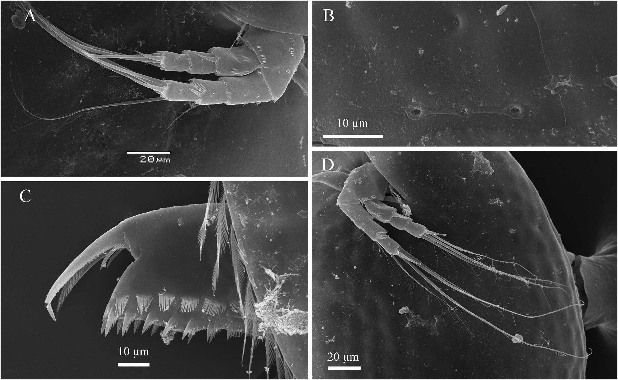
Valves. Ventral margin with 30–40 setae, anteriormost ten setae short ( Fig. 2B View FIGURE 2 ), not forming a group separated from setae in middle of margin, distal setae of moderate length ( Fig. 2C View FIGURE 2 ). Postero-dorsal angle ( Fig. 2D View FIGURE 2 ) bears about 70 short, thin, hair-like setulae of similar length, not organized in groups. A row of about 80–100 thin setulae of variable length along posterior margin at inner side of valve. Valve looks smooth by optical microscopy, but SEM examination reveals a weakly developed linear sculpture.
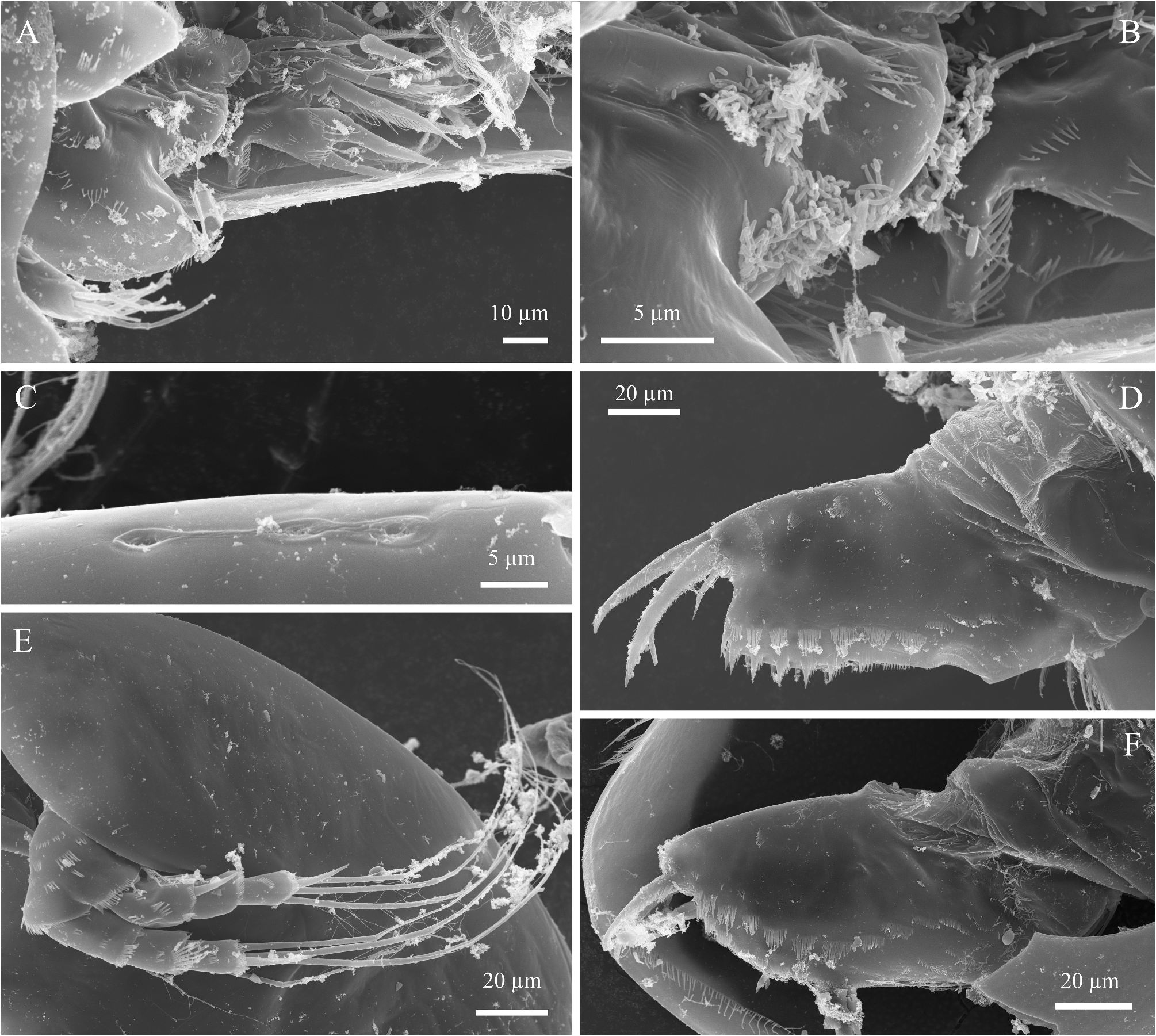
Head of moderate size, oblique, triangular-round in lateral view; rostrum short, pointing downward. Compound eye larger than ocellus. Distance from tip of rostrum to ocellus in adults 1.5 times greater than that between ocellus and eye. Head shield ( Fig. 2E View FIGURE 2 ) with a maximum width behind mandibular articulation, without sculpturing; rostrum short, broadly rounded; posterior portion of head shield triangular with obtuse apex. Three connected major head pores of similar size, middle pore located closer to posterior pore, PP less than 0.5 IP ( Figs. 2F View FIGURE 2 – 4C View FIGURE 4 ). Lateral head pores minute, located at about 0.8 IP distance from midline, at the level of anterior major head pores.
Labrum ( Figs. 2G–H View FIGURE 2 , 4A–B View FIGURE 4 ) of moderate size; labral keel narrow (height about 2 width), with a rounded apex; anterior margin of keel convex, posterior margin with two clusters of short setulae, setulae in anterior cluster pointing almost forward. Posterior portion of labrum under optical microscope appears to be covered by thin setulae, but SEM examination ( Fig. 4A–B View FIGURE 4 ) reveal that these ‘structures’ are in reality epibiotic bacteria.
Thorax twice longer than abdomen, dorsal surface of abdominal segments not saddle-shaped.
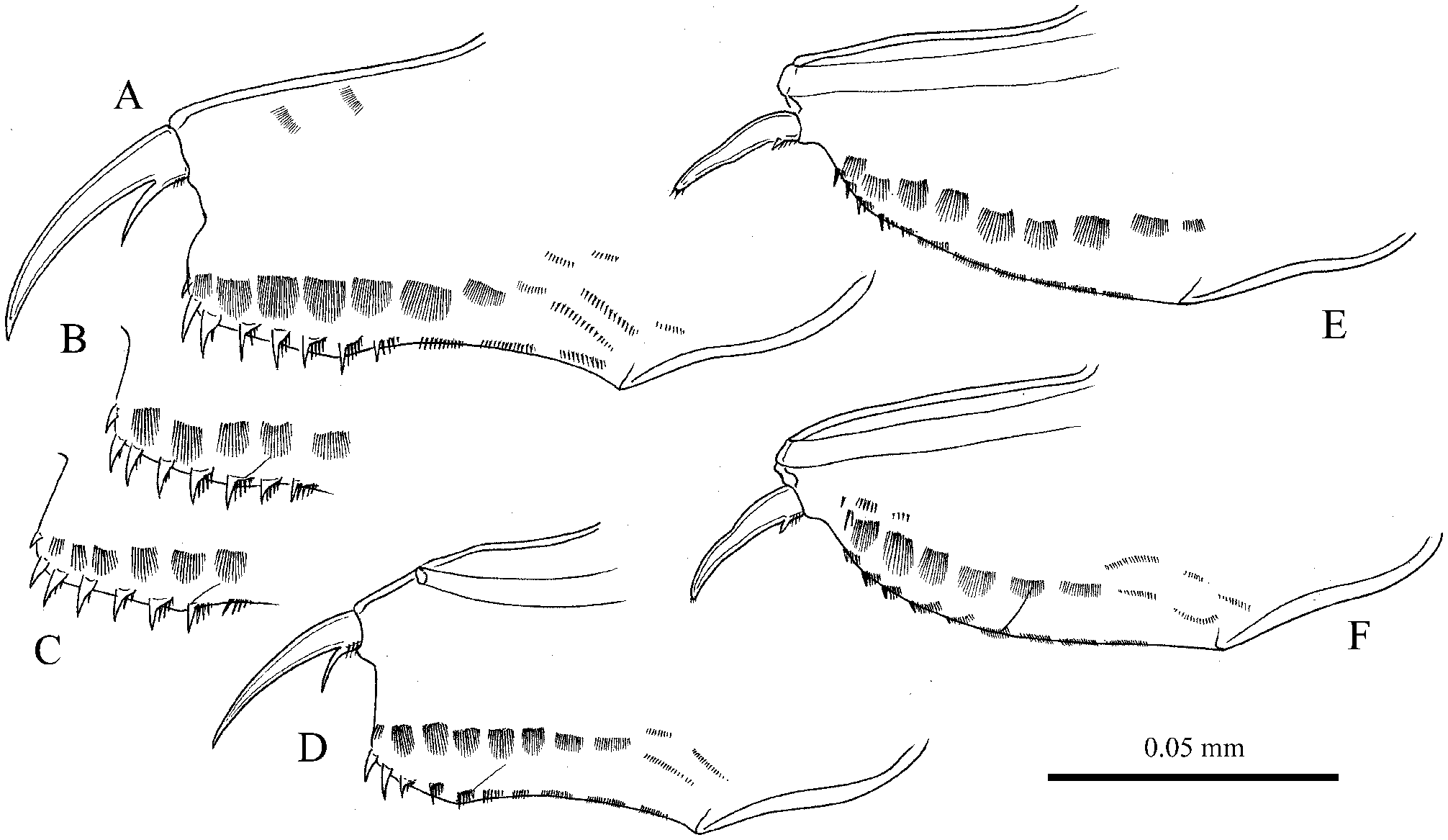
Postabdomen ( Figs. 4D View FIGURE 4 – 5A View FIGURE 5 ) short, of moderate width, slightly narrowing distally, with a rounded, not prominent distal angle, length about 2.5 height. Ventral margin straight. Base of claws separated from distal margin by a weak incision. Distal margin straight to weakly convex, distal angle rounded, not protruding before the base of claw. Dorsal margin with distal part 1.8–27 times longer than preanal one, with postanal portion 1.3–1.5 times shorter than anal. Postanal portion of distal margin straight, anal portion weakly concave. Preanal angle well-defined, but not protruding, postanal angle weakly defined. Postanal margin ( Figs. 5B–C View FIGURE 5 ) with 6–7 marginal denticles, decreasing in size basally, 2–3 distalmost denticles robust, others have broad bases, bearing long spinules anteriorly; length of distal denticles equal to the width of the postabdominal claw base. Anal margin with four groups of short setulae. Eight–nine lateral fascicles of setulae, six–seven distalmost fascicles wide, consisting of thin setulae, with longest setulae in the middle, slightly shorter than longest marginal denticles. Several additional fascicles above the main row in anal portion. Postabdominal claw of moderate length, as long as preanal portion of postabdomen. Basal spine short, curved, about 0.2 of length of claw.
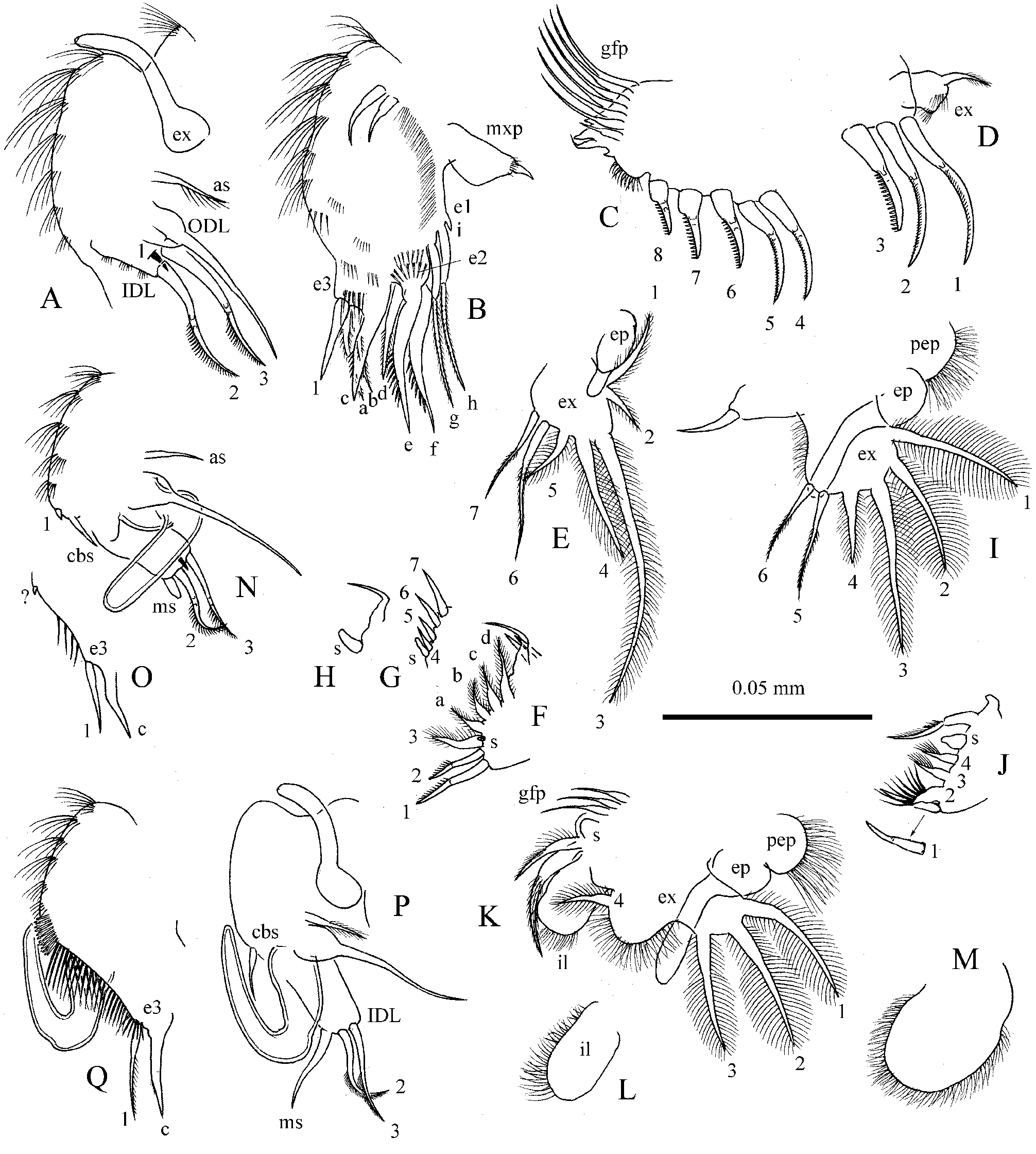
Antennule ( Fig. 2I View FIGURE 2 ) of moderate size, length about 2.5 width, with four clusters of thin setulae at anterior face, setae of two central clusters long. Antennular sensory seta slender, three times shorter than antennule, arising almost at the middle of antennule. Nine terminal aesthetascs, two longest of them about 2/3 length of antennule.
Antenna ( Figs. 2J View FIGURE 2 – 4E View FIGURE 4 ) relatively short. Antennal formula, setae 0–0–3/1–1–3, spines 1–0–1/0–0–1. Basipodite segment robust, branches of moderate length and width, proximal segments of both branches 1.5 times longer than others, middle segments slightly shorter than apical. Seta arising from basal segment of endopodite thin, not reaching the end of endopodite. Seta arising from middle segment of endopodite of similar size with shortest apical setae. Spine on basal segment of exopodite as long as middle segment. Apical spines slightly longer than apical segments.
Thoracic limbs: Six pairs.
Limb I ( Figs. 6 A–B View FIGURE 6 ) of moderate size. Epipodite with long process 3–4 times longer than exopodite itself. Accessory seta about 1/3 length of ODL seta, unilaterally armed by thick setulae. IDL with three setae and two–three clusters of small setulae, setae 2 and 3 only slightly shorter than ODL seta, armed with thin setulae in distal part, seta 1 very short, located laterally. Endite 3 with four setae, inner seta (1) shorter than setae a-c. Endite 2 with three setae (d–f), setae e–f of similar length, 1.5 shorter than limb itself. Endite 1 with two 2-segmented setae (g-h), both setulated in distal part, and very small, rudimentary seta i. No inner setae on edites 1–2. Six-seven rows of thin long setulae on ventral face of limb. Two ejector hooks, one of them larger than the other. Maxillar process elongated, with a short seta.
Limb II ( Figs. 6 C–D View FIGURE 6 ). Exopodite elongated, with a setulated seta as long as exopodite. Eight scraping setae (1–8), armed with small spinules, increasing in length distally, scraper 3 thicker than neighbors and armed with thicker spinules. Distal armature of gnathobase with four elements. Filter plate with seven setae, the posteriormost three times shorter than others.
Limb III. Epipodite oval, with a short process; exopodite trapezium-shaped, with seven setae ( Fig. 6E View FIGURE 6 ). Seta 3 being longest; setae 4 and 6 about 1/2 length of seta 3; setae 1 and 7 about 1/3 length of seta 3; seta 5 about 1/4 length of seta 3; seta 2 short. Setae 6–7 armed with short setulae in distal portion, all other setae plumose. Distal endite ( Fig. 6F View FIGURE 6 ) with three anterior setae (1–3), two distalmost members slender, sharp, with distal parts unilaterally armed with sharp denticles, seta 1 significantly longer than seta 2; basalmost seta (3) as long as seta 2, broad, bilaterally armed with setulae. Basal endite ( Fig. 6G View FIGURE 6 ) with four anterior stiff setae, increasing in size toward the base; a small sensillum near the base of distalmost seta. Four soft posterior setae increasing in size basally (a–d). Gnathobase not clearly separated from basal endite. Distal armature of gnathobase ( Figs. 6 G–H View FIGURE 6 ) with four elements: an elongated, cylindrical sensillum; thin, bent seta; others two represented by sharp spines. Filter plate III with seven setae.
Limb IV ( Figs. 6 I–J View FIGURE 6 ). Preepipodite setulated, epipodite with a process two times longer than exopodite itself. Exopodite subquadrangular, with six setae. Seta 3 longest, setae 1–2 slightly shorter than seta 3, setae 4, 5 and 6 of 1/3, 2/3 and 1/2 length of seta 3, respectively. Setae 1–4 plumose, setae 5–6 with short setulae in distal portion. Inner-distal portion of limb IV with four setae and a small cylindrical sensillum; seta 1 slender, sharp, first flaming-torch seta (2) broad, with 7–8 thick setulae; two other thin, slender, with thin hair-like setulae. Three soft setae of similar size. Gnathobase with a 2-segmented seta, and a small hillock distally. Filter plate with five setae.
Limb V ( Fig. 6K View FIGURE 6 ). Preepipodite setulated, epipodite with process 2–2.5 times longer than exopodite itself. Exopodite divided into two lobes, incursion between lobes very deep, with four plumose setae. Setae 1–3 long, subequal in length, slightly decreasing in size basally, seta 4 short, 2.5 times shorter than seta 1. Inner limb portion, il, ( Fig. 6L View FIGURE 6 ) as a small oval lobe, with setulated inner margin.At inner face, two setae, first seta about 3/4 length of exopodite seta 1, other seta 1.5 times shorter. Filter plate with three setae, a large semicircular sensillum located near it.
Limb VI ( Fig. 6M View FIGURE 6 ) as a large, elongated, rounded lobe with a setulated margin, as long as exopodite V, length about 2 widths.
Ephippial female ( Figs. 2K View FIGURE 2 , 3D–E View FIGURE 3 ) with body slightly higher than parthenogenetic female, dorsal margin without a depression between valves and head shield. Ephippium light yellow-brown in preserved specimens, with weakly developed egg locule, covered by weak polygonal sculpture, the latter slightly more pronounced than that on valves of parthenogenetic specimens.
Male. General. Juvenile male of instar II ( Fig. 2L View FIGURE 2 ) similar in shape to a juvenile female. In adult male, body ( Figs 2N View FIGURE 2 , 3F–G View FIGURE 3 ) low oval, with a maximum height at the middle, height/length ratio about 0.5.
Postabdomen in juvenile males ( Fig. 5D View FIGURE 5 ) similar to that of female, but shorter, gonopore located laterally near ventral margin, at 2/3 distance from the base. Postanal denticles same as in female. In adult male, postabdomen ( Figs. 4F View FIGURE 4 , 5E–F View FIGURE 5 ) short, moderately wide, weakly narrowing distally in anal portion, and strongly narrowing distally in postanal portion. Postanal margin of postabdomen evenly comes to the base of claws, no distinct distal margin and dorsodistal angle. Length about 2.5-2.7 heights. Ventral margin straight. A sperm duct opens at the end of postabdomen, forming small protrusion above the base of claws. Dorsal margin weakly convex in postanal portion and almost straight in anal one. Preanal and postanal angles not defined. Preanal margin almost straight. Distal portion of postabdomen two times longer than preanal one, anal and postanal portions of similar length. Postanal margin with clusters of setulae, in distalmost 4–5 clusters ditalmost setula thick, spine-like. Lateral fascicles of setulae as in female. Postabdominal claw weakly curved, shorter than in female, much shorter than preanal margin of postabdomen, with blunt tip, bearing cluster of setulae. Basal spine very short.
Antennule in juvenile male of instar II ( Fig. 2M View FIGURE 2 ) similar to that of female, with 9 terminal aesthetascs, with very short anlage of male seta. In adult male ( Fig. 2P View FIGURE 2 ) antennule slightly broader than in female, with 12 long terminal aesthetascs, longest aesthetasc about 2/3 length of antennule. Male seta located at 2/3 distance from the base, about 1/3 length of antennule.
Thoracic limb I. In juvenile male of instar II ( Figs. 6 N–O View FIGURE 6 ) limb with a curved copulatory hook, about 2/3 length of limb itself, with anlage of copulatory brush seta and smaller triangular seta, absent in adult male, about it. IDL with an anlage of male seta, IDL setae 1–3 similar to these of female. Ventral limb face under the copulatory brush seta with 4 moderately long setulae. Inner seta (1) of endite 3 same as in female. In adult male ( Figs. 6P–Q View FIGURE 6 ) limb with a U-shaped copulatory hook, 1.5 times shorter than limb itself. IDL seta 1 absent, IDL setae 2 and 3 of similar size, much thinner and shorter than in female; male seta almost straight, as long as setae 2–3. Copulatory brush seta about 1/2 length of IDL setae 2–3. Ventral face of the limb under the copulatory brush with a double row of about 30 long stiff setulae, decreasing in length distally. Inner seta (1) of endite 3 longer and thinner than in female, as long as seta c, unilaterally armed with long setulae in distal portion.
Size. In studied material, adult female length 0.37–0.41 mm, height from 0.24–0.27 mm; according to Megard, length of adult female up to 0.43 mm.Adult male length 0.34–0.35 mm, height 0.2–0.21 mm; juvenile male of instar II length 0.3–0.31 mm, height about 0.18 mm.
Differential diagnosis. P. barbulata differs from P. arvensis in: (1) three main head pores and (2) in labral keel without a prominence on anterior margin. From P. werestschagini in: (1) a smaller size, (2) smaller IP/PP ratio, and (3) shape of both male and female postabdomen. P. barbulata is habitually similar to P. guttata and P. julietae sp. nov., but differs from them in: (1) head shield with well-defined posterior angle, (2) female postabdomen with right, not prominent distal angle, (3) presence of rudimentary seta i on endite 1 of thoracic limb I, and (4) male postabdominal claw with a blunt apex armed with small denticles.
Distribution and ecology. P. barbulata is known from North-West U.S.A. and South-West Canada. In U.S.A., it was recorded from Wyoming, Montana, and Minnesota ( Megard, 1967; our data). In Canada, it was found predominantly in Alberta and British Columbia, and once in Ontario; the species is always encountered on rocky-sandy shores ( Chengalath 1987). Ecology of the species is poorly studied. According to Williams (1982), in Itasca lake, Minnesota it is the only species of Chydoridae to attain a winter density greater than its summer peak density. According to Whiteside et al. (1980), it is a pioneer species in moraine lakes of Yukon, suggesting the species can inhabit cold ultra-oligotrophic lakes.
No known copyright restrictions apply. See Agosti, D., Egloff, W., 2009. Taxonomic information exchange and copyright: the Plazi approach. BMC Research Notes 2009, 2:53 for further explanation.
|
Kingdom |
|
|
Phylum |
|
|
Class |
|
|
Order |
|
|
Family |
|
|
Genus |
Prendalona barbulata ( Megard, 1967 )
| Sinev, Artem Y., Sousa, Francisco Diogo Rocha & Elmoor-Loureiro, Lourdes M. A. 2023 |
Alona
| Baird 1843 |
Alona
| Baird 1843 |