
Spialia diomus Hopffer, 1855
|
publication ID |
https://doi.org/ 10.11646/zootaxa.4173.4.1 |
|
publication LSID |
lsid:zoobank.org:pub:3E955EB2-79DE-462C-B3EE-E4AF334D1F61 |
|
DOI |
https://doi.org/10.5281/zenodo.5632222 |
|
persistent identifier |
https://treatment.plazi.org/id/B14087C8-FFB5-924B-16BA-FF2CFC250585 |
|
treatment provided by |
Plazi |
|
scientific name |
Spialia diomus Hopffer, 1855 |
| status |
|
Spialia diomus Hopffer, 1855 View in CoL
This species was placed in the spio group by De Jong (1978). Hopffer (1855) described Pyrgus diomus from material collected by the German naturalist Wilhelm Peters in Mozambique in 1842. Butler (1899) described Pyrgus machacoana from Machakos , Kenya, collected by R. Crawshay in June 1898 ; it is now recognised as a synonym of S. diomus . Wallengren (1863) described Syrichtus ferax from material collected by Johan Wahlberg in the vicinity of the Kuiseb River in Namibia, by implication near the coast, probably about April 1854 ( Craig & Hummel 1994, pp. 152–153). Based on Evans (1937), Spialia ferax has been treated as the southern subspecies of S. diomus , found from Zambia and to the south, while what was treated as nominate S. diomus is found in sub- Saharan Africa as far south as Zambia and northern Mozambique , as well as Yemen ( De Jong 1978, Ackery et al. 1995). In the discussion section at the end of this account of S. diomus , I show that the two should be treated as separate species.
I have encountered S. diomus (Figure 11.1–2) in the savannah and dry bush of south and south-east Kenya. However De Jong (1978, Map 5) shows records from central and western Kenya as well. I agree with De Jong that this species is associated with open ground—more so than S. spio .
Adult behaviour. In January 1991 I found a male feeding at fresh elephant dung in Tsavo East, near Manyara Gate, and in May 1994 another male feeding at mud where cattle had been on the Magadi Road.
Food plants. Spialia diomus and S. ferax have similar lists of Malvaceae food plants ( Table 4), although not as extensive as that of S. spio ( Table 3). In compiling Table 4, I have assumed that the inclusion of Sida sp. and Waltheria sp. by Henning et al. (1997) and Woodhall (2005) as food plants of S. ferax are based on East African records for S. diomus , although these genera may well be used by S. ferax too. I do not know the origin of Larsen’s (2005) record of Triumfetta rhomboidea .
I have found caterpillars of S. diomus feeding on two plants in Kenya: Sida ovata (Figure 7.1) and Waltheria indica , although in captivity S. rhombifolia was also acceptable. This is the origin of Larsen’s (1991) record of Waltheria as a food plant genus. I have also found caterpillars on W. indica in Rwanda (MJWC 90/200) and Cotonou, Bénin (MJWC 89/207 A –B). On the latter occasion caterpillars of S. spio were also found on the same plants (MJWC 89/207C–D).
Ovum. Ova are laid on the upper surface of the young leaves. R. Crawshay (in Butler 1899) describes them as ‘bluish emerald-green and pale grass-green’. I have examined eclosed ova from Kenya (MJWC 88/16, 91/55). The hatching caterpillar eats only the top portion of the egg shell (Figure 11.3), so that the eclosed ovum appears barrel shaped, although the upper surface is probably rounded. There are between 19 and 24 ribs, which resemble strings of lumps or nodules (note the profiles at the top of Figure 11.3); most of these ribs are continuous from base to where the caterpillar has consumed the upper part, but a few join or split from neighbouring ribs. The space between the ribs is smooth and shiny, with about 15 fine transverse ribs.
Species Location Reference
Hermannia spp. East Africa Sevastopulo 1975, Kielland 1990, Larsen 1991, 2005 Hibiscus sp(p). East Africa Sevastopulo 1975, Larsen 1991, 2005 Pavonia sp(p). East Africa Larsen 1991, 2005
Pavonia burchellii ?East Africa Kielland 1990
(= macrophylla)
Sida sp(p). East Africa Sevastopulo unpublished, Le Pelley 1959, Sevastopulo 1975, Larsen
1991, [Henning et al. 1997], Larsen 2005, [Woodhall 2005] Triumfetta sp(p). East Africa Van Someren 1974, Sevastopulo 1974, 1975 Triumfetta rhomboidea [Afrotropical] Larsen 2005
Waltheria sp. [ Kenya] Larsen 1991, [Henning et al. 1997], Larsen 2005, [Woodhall 2005] Spialia ferax
Hermannia spp. South Africa Heath et al. 2002
Hermannia comosa (= camosa) South Africa Murray 1959, Dickson & Kroon 1978, Pringle et al. 1994, Henning et
al. 1997, Woodhall 2005
Hermannia cuneifolia South Africa Murray 1959, Dickson & Kroon 1978, Pringle et al. 1994, Henning et (= pallens,= pallius, = pollens) al. 1997, Woodhall 2005
Hermannia depressa South Africa Woodhall 2005
Hermannia diffusa (= pilosula) South Africa Murray 1959, Dickson & Kroon 1978, Pringle et al. 1994, Henning et
al. 1997, Woodhall 2005
Hermannia incana South Africa Murray 1959, Dickson & Kroon 1978, Pringle et al. 1994, Henning et (= candicans, = candicamus) al. 1997, Woodhall 2005
Hibiscus aethiopicus South Africa Dickson & Kroon 1978, Pringle et al. 1994, Henning et al. 1997, Heath
et al. 2002, Woodhall 2005
Pavonia burchellii South Africa Murray 1959, Dickson & Kroon 1978, Pringle et al. 1994, Henning et (= macrophylla) al. 1997, Heath et al. 2002, Woodhall 2005
Leaf shelters. T he mature caterpillars pull several of the small leaves of S. ovata together to form a shelter. On Waltheria indica the caterpillars fold together the two halves of a leaf as the leaves are larger and stiffer than those of S. ovata .
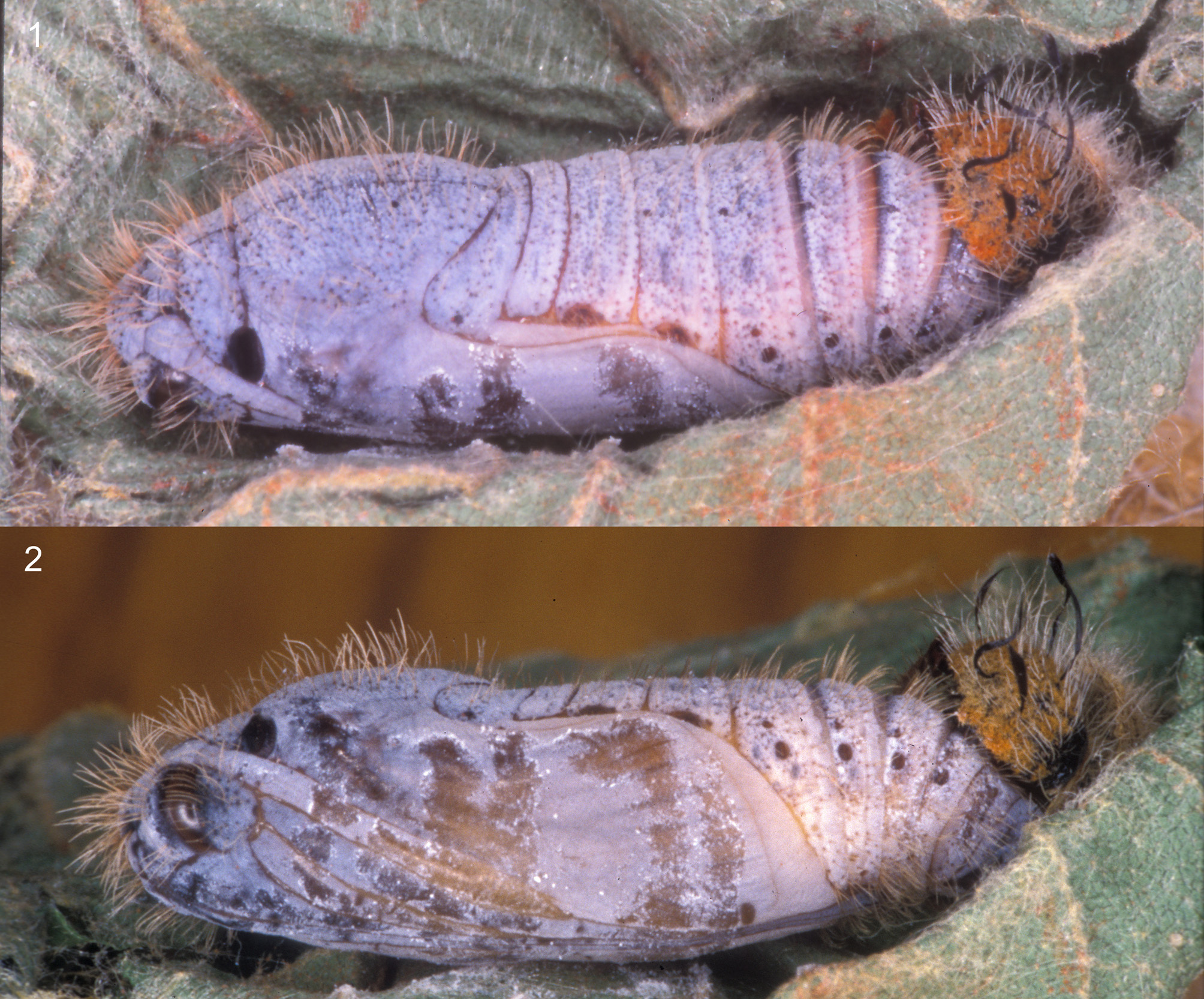
Caterpillar. The mature caterpillar (Figure 12.2–3) is very distinctive, with its head marked in dull orange and green and conspicuous black twisted, linear setae. Late instar caterpillars and pupae from Rwanda ( MJWC 90 /200) and Bénin ( MJWC 89 /207 A –B) do not differ from those described below from Kenya .
Final instar. The caterpillar grows to about 20mm (Figure 12.2–3). Head 2.5 x 2.75mm wide x high (n=3); rounded, hardly indent at vertex; dark brown, shiny, rugose, but hardly visible except narrowly on posterior margin. Most of the head is covered with unusual fan-like setae, having multiple aciculate branches, flattened against the head, mostly entangled with adjacent setae; those adjacent to the epicranial suture, the adfrontals, laterally and dorsally are dull orange, while the remainder of the epicranium and the clypeus are light orange, but in combination with other setae, and in contrast to the dull orange setae appear greenish; the orange stripe down epicranium and on adfrontals appears as an inverted Y on the face. Pale, erect, simple setae of 0.3–0.5mm down centre of face and above mouthparts; longer (1.1–1.2mm) pale, erect, flexible, slightly barbed setae, some dark distally, on the rest of the epicranium; amongst these some longer (1.6–1.7mm) erect, flexible, dark, barbed setae; laterally six very long (2.1mm) erect, twisted linear ribbon-like black setae (not completely parallel side, but nearly so and 0.2mm wide); in some individuals the long slightly barbed setae are almost entirely pale. T1 with long, pale, erect setae; the shorter ones simple, and the longer ones slightly barbed, some of which are dark, or dark distally; T1 posterior third black with broad white dorsal line and white dorsolateral patch; T1 anterior two-thirds orange, white dorsal line, laterally a dark longitudinal line and pale ventral to this; one very long, dark, robust, erect, flexible barbed setae dorsolaterally near anterior margin; a small dark, shiny, slightly domed plaque subdorsally near anterior margin; spiracle dark; legs brown. Body dull blue-green-white; T2– A 1 with very small round black plaques subdorsally and laterally; A 2– A 7 larger black plaques subdorsally, laterally and ventrolaterally; A 8– A 9 small black plaques laterally and ventrolaterally; body covered with fine white, simple setae; spiracles pale, except that of A 9 orange; legs concolorous.
Penultimate (n-1) instar. This is generally similar to the final instar; head 1.83 x 1.87mm wide x high (range 1.69–2.08 x 1.73 x 2.16, n=8); fan-like setae not as dense, paler, and less contrasting; other setae similar but shorter and most are pale; linear setae up to 1.2mm long and 0.1mm wide. T1 as final instar.
Instar n-2. The head (Figure 12.1) is similar in shape to those of the later instars, 1.52 x 1.524mm wide x high (range 1.37–1.62 x 1.45–1.61, n=3); dark brown, shiny, irregularly reticulated; scattered pale fan-like setae, smaller than those of later instars, and only partially covering the surface; scattered short, pale, erect, simple setae; longer, slightly barbed, erect, flexible setae laterally, some pale and some dark; at least five long black twisted linear setae on each side of head, but they seem to be easily abraded. It can be seen in Figure 12.1 that the body is pale green, with a darker dorsal line, and the black plaques described under the final instar above are present as very small dots.
Instar n-3. Similar to instar n-2; head measures 1.11 x 1.16mm wide x high (range 1.04–1.18 x 1.10–1.20, n=4); the black twisted linear setae are 0.4–0.6mm long.
Pupa. In captivity, the pupa ( Figure 13 View FIGURE 13 ) is formed in the last leaf shelter. The pupa is rather uniformly brown, thinly and evenly covered with white waxy bloom; long, pale brown, erect, simple setae except on the appendages; the proboscis projects slightly beyond the wing cases for up to one abdominal segment; the spiracles A 2 – A 3 are normally visible or partly visible. One pupa had died after the butterfly formed up within ( MJWC 88 /79C); white fungal hyphae had grown out of the spiracles A 2 – A 7, indicating that all these have openings, including those of A 2 – A 3 which in many species are covered by the wings, whereas that of A 8 probably does not have an opening (Scoble 1995). The pupa shown in Figure 13 View FIGURE 13 has dark brown-black rounded plaques present subdorsally on the anterior margin of A 2 – A 6, a smaller dark plaque on the anterior margin of A 4 – A 7 just above the level of the spiracle. Seven other pupae were examined, but only one had visible plaques, which were subdorsally on the anterior margin of A 3 – A 8, but partially obscured by the white waxy powder. The spiracle T1 is similar to those of S. colotes ; dark brown; more or less straight anterior margin; sometimes paler on anterior margin; they are quite variable in size: 0.43 x 0.73 x 0.28mm wide x high x thick on posterior margin (range 0.32–0.50 x 0.56–0.90 x 0.24–0.30, n=4). Adults emerge after 9–19 days in Nairobi, or as little as 8 days at the coast (Sevastopulo unpublished).
Natural enemies. I have reared a larval-pupal tachinid parasitoid, Thecocarcelia latifrons , from caterpillars collected on Sida ovata at MacKinnon Road (88.79), and on Waltheria indica at Cotonou, Bénin ( MJWC 89 / 207 A) . The tachinid larva emerges from the host pupa to form a puparium (sometimes in the pupal chamber) about 6 days after pupation; the adult tachinid then emerges from the puparium after a further 21 days or so. Thecocarcelia latifrons is widespread in Africa; it is also recorded as a pupal parasitoid of S. ferax in South Africa (Dickson & Kroon 1978, Plate 15). In Kenya, it also attacks S. zebra bifida Higgins and Gomalia elma (Trimen) . A second species, T. incedens (Rondani) , is also recorded from S. ferax in South Africa (Dickson & Kroon 1978, Plate 15). See also comments under S. spio .
Discussion. Although Higgins (1924) treated S. ferax and S. diomus as separate species, Evans (1937) treated ferax as a subspecies of S. diomus . This was followed by de Jong (1978), who believed that ‘there is no need for a specific separation as there is no evidence for geographic overlap of these forms’. In his work on African Hesperiidae, T.B. Larsen (unpublished) planned to treat ferax and diomus as separate species, based on significant differences in wing patterns, the shape of the valves, a zone where neither occurs, and no signs that a cline is involved.
In this context a comparison of the early stages is helpful. My observations and images are similar to the detailed life history illustrated by G.C. Clark (in Dickson & Kroon 1978, Plate 15), and without doubt they are very closely related. I see no clear differences between the ova. The final instar caterpillars have several important features in common, in particular the setae of the head, including the long black linear scales and the fan-like scales covering the face, and the markings of T1. Clark does not illustrate the face in frontal view; it seems rather unlikely that he would not have done so if it were patterned as strongly and distinctively as that of diomus (Figure 12.3). In addition, the body of ferax is dark dull green with what appear to be dark setae in his Plate 15.12 of the whole caterpillar, but white in his Plate 15.13 of ‘segment 7’, whereas that of diomus is whitish blue-green with pale setae. The conspicuous small dark plaques of the body of diomus are just visible on as the subdorsal row on some segments of Clark’s Plate 15.12 and the lateral row in his Plate 15.13, but they are very inconspicuous. The pupae of both species are covered with white waxy powder, but the painting of ferax is completely white, and does not show the small brown plaques on the abdomen, although these are not conspicuous in S. diomus , and Clark’s painting is not at magnification, so this difference may not be significant.
The photograph of the final instar caterpillar in Henning et al. (1997, p. 113) is also relevant. The body is dull brown-green with a darker dorsal line (matching Clark’s painting) and white dots arranged in longitudinal lines (not apparent in Clark’s painting of the whole caterpillar, but in line with his detailed image of segment A 7) and white setae (dark in Clark’s painting of the whole caterpillar, but in line with his detailed image of segment A 7); the division of the T1 into black and orange-brown areas is hardly discernable; the long black setae of the head appear simple rather than linear (possibly the linear setae have been abraded) and there is a slight differentiation on the epicrania into brown areas on each side of the face and a darker stripe down the epicranial suture (similar to the pattern observed in S. diomus but much darker).
The slightly different food plant lists ( Table 4) may be significant, but further observations would help to assess if these differences are real. On balance, particularly based on the detail so clearly shown in Clark’s work, I conclude these differences offer significant support to Larsen’s view that ferax and diomus should be treated as separate species, and so I reinstate S. ferax stat rev. as a valid species.
No known copyright restrictions apply. See Agosti, D., Egloff, W., 2009. Taxonomic information exchange and copyright: the Plazi approach. BMC Research Notes 2009, 2:53 for further explanation.
|
Kingdom |
|
|
Phylum |
|
|
Class |
|
|
Order |
|
|
Family |
|
|
SubFamily |
Pyrginae |
|
Tribe |
Carcharodini |
|
Genus |
|
Kingdom |
|
|
Phylum |
|
|
Class |
|
|
Order |
|
|
Family |
|
|
SubFamily |
Pyrginae |
|
Tribe |
Carcharodini |
|
Genus |
|
Kingdom |
|
|
Phylum |
|
|
Class |
|
|
Order |
|
|
Family |
|
|
SubFamily |
Pyrginae |
|
Tribe |
Carcharodini |
|
Genus |