
Kanuites lewisae, Dehghani & Werdelin, 2008
|
publication ID |
https://doi.org/ 10.5252/geodiversitas2019v41a6 |
|
publication LSID |
urn:lsid:zoobank.org:pub:619EB4F8-90CD-4559-8B78-2BA79210F73B |
|
DOI |
https://doi.org/10.5281/zenodo.3704564 |
|
persistent identifier |
https://treatment.plazi.org/id/B27C87E2-3546-FF8A-B12A-F9A2FDE08F9C |
|
treatment provided by |
Valdenar |
|
scientific name |
Kanuites lewisae |
| status |
|
cf. Kanuites lewisae View in CoL View at ENA
MATERIAL EXAMINED. — KNM-FT 3369, left m1; KNM-FT 3370, right hemimandible with m1; KNM-FT 3371, left dp4; KNM-FT 3876, right m1; KNM-FT 8744A-B, juvenile mandible with p1 and dp2-dp4, left dp3; KNM-FT 8745, partial juvenile cranium with right P1 and dP2-dP4, left dP3-dP4; KNM-FT 8750, partial juvenile cranium with right P1 and dP2-dP4, left P1 and dP3-dP4; KNM-FT 15093, left m1.
DESCRIPTION
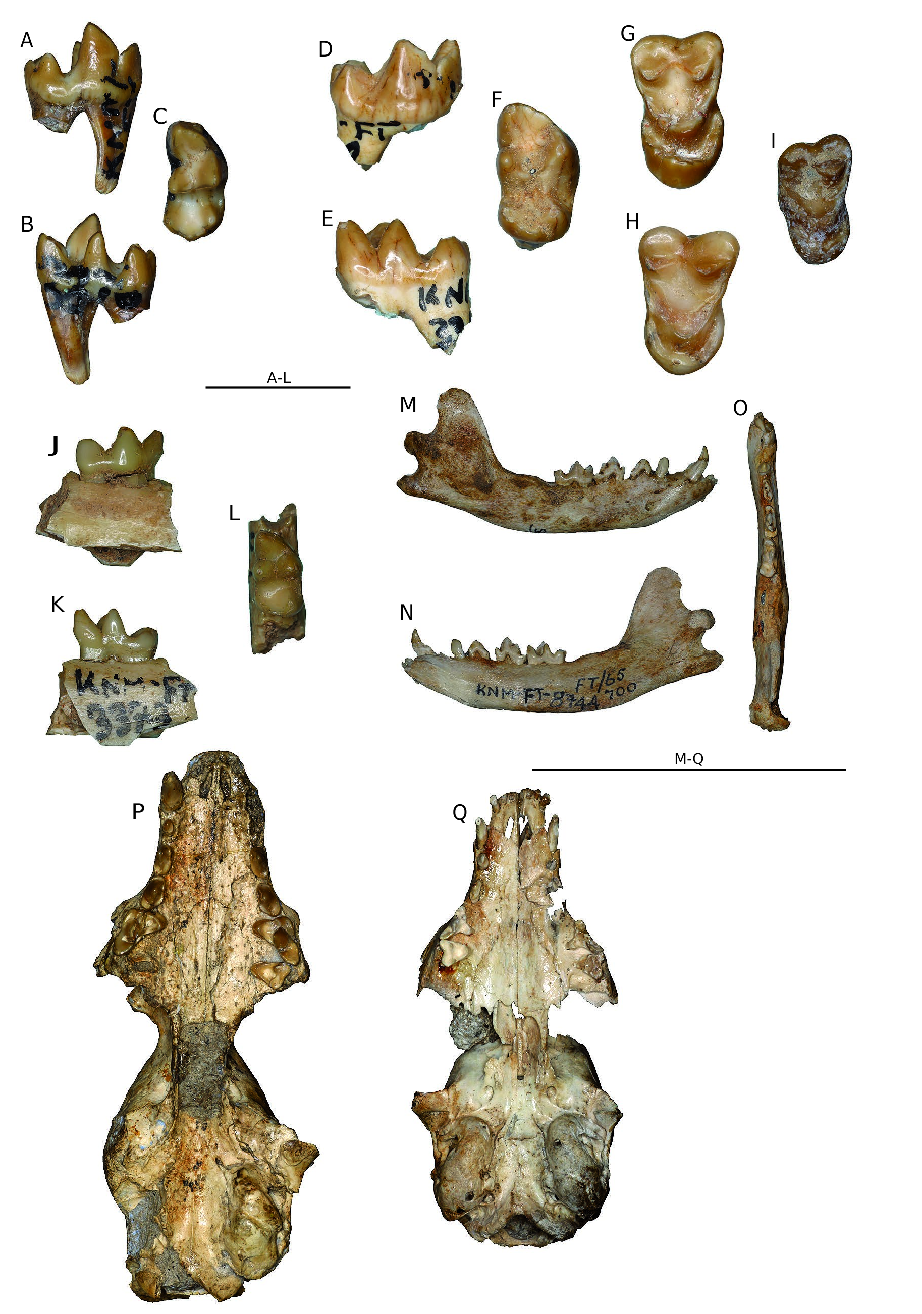
KNM-FT 3876 ( Fig. 4 View FIG A-C)
This is the best preserved of the lower carnassials and its description will also serve for KNM-FT 3369 and KNM-FT
15093, as well for as the m1 of KNM-FT 3370. The tooth is short and broad with a trigonid that accounts for about 60% of the total length of the tooth. The protoconid is the tallest cusp by far, whereas the paraconid and metaconid are of similar height. The angle metaconid-paraconid-carnassial notch is about 63°. In occlusal view the paraconid is slightly larger than the protoconid and both are considerably larger than the metaconid. The central valley is wide but not flat in the middle. The angle between pre- and postprotocristids is about 92°. The talonid is about equal in width to the trigonid. The talonid has three distinct cusps: a large hypoconid that is connected to the trigonid by a weakly developed cristid obliqua, an entoconid that is nearly the same size as the hypoconid, and a hypoconulid that is low situated at the distalmost point of the tooth.
Measurements. KNM-LT 3876.: Lm1 8.1; Wm1 4.1; Ltm1 4.9. KNM-FT 3369 Lm1 8.3; Wm1 3.9; Ltm1 5.2. KNM- FT 15093: Lm1 7.5; Wm1 3.7; Ltm1 4.5.
KNM-FT 3370
Apart from the slightly damaged m1 (which is accounted for under the m1 description above) this specimen also includes the hemimandible and a very damaged p4. The corpus is long and low with its deepest part beneath m1. There is one discernible mental foramen, located near the anterior alveolus for p2. The alveoli for p2 and p3 are separated by short diastemas, but no alveolus for p1, if present, can be seen. The alveolus or alveoli for m2 also cannot be seen. The masseteric fossa ends near the inflection point between corpus and ascending ramus. The latter is low and has a squared-off rim. The condyle is substantial and set somewhat obliquely to the anteroposterior axis of the corpus. The angular process is large and extends nearly as far posteriorly as the condyle.
Measurements. Lm1 6.7; Wm1 3.8; Ltm1 4.1.
KNM-FT 8744A, B ( Fig. 4 View FIG M-O)
These hemimandibles have long and slender corpuses. There is a large mental foramen beneath dp2, near the ventral edge of the corpus. Smaller foramina are present anteroventral to the large one. The symphysis is long, slender, and low, reaching the middle of dp2. The ascending ramus is slender, with a rounded rim (unlike the squared-off rim described for the adult hemimandible above). The p1 is more or less triangular in occlusal view, with the apex facing mesially, and is about equal in length and width. The dp2 is a small, slender tooth with its apex above the mesial root. There are no mesial or distal cusps. The greatest width is distal to the main cusp. The dp3 is also slender, with its widest part distally, where the tooth widens into a short shelf. The mesial accessory cusp has a marked mesial lean. The distal accessory cusp is large and appressed to the main cusp. The dp4 has a trigonid that forms about 60% of the length of the tooth. The paraconid is the largest trigonid cusp and is situated mesially, where it cups the mesial margin of the tooth. The protoconid is the tallest trigonid cusp. It is crest-like and located at the buc- cal edge of the tooth. The metaconid is of about the same height as the protoconid, but is the smallest of the three cusps. It is located at the distolingual corner of the trigonid, well separated from the protoconid. The talonid is slightly broader than the trigonid. The cristid obliqua is prominent and leads to a moderately tall hypoconid. The entoconid is of about equal height to the hypoconid, and there is a small distal hypoconulid.
Measurements. Ldp2 3.6; Wdp2 1.4; Ldp3 5.2; Wdp3 2.1; Ldp4 6.5; Wdp4 2.7; Ltdp4 3.9.
KNM-FT 3371
A dp4 that is closely similar to, but larger than, the one described above from KNM-FT 8744A.
Measurements. Ldp4 8.0; Wdp4 3.9; Ltdp4 5.3.
KNM-FT 8745, KNM-FT 8750 ( Fig. 4Q View FIG )
Two juvenile crania that are closely similar, especially dentally, and therefore will be described together. Some incisors are preserved in KNM-FT 8745, but their crowns are damaged and provide little or no useful information beyond being typical small carnivore incisors. The same can be said of the upper canine, which is long, slender, and relatively straight. The P1 is pear-shaped, with its widest point close to the distal end of the tooth. It is separated from the canine by a short diastema that is half the length of the diastema between P1 and DP2. The dP2 is long and slender. Its apex lies anterior to the middle of the tooth. There are no accessory cusps either mesially or distally. The dP3 has a crescent-shaped preparastyle situated at the mesialmost point of the tooth. The parastyle is small and located mesiolingual to the paracone. The paracone is tall and sharp and connects to the metastyle by a shallow notch. There is a distinct metacone close to this notch. The protocone is low but broad and set lingual to the apex of the paracone. The dP4 is triangular with a concave lingual border. There is a stylar cusp at the mesiobuccal corner of the tooth. The stylar shelf is wide. The paracone is low and crescentic and the metacone set near the distobuccal corner of the tooth. The protocone is large and set lingually, separated from the paracone and metacone by a wide shelf.
The cranium will not be described here. In the absence of suitable comparative material, it is difficult to evaluate any characteristics of the sutures and other structures. The auditory bullae are intact, so their internal structure cannot be evaluated at present.
Measurements. KNM-FT 8745: LP1 2.3; WP1 1.6; LdP2 3.6; WdP2 1.5; LdP3 6.6; WadP3 3.8; WbldP3 2.1; LpdP3 2.3; LmdP3 2.7; LdP4 4.6; WdP4 4.7. KNM-FT 8750: LdP2 3.5; WdP2 1.6; LdP3 6.4; WadP3 3.8; WbldP3 1.8; LpdP3 2.4; LmdP3 2.6; LdP4 4.5; WdP4 4.5.
DISCUSSION
Kanuites lewisae was described on the basis of the well-preserved cranium KNM-FT 8747 ( Dehghani & Werdelin 2008). The
material definitively assigned to K. lewisae above is identical with the original hypodigm of the species, identified due to overlap with the dentition of the holotype cranium. This material did not include any of the lower dentition, making any identification of such elements tentative, as is the case with the juvenile specimens. The main argument for the identifications is size. Leaving the very small m1 KNM- FT 3373 (see below) aside, there are two sizes of lower m1 that might fit with the K. lewisae upper dentition. Of these the m1s KNM-FT 3360 and KNM-FT 3368 are, barring very odd dental proportions, too large to match the P4 of K. lewisae . A similar argument, if more tenuous, applies to the deciduous dentition. In addition to these arguments by size it is also possible to make an argument by commonality, i.e., that the most common elements in each dental region are likely to belong to the same taxon. In this case the two arguments coincide. If correct, this makes K. lewisae the best characterized Miocene viverrid in Africa.
Kanuites bears similarities to both Viverridae and Hyaenidae . Among the latter it is broadly similar to species of Protictitherium in size. This genus is known from the early and middle Miocene in Eurasia and the middle Miocene of Africa ( Werdelin & Solounias 1991; Werdelin & Peigné 2010). African finds come from two North African sites (Beni Mellal, Morocco; Beglia Formation, Tunisia) and a single find in eastern Africa (Namurungule Formation, Kenya). The two North African sites are similar in age to Fort Ternan, whereas the Kenyan find is early late Miocene in age. None of the finds were originally described as Protictitherium.
I will here focus on the find from the Beglia Formation, originally described by Kurtén (1978) as Tungurictis punica, but reassigned to Protictitherium punicum by Werdelin & Solounias (1991). The specimen consists of a maxilla fragment with P3-M2 and thus overlaps with the type specimen of Kanuites lewisae in both age and representation. It should be noted that where overlap in representation occurs the P. punicum specimen is closely similar to the Beni Mellal material assigned by Ginsburg (1977) to Ictitherium cf. arambourgi as far as can be determined from photographs in the latter publication. Comparison between P. punicum ( Kurtén 1978: figs 1, 2) and the type specimen of K. lewisae ( Fig. 4P View FIG ; Dehghani & Werdelin 2008: fig. 1) shows some general similarities in size and structure but considerable differences in detail. The P3 is similar in size and proportions, but the P. punicum specimen has a hyaenid-like cingulum surrounding the crown, whereas in K. lewisae the cingulum is absent buccal and the lingual cingulum bulges further lingually than in P. punicum. The morphology of the P 4 in P. punicum is typically hyaenid, with paracone and metastyle longer and more slender than in K. lewisae , whereas the P4 protocone of P. punicum is short and has its distal connection to the paracone near the anterior end of the latter cusp. In K. lewisae the protocone reaches distally to about the middle of the paracone. The M1 of P. punicum is mesiodistally foreshortened and its mesial and distal margins are curved. In K. lewisae the mesial and distal margins are straight and the tooth overall longer mesiodistally compared to buccolingual breadth. Thus, K. lewisae differs from the penecontemporaneous P. punicum in a number of characters that precludes referral to Hyaenidae . Referral to Herpestidae is unlikely for reasons discussed in Dehghani & Werdelin (2008) and therefore Viverridae remains as the likely family assignment for Kanuites lewisae .
Subfamily designation for Kanuites is difficult but it is here referred to Paradoxurinae for the following reasons. It shows some overall similarities to Kichechia zamanae, assigned to Paradoxurinae by Morales & Pickford (2011) in molar shape. It is less hypercarnivorous, with short, relatively broad carnassials and molars, than known African Viverrinae . Most importantly, the M1 of K. lewisae shows the presence lingually of a cingulum surrounding the protocone, as well as overall occlusal surface symmetry. I interpret this as a less derived version of the condition seen in the M1 of cf. Tugenictis, described below. The cingulum is also seen in Kichechia, but not in Viverrinae that I have observed.
No known copyright restrictions apply. See Agosti, D., Egloff, W., 2009. Taxonomic information exchange and copyright: the Plazi approach. BMC Research Notes 2009, 2:53 for further explanation.