
Mycomelichares polypori Mašán & Joharchi, 2021
|
publication ID |
https://doi.org/ 10.11646/zootaxa.4980.1.10 |
|
publication LSID |
lsid:zoobank.org:pub:012A0F7E-1DC9-4A74-9FE6-4ACCF1436E78 |
|
DOI |
https://doi.org/10.5281/zenodo.4944959 |
|
persistent identifier |
https://treatment.plazi.org/id/B3384A3B-FFC7-9D0B-1FB2-1542FDABFBD3 |
|
treatment provided by |
Plazi |
|
scientific name |
Mycomelichares polypori Mašán & Joharchi |
| status |
sp. nov. |
Mycomelichares polypori Mašán & Joharchi sp. nov.
( Figs 1–17 View FIGURES 1–6 View FIGURE 7 View FIGURE 8 View FIGURES 9–17 )
Type material examined
Holotype female: SW Slovakia, Little Carpathians Mountains, Pernek Village, Mäsiarsky Ostrovec Forest, on Polyporus squamosus (Huds.) Quélet (Fungi: Basidiomycota, Polyporales ) growing on beech stump, beech forest, 470 m a.s.l., June 26, 2019. Paratypes: 28 females, with the same data as for holotype; one female, with the same collection site as for holotype, under elytra of Triplax sp. ( Coleoptera , Erotylidae ) found on Hydnum sp. (Fungi: Basidiomycota, Cantharellales ), June 6, 2019; three females, with the same collection site as for holotype, on Triplax lepida (Faldermann) found on P. squamosus , July 2, 2019; ten females, with the same collection site as for holotype, on Triplax rufipes (Fabricius) found on P. squamosus , July 2, 2019; seven females, SW Slovakia, Little Carpathians Mountains, Bratislava Capital, Horský Park, on P. squamosus growing on logs of Prunus avium , broadleaved forest, 230 m a.s.l., June 21, 2019; two females, SW Slovakia, Rusovce Village, Rusovecký Park, under elytra of Triplax sp. found on unidentified wood decaying fungus, flood-plain forest, 140 m a.s.l., April 25, 2019; one male, the same collection site as previous record, on P. squamosus , May 4, 2019; three females, SW Slovakia, Považský Inovec Mountains, Hrádok Village, Hrádocká Dolina Valley, on Triplax sp. found on P. squamosus , broadleaved forest, 410 m a.s.l., August 2, 1997. All specimens are deposited in the Institute of Zoology, Slovak Academy of Sciences, Bratislava, Slovakia.
Diagnosis (Adults)
Idiosoma widely oval, with legs shorter than dorsal shield. Dorsal shield normally with 32–33 pairs of setae, 18–19 pairs on podonotal (j1–j6, z2, z4–z6, s1–s6, r3, and r4; r6 often on soft integument in female) and 14 pairs on opisthonotal portion (J1–J5, Z1–Z5, S2–S5); setae z1, z3, r2, r5, and S1 not expressed, absent; setae of R- setal rows completely situated on soft integument close to posterolateral margins of the shield in female, or fully reduced and absent in male; male dorsal setae conspicuously longer than those in female. Female tritosternum robust, brushshaped, with shortened, thickened and densely pilose laciniae; male tritosternum relatively small, with short base, thin and sparsely pilose laciniae. In female, metasternal setae (st4) and associate lyrifissures (iv3) situated on soft integument. Peritremes shortened, with anterior ends reaching the level between setae s1 and s2. Anus small. Ventral surface of hypostome normally with nine transverse rows of denticles (not discernible in male); anteriormost rostral setae h1 longest, thickened basally and slightly spatulate distally in female (not modified in male); other setae on hypostome needle-like. In female, chelicera with relatively shorter and wider digits, and robust and sharp denticles; fixed digit with usually 5–7 denticles in addition to bidentate terminal hook; male spermatodactyl narrowed terminally, nearly as long as fixed digit.Anterior margin of epistome denticulate (female) or smooth (male). Gnathosomal corniculi horn-like and well spaced. Palp apotele 2-tined. Ventral surface of genu III with one ventral seta (pv absent); some of dorsal setae on trachanters and femora thickened, spur-like; male leg II not armed by spurs.
Description (Adults)
Female. Dorsal idiosoma ( Figs 1 View FIGURES 1–6 , 7 View FIGURE 7 ). Idiosoma widely oval to round (especially in mature specimens with egg), up to 640 μm in length and 500 μm in width, often not completely covered by dorsal shield; the shield widely oval, usually with lateral margins irregularly formed, 480–570 μm long and 345–390 μm wide (n=15), completely and densely reticulate, bearing 32–33 pairs of setae of which 18–19 pairs on podonotal portion (j1–j6, z2, z4–z6, s1–s6, r3, and r4; r6 often on soft integument in female) and 14 pairs on opisthonotal portion (J1–J5, Z1–Z5, S2–S5); setae z1, z3, r2, r5, and S1 absent. Except setae Z5 (49–58 μm), all dorsal shield setae similar in size and form, simple, needle-like and short; lengths of some selected setae as follows: j1 25–30 μm, j5 14–18 μm, r3 28–34 μm, J1–J3 18–25 μm, J4 24–29 μm, J5 6–11 μm, Z1–Z4 24–31 μm, S2– S4 25–33 μm, S5 24 –30 μm.
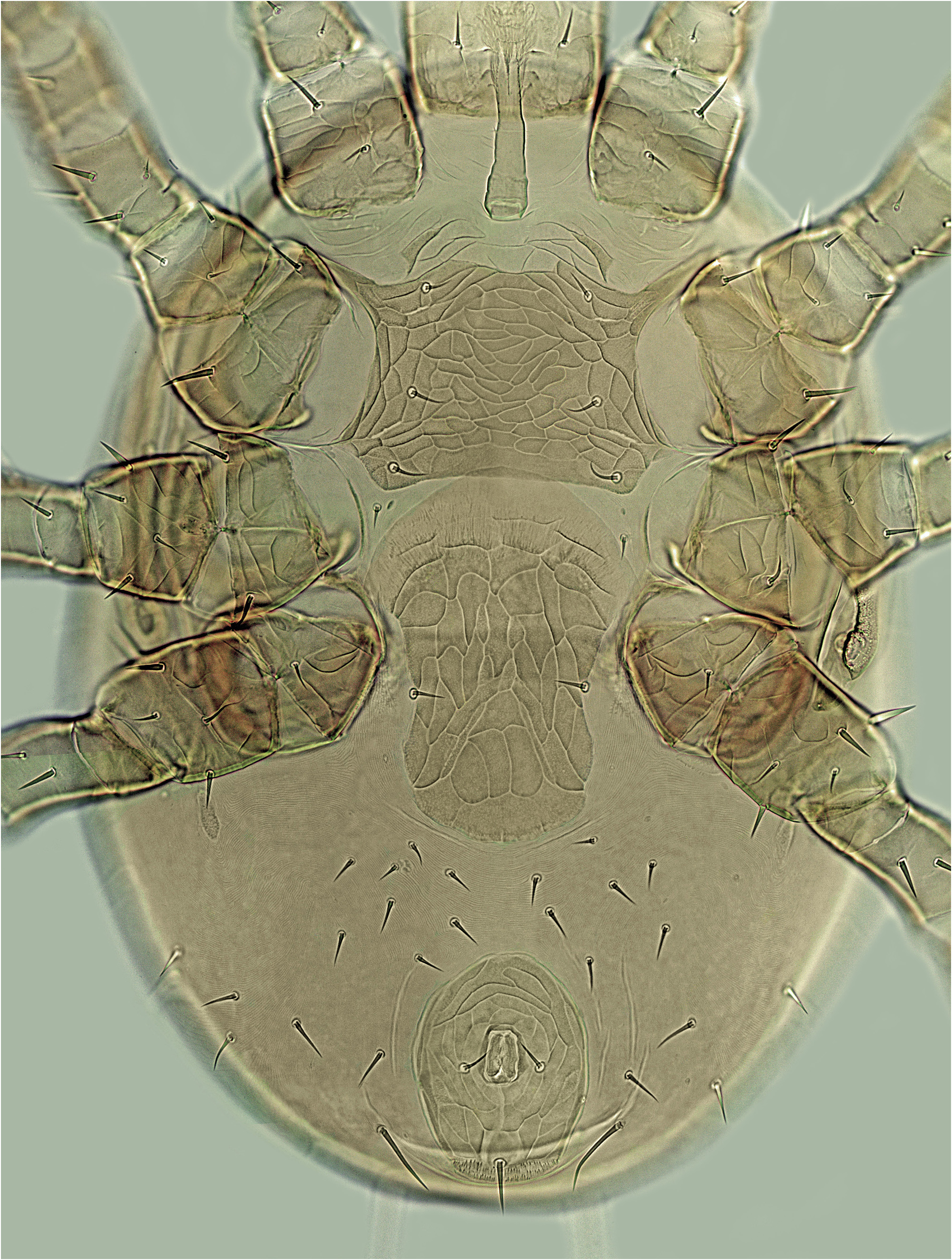
Ventral idiosoma ( Figs 2 View FIGURES 1–6 , 8 View FIGURE 8 ). Tritosternum robust, with columnar base and thickened laciniae, laciniae proximally fused, distally with dense pilosity, brush-shaped ( Fig. 12 View FIGURES 9–17 ). Presternal region transversely striate, weakly sclerotised, with a pair of elongate, obliquely oriented and indistinct platelets. Sternal shield wider than long, 84–98 μm long and 115–130 μm wide between coxae II, densely reticulate throughout except for very narrow posteriormost surface; anterior margin with slight medial concavity, posterior margin straight or slightly concave; shield with two pairs of lyrifissures (iv1, iv2) and three pairs of similar setae (st1 12–16 μm, st2 14–18 μm, st3 16–21 μm). Metasternal setae (st4) shortest of those on ventrum, 9–13 μm long, together with associated lyrifissures (iv3) situated on soft integument (metasternal platelets absent). Endopodal plates between coxae III–IV narrow. Epigynal shield oblong, relatively wide (especially in anterior portion), moderately constricted at level of genital setae (st5), tongue-shaped, 170–185 μm long and 82–92 μm wide (st5–st5 75–82 μm), hyaline and widely convex anteriorly (hyaline part reaching only slightly beyond posterior margin of sternal shield), rounded posteriorly, bearing one pair of setae and a pattern of reticulation; genital lyrifissures (iv5) situated on soft integument behind st5. Peritremes short and relatively wide (mainly close to stigma), with anterior ends reaching anterior margin of coxae I; peritrematal shields reduced to narrow plate fused to outer medial section of peritremes, and short poststigmatic parts tapered terminally and narrowly fused to exopodal plates. Soft integument behind coxae IV with a pair of small and suboval metapodal platelets; four pairs of very weakly defined narrow sclerites present close to posterior margin of epigynal shield (three pairs), and behind coxae IV (one pair). Anal shield suboval to ovoid, oblong, rounded anteriorly, convex or almost straight posteriorly, 97–112 μm long and 75–89 μm wide, with reticulate surface, three circum-anal setae, and a pair of marginal gland pores (gv3) at level of posterior margin of anal opening; postanal seta almost twice longer than adanals (pa 25–30 μm, ad 14–18 μm); anus small, 23–28 μm long and 15–19 μm wide, with anteromedial to almost central position on the shield. Soft lateral and opisthogastrict integument bearing normally 16 pairs of setae (JV1–JV5, ZV1, ZV2, ZV4, ZV5, SV2, UR, R1–R5), submarginal setae at level of coxae IV absent; opisthogastric setae simple and needle-like (JV1 11–16 μm, JV2 14–19 μm, JV5 35–44 μm).
Sperm induction system ( Fig. 17 View FIGURES 9–17 ). Coxae III associated with thin and long tubular section, other components of sperm system not discernible.
Gnathosomal structures ( Figs 3, 4, 6 View FIGURES 1–6 , 9–11, 13, 14 View FIGURES 9–17 ). Anterior margin of epistome normally narrowly convex, irregularly denticulate, sometimes with inconspicuous central apex formed by larger or isolated denticle; sometimes epistome subtriangular, with well-tapered apex, rarely tricuspidate ( Figs 4 View FIGURES 1–6 , 11, 13, 14 View FIGURES 9–17 ). Venter of hypostome with widened longitudinal furrow and increased number of nine transverse rows of denticles, occasionally with five or up to 11 rows of denticles, each row with multiple denticles; corniculi horn-like, well spaced, more or less convergent to each other, not reaching apices of internal malae ( Figs 3 View FIGURES 1–6 , 9 View FIGURES 9–17 ). Anterior rostral setae (h1) thickened, with distal spatulate portion, other rostral setae simple and needle-like (h1 32–37 μm, h2 11–17 μm, h3 17–22 μm, pc 16–21 μm). Median article of chelicera 72–87 μm long; cheliceral denticles well-developed, robust; fixed digit normally with six denticles (rarely five or seven) in addition to terminal bidentate hook, movable digit with three denticles and terminal hook ( Figs 6 View FIGURES 1–6 , 10 View FIGURES 9–17 ).
Legs. All legs with well-developed pretarsus and ambulacral apparatus (including pulvillus and two claws), shorter than dorsal shield: legs I 390–430 μm, legs II 320–345 μm, legs III 340–365 μm, legs IV 425–460 μm long. Leg chaetotaxy pattern as follows: leg I – coxa (2), trochanter (6), femur 2 3/1, 2/2 2 (12), genu 2 3/2, 3/1 2 (13), tibia 2 3/2, 3/1 2 (13); leg II – coxa (2), trochanter (5), femur 2 3/1, 2/2 1 (11), genu 2 3/1, 2/1 2 (11), tibia 2 2/1, 2/1 2 (10); leg III – coxa (2), trochanter (5), femur 1 2/1, 1/0 1 (6), genu 2 2/1, 2/0 1 (8), tibia 2 1/1, 2/1 1 (8); leg IV – coxa (1), trochanter (5), femur 1 2/1, 1/0 1 (6), genu 2 2/1, 3/0 1 (9), tibia 2 1/1, 3/1 2 (10). Leg setae smooth and mostly needle-like; some dorsal setae enlarged, spiniform (pd on trochanter I; ad1, pd1, pd2 on femur I and II; ad1 on femur III; pd2 on genu III; ad1 on femur IV); some setae minute (av, pv2 on trochanter I; av, ad2, ad3 on femur I and II; av2 on genu I; av on genu II and tibia II; pd and pl on femur IV). No macrosetae on tarsi I–IV present.
Male ( Figs 5 View FIGURES 1–6 , 15, 16 View FIGURES 9–17 ). Dorsal shield widely oval, 425 μm long and 318 μm wide; shield with similar chaetotaxy to female, except absence of posteromarginal setae (R1–R5), completely and very weakly reticulate, slightly expanded posterolateraly and covering narrow lateral strips of ventral idiosoma (setae Z5, S4 and S5 with ventral position); most of dorsal setae longer as in female: j1 24–28 μm, j5 27–32 μm, podonotal setae with medial position 36–45 μm, podonotal setae with marginal position 45–50 μm, opisthonotal setae with medial position 39–42 μm, opisthonotal setae with marginal position 48–62 μm. Tritosternum with columnar base and two laciniae; base short, narrowed distally and denticulate laterally; laciniae thin, sparsely and shorty pilose ( Fig. 16 View FIGURES 9–17 ). Venter probably with two separate plates, sternogenital and ventrianal shield (their adjacent margins not clearly visible due to surface invagination), both very delicately reticulate; sternogenital portion with three pairs of lyrifissures and five pairs of setae: st1 19–20 μm, st2 15–17, st3 19 μm, st4 16–18 μm, st5 26–28 μm; ventrianal shield with five pairs of setae in addition to three circum-anal setae: JV1 15 μm, JV2 33–35, JV3 24–27 μm, JV4 21–30 μm, ZV2 25–30 μm; ad 16–17 μm, pa 35 μm. Opisthogastric soft integument with only two pairs of setae: JV5 54 μm, ZV4 27–29 μm. Peritrematal shields with posterior ends connected to exopodal shields, free from anterolateral corners of ventrianal shield; relative length of peritremes similar to that of female.
Gnathosoma similar to that of female, ventral hypostome with transverse rows weakly defined and very finely denticulate. Epistome regularly convex, with smooth margin ( Figs 5 View FIGURES 1–6 , 15 View FIGURES 9–17 ). Chelicera with distally narrowed spermatodactyl, spermatodactyl about as long as fixed digit; cheliceral digits with the same number of smaller denticles as in female. Rostral setae uniform, similar in lengths: h1 22–23 μm, h2 15–16, h3 16–18μm, pc 16–17 μm.
Sexual dimorphism of leg setae not marked. Lengths of legs as follows: legs I 345–355 μm, legs II 290–300 μm, legs III 305–310 μm, legs IV 395–400 μm.
Etymology
The name of this species refers to its specific association with the polypore bracket fungus, dryad’s saddle Polyporus squamosus (classified by some mycologists in the genus Cerioporus P. Micheli ex Adans. ).
Ecological notes
Mycomelichares polypori appears to be a specialised mycetobiont exclusively colonising the fruiting bodies of Polyporus squamosus , where it occurs inside the large pores of the active hymenophore. This polypore fungus has a widespread distribution, being found in North America, Australia, Asia, and Europe, where it causes a white rot in the heartwood of living and dead hardwood trees. Mycomelichares polypori is common and abundant species often with several dozens of mites in a single fungus, and with well-developed phoretic activity on erotylid beetles of the genus Triplax . In Slovakia, it is distributed mainly in lowlands and lower mountainous areas, and it was not found on the host fungi collected in higher mountainous habitats, or on dried out or rotting fruiting bodies of the old fungi. Mites can be extracted directly from fruiting bodies using forceps, but it was necessary to wait a while after collecting the mushrooms until the mites began to climb out of the hymenophoral pores and move freely around the fruiting body. I could collect many individuals directly from dark clothing when they left the fungus, and on which they contrasted significantly like bright dots.
Bracket fungi provide a microhabitat for a wide variety of mite groups. The Mesostigmata in these fungi are mostly predators, but a few genera include truly mycophagous species ( Walter & Proctor, 2013). These fungus-feeding or fungus-inhabiting species have developed some striking adaptations for life in fungal spore-tubes ( Lindquist, 1995), especially such as strongly narrowed and elongated body. Interestingly, this adaptation did not occur in Mycomelichares polypori , probably because the tubes of the host fungus are very wide and spacious in comparison with all other basidiomycete bracket fungi.


No known copyright restrictions apply. See Agosti, D., Egloff, W., 2009. Taxonomic information exchange and copyright: the Plazi approach. BMC Research Notes 2009, 2:53 for further explanation.