
Hedya tritofa, Alipanah, Helen & Baixeras, Joaquín, 2011
|
publication ID |
https://doi.org/ 10.5281/zenodo.201706 |
|
DOI |
https://doi.org/10.5281/zenodo.6190563 |
|
persistent identifier |
https://treatment.plazi.org/id/B66B87BA-FFDC-FFED-FF29-FE39FB83277E |
|
treatment provided by |
Plazi |
|
scientific name |
Hedya tritofa |
| status |
sp. nov. |
Hedya tritofa View in CoL sp. n.
Holotype: 3, Siāhkal- Deylamān Rd., Tootki vill. (Gilān Prov.), N 37° 3ˏ 37.74˝, E 49º 50´56.41˝, 464 m, 22.vii.2010, Ālipanāh leg. (GS: HA 1170). Deposited in IRIPP.
Paratypes: 13, Rāmsar, Rāmsar- Javāherdeh Rd. (km 6) (Māzandarān Prov.), N 36° 54ˏ 29.3˝, E 50° 35ˏ 13.2˝, 554 m, 23.vii.2007, Ālipanāh, Zahiri leg. (GS: HA 928); 1 3, Bandar Anzali, Punel, 30 km S Asālem (Gilān Prov.), 250 m, 12.viii.1974, Mirzāyāns, Ilkhāni leg.; 4 3, Rāmsar, Eshkatechāl (Māzandarān Prov.), 1200 m, 28.v.2003, Gilāsiān, Nematiān leg.; 2 3, Tang-e Gol, Golestān National Park (Golestān Prov.), N 37° 22ˏ 14.7˝, E 55° 56ˏ 00.0˝, 718 m, 30.viii.2009, Ālipanāh, Buszko leg.; 1 Ƥ, Behshahr, Duk forest (Māzandarān Prov.), 840 m, 23.vii.1977, Pāzuki, Mortazavihā leg.; 1 Ƥ, Nekā (Māzandarān Prov.), N 36° 30ˏ 16.7˝, E 53° 23ˏ 27˝, 527 m, 30.ix.2007, Ālipanāh, Buszko, Zahiri leg.; 2 3, 3 ƤƤ, Siāhkal- Deylamān Rd., Tootki vill. (Gilān Prov.), N 37° 3ˏ 37.74˝, E 49º 50´56.41˝, 464 m, 22.vii.2010, Ālipanāh leg. All material deposited in IRIPP.
Description. Male (Fig. 1). Head: Scales slightly erect, dark brown, with some dark iridescence on vertex and crown; smaller scales on fronto-clypeus concolorous; labial palpus brown or dark brown laterally, with length less than twice width of compound eye; proboscis developed, unscaled; antenna dorsally brown, ventrally cream, with short, dense ventral cilia; ocelli and chaetosemata well developed. Thorax: Scaling smooth, dorsally glossy dark brown to somewhat iridescent, progressively paler toward ventral areas; legs unmodified, brown dorsally, paler ventrally, concolorous with thorax; hair pencil from metatibia base. Forewing length 6.7–8.3 mm (= 7.2 mm, n= 11); upperside with basal, subbasal and median fasciae almost fused (Figs. 1, 3), with small but distinctive group of dark scales centered on base of M1 and M2 slightly distal to median fascia; distal one-half of wing almost white; postmedian fascia represented by small group of dark scales on margin where R1 meets costa, preterminal fascia represented by similar fragment where R3 meets costa; postmedian or preterminal fragments not expressed on termen or inner margin of wing; an irregular patch of scattered dark scales between R4 and M2, sometimes extending distally; a thin band of scales along margin from R4 (costal) to M1 (terminal); strigulae 1 to 4 indistinct, those between Sc and R4 fused into conspicuous white costal marks between veins, concolorous with adjacent wing surface; terminal strigulae absent; fringe mostly brownish, creamy-white toward tornus; underside light brown, much paler toward termen, with overlapping area slightly iridescent; strigulae detectable on costa from Sc to R4. Hindwing with upperside greyish brown, underside lighter; fringe concolorous with wing; anal roll simple, extending less than one-half length of 3A. Abdomen: Cream to dirty-cream. Genitalia (Figs. 4, 6) with uncus slender, elongate, hairy ventrally, especially at apex; socii large drooping hairy lobes; anal tube slightly sclerotized laterally (subscaphium), connected basally to base of socii through two slightly sclerotized plates (gnathos); cucullus about one-half length of valve, narrow, length ca. 5 × medial width; ventral lobe and distal margin of cucullus spined; medial surface progressively less hairy toward apical and dorsal areas, apical edge of cucullus with sparse setae; neck of valva almost naked, sometimes with few scattered, variably spinulose setae on the ventral edge; basomedial surface of valva with three spine clusters: one (Spc1) on large prominent lobe at distal part of basal excavation and two (Spc2 and Spc4) between Spc1 and ventral margin of neck, central cluster (Spc2) including setae and long spines as well as short more robust spines in distal position; membrane of basal excavation with small group of short hairs (Spc5) on slightly sclerotized plate near distal margin; juxta with dorsolateral processes extended to junction of valva and tegumen (point of articulation with tegumen); phallus stout, slightly curved, distally dentate on left; cornutus not detectable.
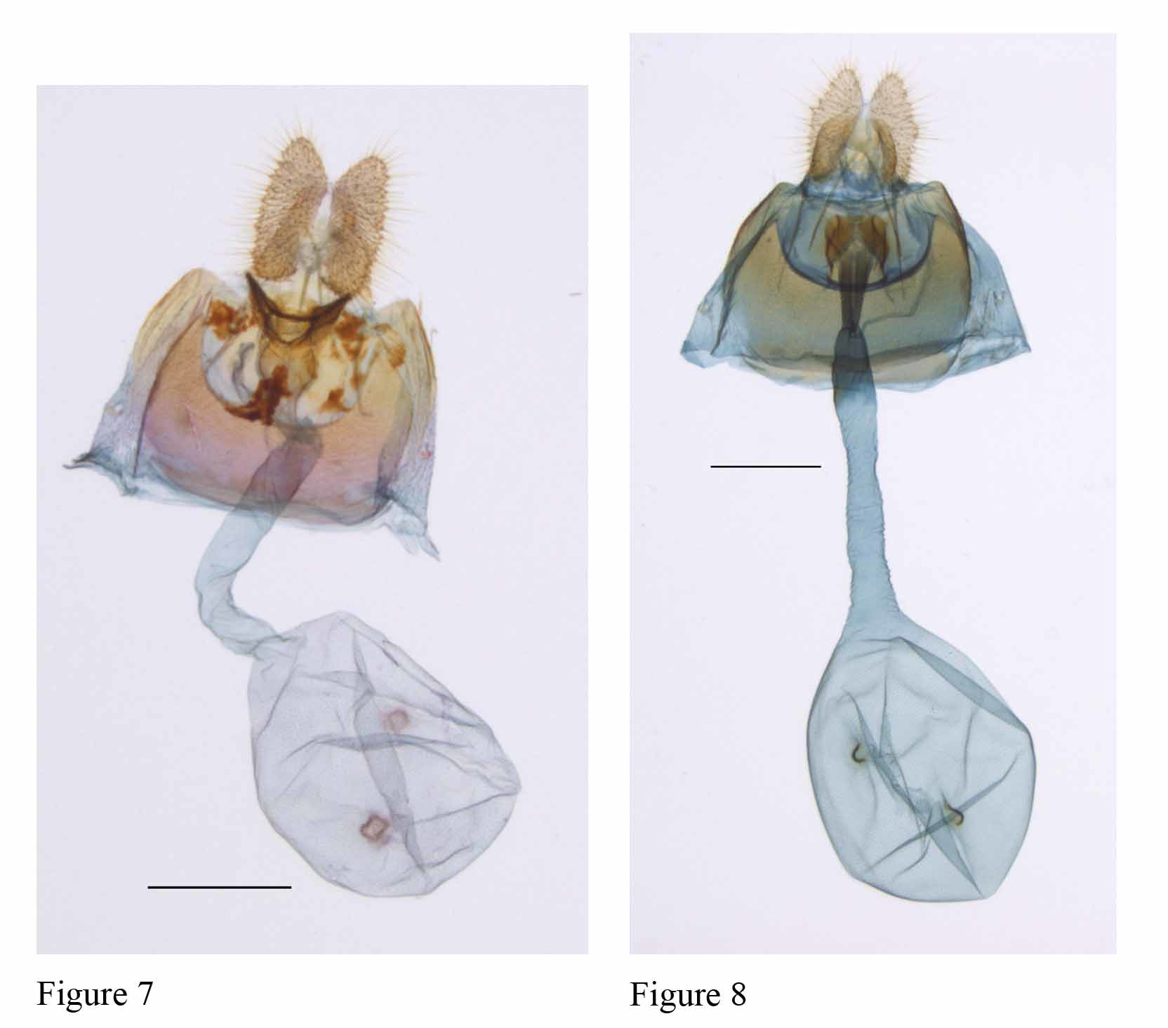
Female. Head and thorax: Essentially as in male, except antennae with ventral cilia sparse, metatibia lacking hair pencil, hindwing without anal roll; forewing length 7.2–5.5 mm (= 6.7 mm, n= 5). Abdomen: Genitalia ( Fig. 7 View FIGURES 7 – 8 ) with posterior margin of seventh sternum broadly excavated, otherwise unmodified; sterigma a protruding FIGURES 1–6. Hedya adults and male genitalia. 1. H. tritofa , male (Paratype, Iran, Māzandarān Prov., Rāmsar, Eshkatechāl, 1200 m, 28.v.2003, Gilāsiān, Nematiān leg.). 2. M. atropunctana , male ( Germany, Wurtemberg, Schwarzwald, Wildbad, 520 m, 5.v.1973, L. Süssner). 3. Forewing pattern interpretation and comparison between H. tritofa (a: Siāhkal- Deylamān Rd., Tootki vill. (Gilān Prov.), N 37° 3ˏ 37.74˝, E 49° 50ˏ 56.41˝, 464 m, 22.vii.2010, Ālipanāh leg.) and M. atropuntana (b: same specimen as Figure 2) (yellow lines indicate limits between fascial and interfascial areas; broken lines indicate ill-defined limits; red lines indicate distal course of major veins; numbers indicate hypothetical position for pairs of strigulae; bs: basal fascia; sb: subbasal fascia; m: median fascia; pm: postmedian fascia; pt: preterminal; dd: discal dot). 4. H. tritofa , male genitalia (same data as Figure 1, GS: JB 20516). Scale bar = 500 μm. 5. M. atropunctana , male genitalia (same data as Figure 2, GS: JB 20515). Scale bar = 500 μm. 6. Comparison of male genitalia (neck of valva) between H. tritofa (a) and M. atropunctana (b) (same data as Figures 5 and 6) (Spc1 to Spc5: spine clusters 1 to 5; SpSc: saccular spines).
cylindrical aciculate lobe, expanded laterally and ventrally; lamella postvaginalis undeveloped; colliculum bottleshaped, bivalval; ductus bursae long, length more than twice diameter of corpus bursae, slightly twisted, weakly sclerotized distally; ductus seminalis attached from near anterior extremity of colliculum; corpus bursae subspherical with signa represented by two slightly scobinate depressions; apophyses anteriores and posteriores relatively short.
Bionomy. Food plants and early stages unknown.
Distribution. Iran: Mazandaran (Ramsar, Behshahr, Neka), Gilan (Bandar Anzali, Siahkal) and Golestan (Golestan National Park) provinces.
Etymology. The specific epithet is derived from the three spine clusters on the baso-medial surface of the male valve: tritofa (tri = three + tofa = groups of spines).
Diagnosis. Hedya tritofa is similar to Metendothenia atropunctana , M. separatana , and M. inouei ; the four species share the following characteristics: distal one-half of forewing mostly white, with small but distinctive group of dark scales on base of M1 and M2 (Fig. 3); postmedian and preterminal fasciae strongly reduced; and male genitalia symmetrical, with well developed cucullus. Males differ in the number and configuration of spine clusters (Fig. 6) on the baso-medial surface of the valval neck. Cluster Spc3 is absent in H. tritofa (Fig. 6a) but present in M. atropunctana (Fig. 6b) and M. separatana ( Gilligan et al. 2008: 206) . Metendothenia atropunctana and M. separatana have Spc1, Spc2, and Spc4 similarly expressed, Spc4 being hairy, but in M. separatana Spc 1 is developed as a dorsal lobe, Spc2 is reduced but in a homologous position, and Spc3 consists of a group of rather long setaceous spines. This last taxon also has a transverse development at the base of cucullus that is lacking in the other two species. In M. inouei ( Kawabe 1987: 142) , Spc1 is developed from the costa to the ventral edge of valva in such a way that it is difficult to determine whether or not it is the result of fusion with Spc2+Spc4. Females of the four species have an aciculate protruding sterigma, an excavated seventh sternum, and two small scobinate depressions as signa. The new species can be distinguished by the flangelike lobe of the sterigma that extends ventrally and laterally from the ostial margin ( Fig. 7 View FIGURES 7 – 8 ). In M. atropunctana ( Fig. 8 View FIGURES 7 – 8 ) and M. inouei ( Kawabe 1987: 143) the sterigma is less conspicuous and often is referred to as tulip-shaped or heart-shaped ( Diakonoff 1973, Kawabe 1987). In M. separatana the sterigma extends in a lamella antevaginalis ( Gilligan et al. 2008: 259).
No known copyright restrictions apply. See Agosti, D., Egloff, W., 2009. Taxonomic information exchange and copyright: the Plazi approach. BMC Research Notes 2009, 2:53 for further explanation.
|
Kingdom |
|
|
Phylum |
|
|
Class |
|
|
Order |
|
|
Family |
|
|
Genus |