
Gieysztoria atalaya, Brusa, Francisco, Damborenea, Cristina & Noreña, Carolina, 2008
|
publication ID |
https://doi.org/ 10.5281/zenodo.183668 |
|
DOI |
https://doi.org/10.5281/zenodo.5618718 |
|
persistent identifier |
https://treatment.plazi.org/id/B67787C9-3A27-FFF9-FF7D-F85F6F8DF9E5 |
|
treatment provided by |
Plazi |
|
scientific name |
Gieysztoria atalaya |
| status |
sp. nov. |
Gieysztoria atalaya View in CoL n. sp.
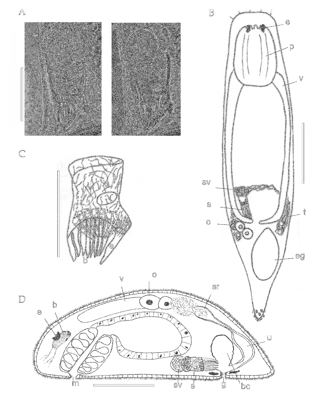
Figures 1 View FIGURE 1 , 7 View FIGURE 7 C–F
Material. Holotype: one specimen in toto mounted in polyvinyl–lactophenol MLP 5420, Atalaya 21–02–03.
Paratypes: Four specimens, sagittally cut MLP 5340, 5341, Atalaya 05–03–01, 020202. One specimen in toto mounted in polyvinyllactophenol, MLP 5421, Atalaya 10–04–03.
Other material: Fifteen specimens, sagittally cut MLP 5342, 5343, 5344, 5723, 5724, Atalaya 05–03–01, 020202, 040402, 210203, 100403.
Type locality. Atalaya (35º00’53.6”S – 57º32’3.3”W), Buenos Aires province, Argentina.
Etymology. The species name refers to the type locality.
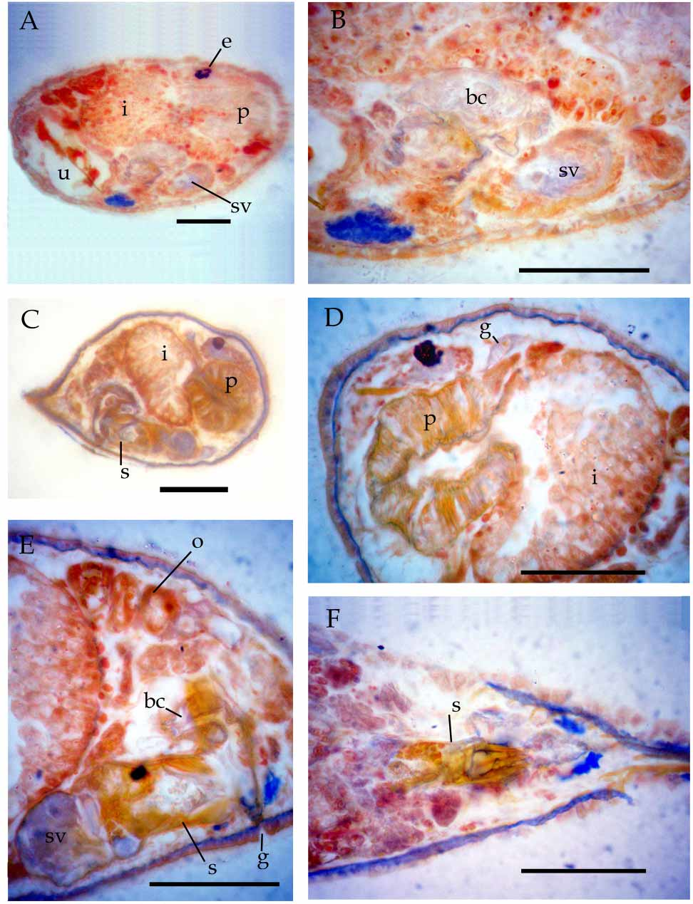
Description. Live adult specimens 430–575 µm long and 95–115 µm wide. Anterior end rounded. Five or six caudal papillae at abruptly tapering posterior end. Rhabdites arranged in groups of 3–4 along body. Rigid cilia at anterior and posterior ends of body longer than those on remaining epidermis.
Pharynx in vivo 130 µm long and 100 µm wide, with four dorsal papillae at anterior margin of pharynx in contrast to other Gieysztoria species.
Broad nerve tracts from brain to anterior region observed in sagittal sections. Conspicuous nucleus evident at peripheral anterior and posterior regions of brain. Black eyes formed by very numerous pigment spheres arranged in a kidneyshaped structure.
Male reproductive system formed by two noncompact testes situated in posterior body region. Spermatozoids observed at periphery of testes. Vasa deferentia projecting from rostral portion of testes toward anterior region and apically or subapically entering seminal vesicle; the latter relatively small and continuous with similarsized prostate vesicle.
Sclerotic stylet approximately 62 µm long and 30 µm wide, with welldefined fibrouslike proximal belt showing a clear window on one side (figure 1A, C). Belt open at side opposite window. Three types of spines originating from belt. A) One spine (spine “a” in figure 1C, length 23 µm) issuing from under window – broad at the base and tapering towards blunt distal end. Spine hollow, situated between the other two groups of spines. B) On one side of stylet, a group of approximately ten thick spines (spines “b” in figure 1C), originating from belt margin, all of similar shape and length. Very thin spines scattered among them. C) On other side of spine “a”, a third group of spines (spines “c” in figure 1C) arranged in several rows forming a “brush”. Approximately 10 spines in proximal row, with broad base and rapidly tapering so as to present triangular shape (figure 1C).
Female reproductive system comprising an ovary located on right side of body and dorsal to intestine. Oocytes arranged in two or three series at proximal part of ovary, and uniserially at distal part (figure 1D). Ovary continued into a short oviduct; oviduct widening to form a seminal receptacle connected to uterus by a long common duct. Uterus with thick walls, curved to open into the genital atrium. Egg in uterus large (greater diameter 152 µm, lesser diameter 93 µm), oval, with operculum at one of its ends. Vitellaria smooth (figure 1B); anteriorly reaching base of pharynx and ventral at this level; becoming dorsal at posterior end, there merging to enter the common duct. Bursa copulatrix with very thick muscular walls, opening into common duct. Gonopore surrounded by a strong sphyncter and cement glands.
Discussion. Based mainly on the configuration and shape of the male stylet, G. atalaya could be compared with three species: Gieysztoria pavimentata ( Beklemischev 1926) Luther 1955 , G. virgulifera ( Plotnikov 1906) Luther 1955 and G. beltrani ( Gieysztor 1931) Luther 1955 . Gieysztoria pavimentata is the species with the most similar stylet to that of the new species. G. pavimentata was considered by Luther (1955) as belonging to the Inaequales group, subgroup Fenestrate. Both species possess a robust spine, but in G. atalaya this spine occupies a central position on the girdle (spine “a” figure 1C) whereas in G. pavimentata it is located on the right side of the stylet (spine “d” of Luther 1955, figure 41 G, H and 42B). In addition to the different location, the central spine of G. atalaya is clearly larger and differs in shape from the rest of spines, which is not the case of spine “d” (after Luther 1955) in G. pavimentata .
Furthermore, the constellation of the smaller spines of G. pavimentata is different from that of G. atalaya . G. pavimentanta bears 10 spines on the girdle edge, followed by 2 or 3 irregular rows of spines on the wall of the copulatory organ. In contrast, the spines of G. atalaya are arranged in two groups, the first group (spines “b”) formed by about 10 spines and the second group (spines “c”) formed by three or four rows of thin spines with similar shape, but different length.
The stylets of Gieysztoria beltrani and G. virgulifera are also similar to that of G. atalaya ; both have a belt with a middle window and a large spine issuing from under it. In G. beltrani the large spine under the window ( Gieysztor 1931; Luther 1955) bears small spines, contrasting with the smooth large spine of G. atalaya . In G. beltrani , the rest of the spines are grouped at the sides of the large spine, with six spines on one side and a variable number on the other. G. v i rg u l i f e r a has two groups of spines at both sides of the smooth large central spine, similarly to the condition in G. atalaya , but the groups differ in number and shape of the spines. In G. virgulifera the first group is formed by 4–7 spines on one side and only two robust, scimitarshaped spines, on the other ( Luther 1955).
| MLP |
Museo de La Plata |
No known copyright restrictions apply. See Agosti, D., Egloff, W., 2009. Taxonomic information exchange and copyright: the Plazi approach. BMC Research Notes 2009, 2:53 for further explanation.
|
Kingdom |
|
|
Phylum |
|
|
Class |
|
|
Order |
|
|
Family |
|
|
Genus |