
Coryphaenoides asper Günther, 1877
|
publication ID |
https://doi.org/ 10.11646/megataxa.3.1.1 |
|
persistent identifier |
https://treatment.plazi.org/id/B711B23F-FF4F-868E-D99D-C37EFEA97A59 |
|
treatment provided by |
Plazi |
|
scientific name |
Coryphaenoides asper Günther, 1877 |
| status |
|
Coryphaenoides asper Günther, 1877 View in CoL
[Japanese name: Zara-dara]
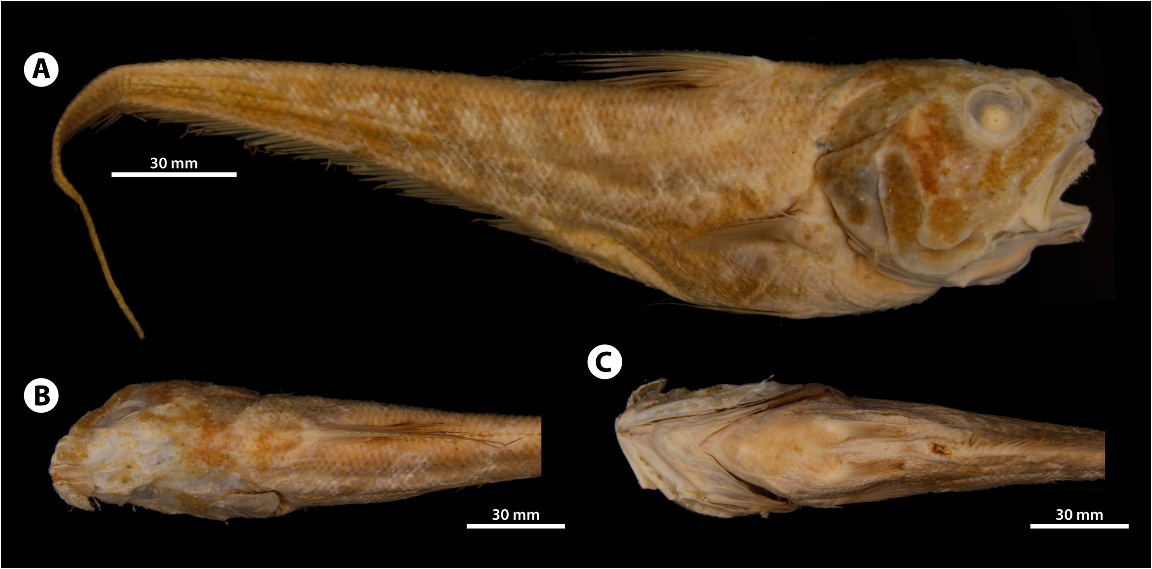
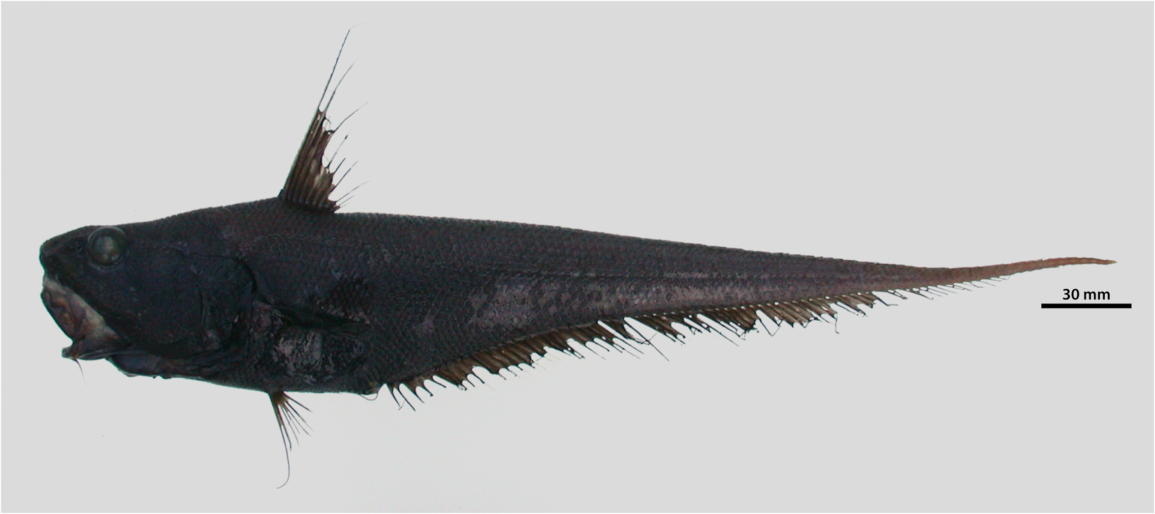
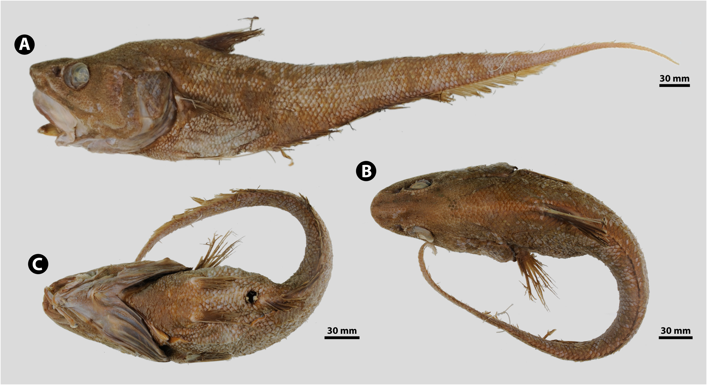
( Figs. 101–102 View FIGURE 101 View FIGURE102 ; Appendix 3-6A)
Coryphaenoides asper G̹nther, 1877 View in CoL :440 [original description; lectotype (designated herein): BMNH 1887.12.7.88, from southeast of Boso Peninsula, Challenger sta. 237, in 1875 ftm (3429 m); in part (see Remarks)]; Okada & Matsubara 1938:449 (in key; Japan); Matsubara 1955:1309 (in key; Japan); Okamura 1970a:136 [brief description after G̹nther (1877, 1887)]; Tominaga & Uyeno 1981:488 (listed; Japan); Okamura 1984b:95, pl. 345, fig. I (compiled); Okamura 1988:95, pl. 345, fig. I (compiled); Nakabo 1993:364 (in key; Japan); Nakabo 2000:428 (in key; Japan); Nakabo 2002:428 (in key; Japan); Nakabo & Kai 2013:504 (in key; Japan); Motomura 2020:39 (listed; Japan).
Macrurus asper View in CoL : G̹nther 1887:137, pl. XXXVI, fig. A [brief description; 1 spec. from “south of Japan ” (= off Boso Peninsula ), Challenger sta. 237] .
Macrourus asper View in CoL : Jordan & Snyder 1901:120 (listed; Japan); Jordan & Gilbert in Jordan & Starks 1904:617 [after G̹nther (1877, 1887)]; Jordan et al. 1913:417 (listed; Japan; new Japanese name: “Zara-dara”).
Coryphaenoides (Coryphaenoides) asper: Okamura 1970b View in CoL : table 1 (listed; Japan).
Diagnosis. Pelvic-fin rays 11–12. Snout bluntly pointed, only slightly protruding beyond upper jaw. Lateral angles of snout armed with small, stout, modified scales. Scales on anterior half of infraorbital ridge slightly thickened. Dorsal contour of head smoothly curved from snout tip to first dorsal-fin origin. Orbit diameter 23% HL. Mouth large, posterior margin of upper jaw extending to below midorbit; upper-jaw length 35% HL; lateral corner of mouth not restricted by skin folds. Outermost gill slit greatly restricted, length 10% HL. Barbel well developed, slender, length 12% HL. Teeth in tapered bands in both jaws, with outer premaxillary series enlarged. Body scales covered with narrowly divergent rows of long, reclined, slender, needle-like spinules; tip of last spinule in each row extending well beyond posterior scale margin; middle row distinctly higher than those of adjacent rows. Transverse scale rows below first dorsal-fin midbase 5. Underside of snout anterior to lateral angles broadly naked; top of snout narrowly naked posterior to leading edges. Height of first dorsal fin 109% HL; second spinous ray serrated along its leading edge; first dorsal-fin rays II,11. Pectoral-fin rays i24. Outermost pelvic-fin ray moderately prolonged, its tip extending well beyond anal-fin origin. Body uniformly dark.
Material examined. 1 specimen. Lectotype of Coryphaenoides asper (designated herein): BMNH 1887.12 .7.88 (65.7 mm HL, 316+ mm TL), southeast of Boso Peninsula , Chiba Pref., Japan, 34.6167ºN, 140.5333ºE, 1875 ftm (3429 m), Challenger sta. 237, trawl, 17 Jun. 1875. GoogleMaps
Counts and measurements. Counts: first dorsal-fin rays II,11; pectoral-fin rays i24; pelvic-fin rays 11–12; gill rakers on first arch (outer/inner) 5–6/10–11, on second arch 8–9/9–11; longitudinal scales 36; transverse scale rows below first dorsal-fin origin 7–8, below first dorsalfin midbase 5, below second dorsal-fin origin 6–7.
The following measurements are in % of HL, followed by those in % of PRL in parentheses: snout length 28 (36); orbit diameter 23 (31); postorbital length 54 (71); postrostral length 76; orbit–preopercle distance 51 (67); suborbital width 15 (20); upper-jaw length 35 (46); length of rictus 29 (38); length of premaxillary tooth band 24 (31); preoral length 13 (17); interorbital width 28 (37); body width over pectoral-fin bases 54 (71); body depth at first dorsal-fin origin 93 (122); body depth at anal-fin origin 77 (101); isthmus–pelvic distance 50 (65); isthmus–anal distance 56 (74); pelvic-fin length 85 (111); pectoral-fin length 62 (81); predorsal length 112 (147); height of first dorsal fin 109 (143); length of first dorsal-fin base 27 (36); interdorsal length 22 (29); length of gill slit 10 (13); length of posterior nostril 7 (9); barbel length 12 (16).
Redescription of lectotype. General features are shown in Fig. 101 View FIGURE 101 . Body deepest at first dorsal-fin origin, gradually tapering to long tail. Trunk short, moderately compressed, width over pectoral-fin bases 1.7 in depth at first dorsal-fin origin. Head large, HL about 4.8 in TL; head bones and flesh moderately firm. Predorsal contour smoothly curved, but slightly elevated anterior to first dorsal-fin origin. Snout short, only slightly protruding beyond upper jaw, length 1.2 times as long as orbit diameter; ventral contour oblique in lateral view; snout low, its tip on horizontal through midorbit. Orbit moderate in size, circular, greatest diameter 2.3 in postorbital length. Interorbital space broad, flat, width 0.8 in orbit diameter. Mouth large, oblique, subinferior, upper-jaw length 0.7 in orbit diameter; posterior margin of maxilla reaching vertical through midorbit; lateral corner of mouth not restricted by lip folds; lips somewhat thin. Suborbital region weakly ridged; upper half almost vertical, lower half moderately inclined mesially. Preopercle large, hind margin slightly inclined from vertical, forming broad lobe posteroventrally; preopercular ridge low, stout, with distinct backward extension at angle. Interopercle mostly hidden behind preopercle. Subopercle and opercle smoothly adjoined, forming posterior margin of gill cover. Gill membranes narrowly connected across, and attached mesially to isthmus, with posterior free fold; gill opening moderately wide, extending forward to below posterior margin of lower jaw. Outermost gill slit greatly restricted by skin folds, length 2.3 in orbit diameter. Lower gill rakers developed into small, low tubercles; upper rakes somewhat pad-like; outer rakers of first arch much smaller than others; all rakers armed with short, slender spines. Barbel well developed, slender, length 1.9 in orbit diameter.
Anus immediately anterior to anal-fin origin; periproct poorly developed. Ventral light organ absent.
Teeth short, slender, conical, in long tapered band in both jaws. Premaxillary teeth with about 7 tooth rows at widest point near symphysis, with about 4 tooth rows posteriorly; outermost series enlarged, incurved; posterior margin of tooth band almost reaching lateral corner of mouth. Mandibular band much narrower than premaxillary band, but teeth much larger than those in premaxillary, with about 5 tooth rows anteriorly, about 2 rows posteriorly; posterior margin of tooth band falling short of lateral corner of mouth; no teeth significantly enlarged.
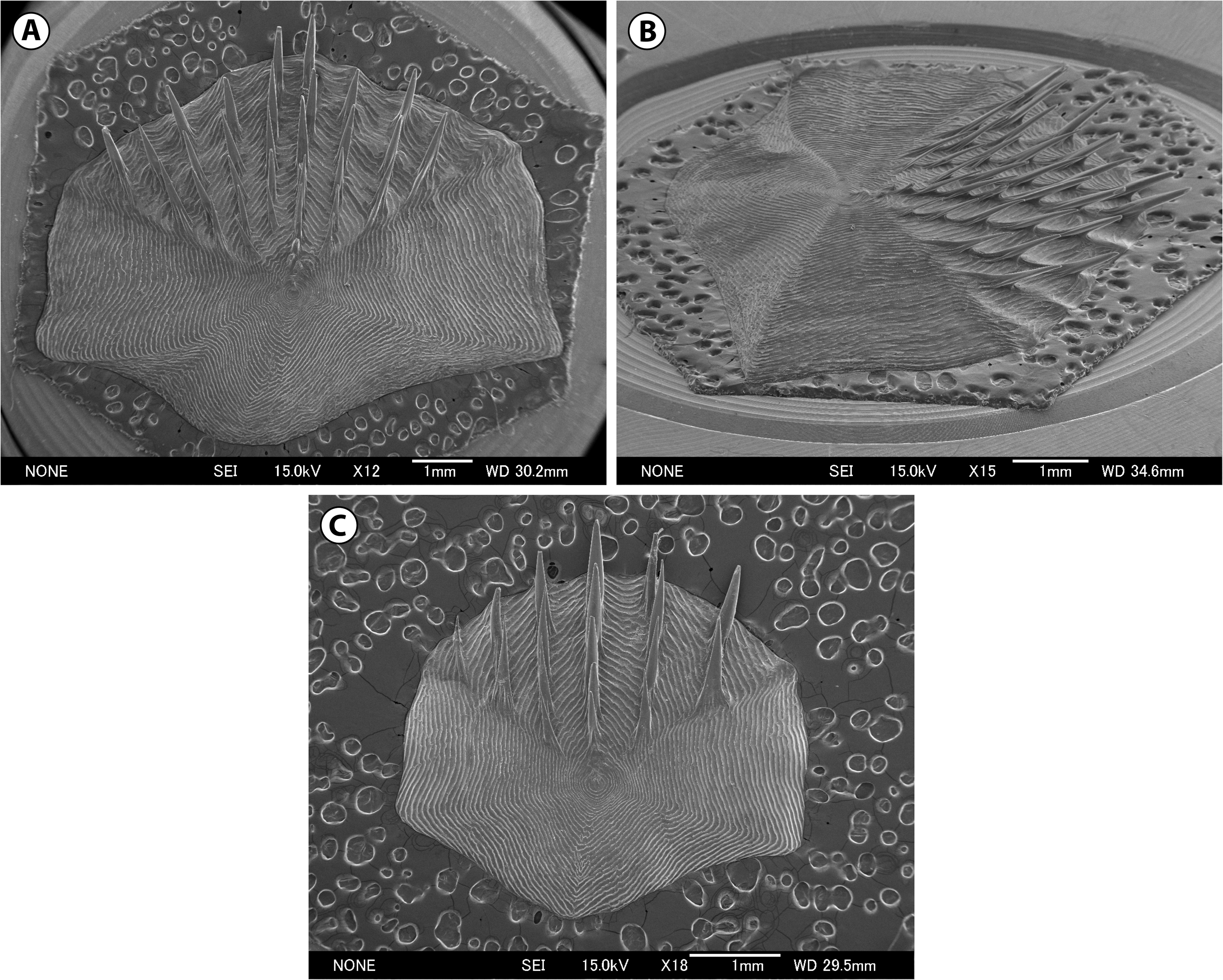
Body scales small, thin, deciduous, covered with narrowly divergent rows of long, slender, reclined, needle-like spinules ( Fig. 102 View FIGURE102 ); those on dorsum below interdorsal space with 3–6 spinule rows; middle row with about 4–5 spinules, distinctly higher than other rows; height of spinules increasing posteriorly, with last spinules extending well beyond posterior scale margin; spinules of each row greatly overlapping, but free from one another; lateral buttresses barely developed; no reticulate structure on scale surfaces. Body completely and uniformly scaled except for fins.
Scales on anterior half of infraorbital ridge slightly thickened. Lateral angles of snout armed with small, but stout, modified scales; presence/absence of terminal snout scute unknown due to poor condition of specimen. Head scales small except for those on opercle and postorbital canal, similar to those on body, but spinules more erect, giving spiny appearance to head surfaces. Head mostly covered with scales. Underside of snout broadly naked; naked area extending posteriorly to below lateral angles of snout. Dorsal surface of head fully scaled, but top of snout narrowly naked posterior to its leading edges. Other naked areas confined to anterior 1/3 of mandibular rami, gular and branchiostegal membranes, and nasal fossa.
No open pores on cephalic sensory canals. Naked area on underside of snout heavily covered with small free neuromasts and short hair-like papillae. Similar neuromasts scattered on snout, interorbital space, suborbital area, mandibular rami, preopercle, and postorbital canal. Lateral line not interrupted throughout.
Origin of first dorsal fin slightly posterior to pelvicfin base; first dorsal fin well developed, height 4.0 times as long as its base length; second spinous ray moderately long, but not especially prolonged, armed along its leading edge with 23 short, sharp, slender denticles; denticles becoming much slender and erect distally; tip of second spinous ray extending to base of 15th second dorsal-fin ray when laid back. Second dorsal fin moderately separated from first dorsal, interdorsal space 0.8 times as long as first dorsal-fin base length. Pectoral fin moderately short, its tip extending to vertical through anal-fin origin; pectoralfin base slightly anterior to pelvic-fin base. Outermost pelvic-fin ray moderately elongate, its tip reaching base of 8th anal-fin ray when laid back. Anal fin well developed, originating slightly anterior to second dorsal-fin origin.
Color when fresh. Unknown.
Color in alcohol ( Fig. 101 View FIGURE 101 ). Head and body dusky brown overall; abdomen and gill cover much darker than surroundings; first dorsal fin mostly blackish (about distal 2/3), but paler basally; second dorsal fin probably blackish originally; pectoral fin uniformly dark; pelvic fin generally pale, but distal margin darker; free portion of outer pelvic-fin ray blackish; basal half of anal fin pale, distal half blackish; oral cavity grayish brown; gill cavity black; gill rakers, arches, and filaments pale brown; barbel and gular membrane pale, but branchiostegal membranes blackish posteriorly; anterior margin of posterior nostril narrowly edged with black; periproct intensely black.
Size. To about 32 cm TL.
Distribution. So far known only from the lectotype collected from off the Boso Peninsula at a depth of 3429 m (Appendix 3-6A). Very rare.
Comments on type specimens. According to the original description, G̹nther (1877) described Coryphaenoides asper based on specimens collected from the “south of the Philippines and Japan ”. However, in the “ Challenger ” Report, G̹nther (1887) listed only a single specimen for this species (BMNH 1887.12.7.88, 65.7 mm HL, 316+ mm TL; Fig. 101 View FIGURE 101 ). Although it is uncertain how many specimens were involved in the original description, it is most likely that the material from the Philippines (originally reported as C. asper ) was re-identified as other species in the “ Challenger ” Report. Among the specimens examined by G̹nther (1887) was only a single specimen of macrourid grenadiers collected from the Philippines (Challenger sta. 214; see also G̹nther 1887:143, pl. XXVI, fig. B), viz., Trachonurus sp., BMNH 1887.12.7.106, 45 mm HL, 246 mm TL (see the Comments on type specimen of T. villosus ). Species of Trachonurus are more or less similar to C. asper in general appearance, and thus the above Trachonurus specimen could have been misidentified as C. asper in the original description of the latter species. In order to avoid further confusion, BMNH 1887.12.7.88 is herein designated as the lectotype of C. asper . Consequently, other specimens (if any exist) used in the original description automatically become paralectotypes of the species.
Remarks. Jordan & Thompson (1914) reported C. asper based on two specimens collected from Misaki (Sagami Bay), Japan. Although their specimens were not found among the ichthyological collection of the Field Museum of Natural History (FMNH) during the author’s visit in 2012, their figure (pl. 38, fig. 2) depicts a typical form of Ventrifossa misakia ( Jordan & Gilbert in Jordan & Starks, 1904). Kuroda (1965) also reported C. asper from Suruga Bay based on three specimens collected by local fishers. Because he referred to a description and figure of “ C. asper ” given by Jordan & Thompson (1914), his materials are likely a misidentification of V. misakia .
The lectotype was the only representative of this rare species until Shao et al. (2008a, 2008b)reported the second specimen from the northern part of the South China Sea off Taiwan. Iwamoto et al. (2009) provided a description of this additional specimen, noting a difference in bodyscale spinulation between the lectotype and their material (spinules in narrowly divergent rows in the lectotype vs. subparallel to slightly convergent rows in the Taiwanese specimen). They also noted that “we cannot evaluate the significance of this difference without additional specimens for comparisons”. Subsequently, Iwamoto et al. (2015) re-identified the Taiwanese specimen as Coryphaenoides sp. cf. asper .
As suggested by previous authors, the additional specimen from Taiwan ( ASIZP 66107 , 92.3 mm HL) differs notably from the lectotype in the morphology of the body scales (see Iwamoto et al. 2009: fig. 3C). In the Taiwanese specimen, the spinules on the scales are arranged in about 15 convergent rows, with the last spinule in each row barely extending beyond the posterior scale margin, whereas those in the lectotype are in five divergent rows and the last spinules extend well beyond the scale margin. The Taiwanese specimen also has much shorter spinules on the scales, which give a smooth texture to the body (vs. spinules distinctly longer in the lectotype, giving a spiny appearance to body surfaces). These differences are notable even considering the Taiwanese specimen is much larger than the lectotype (65.7 mm HL). It further differs from the lectotype in having lower counts of pectoral- (i20–i21 vs. i24) and pelvic-fin rays (10 vs. 11–12) and inner gill rakers on the first arch (7 vs. 10–11). In addition, the head is completely scaled in the Taiwanese specimen, but the lectotype has a narrow naked area on the underside of the snout. These differences further support their specific separation, and the Taiwanese specimen will be officially described by the author in a separate publication as a representative of a new species.
Consequently, C. asper is currently represented by only the lectotype from Japan. Because G ̹nther’s (1877, 1887) description of the species is brief, the specimen is redescribed here for future studies.
Relationships and comparisons. Coryphaenoides asper belongs to the subgenus Coryphaenoides (sensu Iwamoto 1990) . This species is most similar to C. soyoae Nakayama & Endo, 2016 , with which it shares most of the diagnostic characters distinguishing the latter species from its congeners. However, the two species are readily distinguished from each other by scale spinulation: the spinules of C. asper are much longer as compared with C. soyoae , with the middle row enlarged, giving a spiny appearance to the head and body ( Fig. 102 View FIGURE102 ), whereas those of C. soyoae are much shorter, so as to give a smooth texture to the head and body surfaces ( Fig. 133 View FIGURE133 ). In addition, the predorsal profile of C. asper is smoothly curved from the snout tip to the first dorsal-fin origin ( Fig. 101A View FIGURE 101 ), whereas in C. soyoae , there is a pronounced hump over the nape, with a distinct depression above the orbits ( Figs. 131 View FIGURE 131 , 132A View FIGURE 132 ). Other differences include the number of pectoral-fin rays (i 24 in C. asper vs. i19–i 22 in C. soyoae ), orbit diameter (23% HL vs. 20–21%), orbit–preopercle distance (51% HL vs. 49%), height of first dorsal fin (109% HL vs. 87%), pectoral-fin length (62% HL vs. 49%), and length of outer gill slit (10% HL vs. 7–8%), although Nakayama & Endo (2016a) noted the necessity to confirm the meristic and morphometric differences when more specimens become available. Coryphaenoides asper superficially resembles C. asprellus (Smith & Radcliffe in Radcliffe, 1912), C. hextii ( Alcock, 1890) , and C. woodmasoni ( Alcock, 1899) , all known from the Indian Ocean ( C. asprellus is also distributed in the East Indies). However, it chiefly differs from these species in having higher counts of pelvic-fin rays [11–12 vs. 7–10; data for the latter three species are from Shcherbachev & Iwamoto (1995)].

No known copyright restrictions apply. See Agosti, D., Egloff, W., 2009. Taxonomic information exchange and copyright: the Plazi approach. BMC Research Notes 2009, 2:53 for further explanation.
|
Kingdom |
|
|
Phylum |
|
|
Class |
|
|
Order |
|
|
Family |
|
|
Genus |
Coryphaenoides asper Günther, 1877
| Nakayama, Naohide 2020 |
Coryphaenoides (Coryphaenoides) asper
| : Okamura 1970 |
Coryphaenoides asper G̹nther, 1877
| Gunther 1877 |
Macrurus asper
| Goode & Bean 1833 |