
Cephalothrix suni, Chernyshev & Polyakova, 2021
|
publication ID |
https://doi.org/ 10.11646/zootaxa.4908.4.10 |
|
publication LSID |
lsid:zoobank.org:pub:6C3E52A4-1FE3-472D-9724-6BF31441A18A |
|
DOI |
https://doi.org/10.5281/zenodo.4455270 |
|
persistent identifier |
https://treatment.plazi.org/id/602F23A5-7D8A-47BF-AB88-10D40D72C6BA |
|
taxon LSID |
lsid:zoobank.org:act:602F23A5-7D8A-47BF-AB88-10D40D72C6BA |
|
treatment provided by |
Plazi |
|
scientific name |
Cephalothrix suni |
| status |
sp. nov. |
Cephalothrix suni sp. nov.
( Figs 1A, B View FIGURE 1 ; 2 View FIGURE 2 A-I; 3A-I)
urn:lsid:zoobank.org:act:602F23A5-7D8A-47BF-AB88-10D40D72C6BA
syn. Cephalothrix sp. VIE: Chen et al., 2010, tab. 2, N 21.
Cephalothrix suni: Chernyshev, 2016, p. 288 (nomen nudum)
Type material. Holotype No. 40373 ( MIMB), slides with transverse sectionas, Nam Du Island (09°43′ N, 104°23′ E), South China Sea, Vietnam, May 15, 2010, intertidal, calcareous red algae, collected by A. V. Chernyshev GoogleMaps . Paratype No. 40374 ( MIMB), Van Phong Bay (12°34′ N, 109°24′ E), South China Sea, Vietnam, January 13, 2005, intertidal, calcareous red algae, collected by A. V. Chernyshev GoogleMaps .
Other material examined. Three specimens, Van Phong Bay (12°33′ N, 109°23′ E), South China Sea, Vietnam, January 18, 2005, intertidal, calcareous red algae, collected by A. V. Chernyshev GoogleMaps .
GenBank accession numbers. Holotype ( KU 840272 View Materials – Sundberg et al. 2016; see Table 2), paratype ( GU726621 View Materials – Chen et al. 2010).
Etymology. The specific epithet honors Prof. Sun Shichun (Ocean University of China).
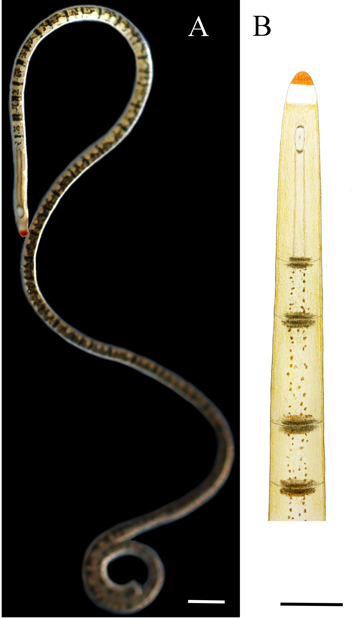
Description. External features. Body 50–80 mm long but less than 1 mm wide. Bluntly rounded head not narrower than trunk; anterior tip of head red (holotype) or orange (paratype), demarcated posteriorly by white band ( Fig 1 View FIGURE 1 ). Eyes absent. Background colour pale yellowish; dark pigment present on ventral side only. Small round mouth rimmed by brown ring with thicker anterior and posterior margins; two thin brown longitudinal stripes connected with in posterior part of the ring and extend to gut region ( Fig. 1 View FIGURE 1 ). Gut region with transverse dark drown bands; very thin transverse ring (epidermal furrow?) runs through each band. Brown spots also arranged between bands, sometimes forming irregular longitudinal stripes. Worms crawl on the dorsal side of their bodies, with the ventral side facing up.
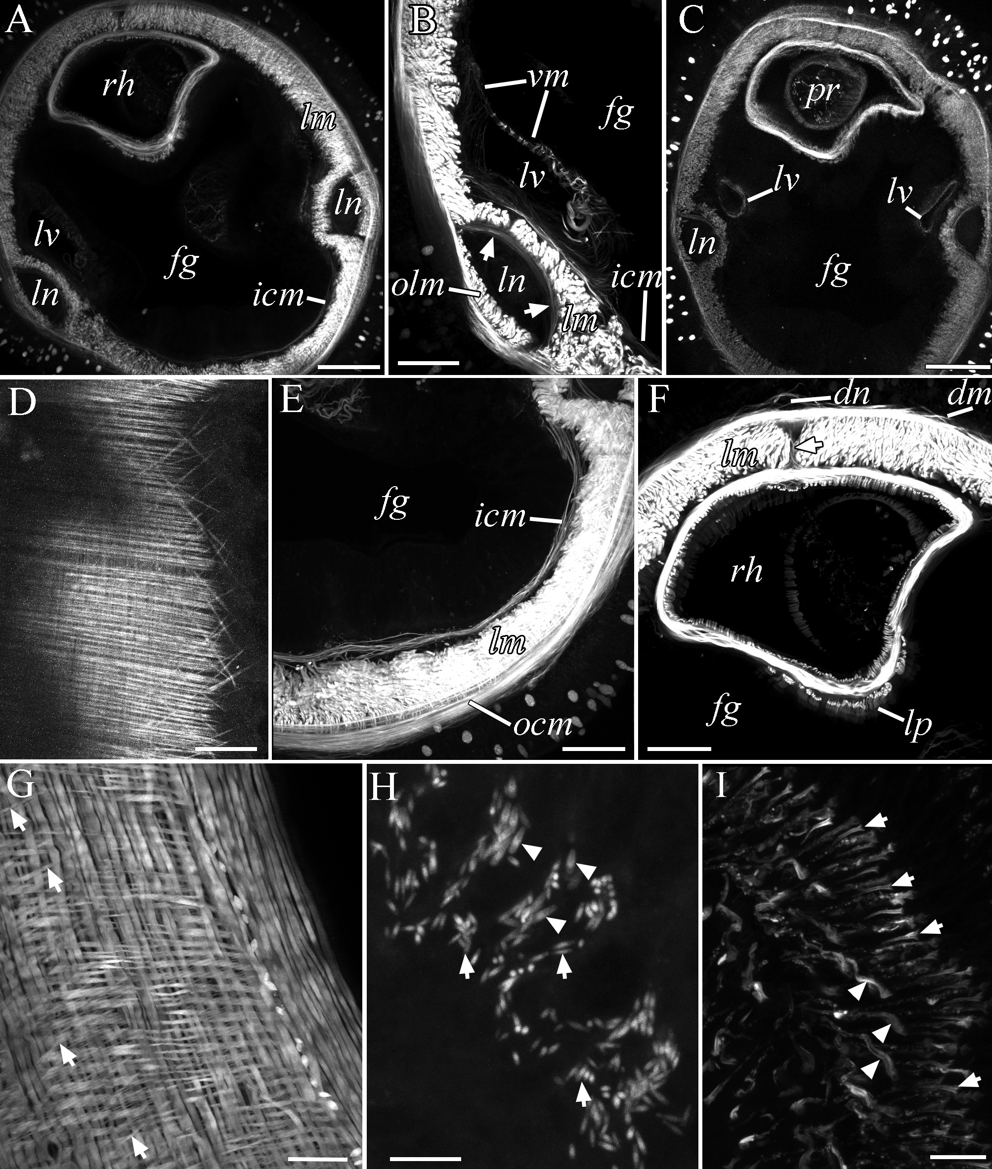
Internal morphology. Body wall: E = 20–26 µm, D = 5–8 µm, OCM = 4–7 µm, LM = 18–35 µm; outer part of LM and inner part of split OCM present ( Figs 2B View FIGURE 2 ; 3 View FIGURE 3 C-E;); in cerebral and foregut regions, crisscrossed DM located between dermis and OCM ( Fig. 2D, F View FIGURE 2 ); in foregut region, dorsal muscular cross present between OCM and rhynchocoel wall ( Fig. 2F View FIGURE 2 ); very thin ICM adjoins ventral wall of anterior foregut portion and connected with musculature of lateral blood vessel ( Fig. 2A, B, E View FIGURE 2 ); posterior foregut portion lacks ICM ( Fig. 2C View FIGURE 2 ). Longitudinal muscle plate between rhynchocoel and digestive tract present ( Fig. 2F View FIGURE 2 ). Proboscis musculature consists of OCM, DM, LM, and very thin ICM ( Fig. 2G View FIGURE 2 ); large pseudocnidae 5.6–6.5 × 1.9–2.5 µm, small pseudocnidae 3.3–4.1 × 0.9–1.3 µm ( Fig. 2H View FIGURE 2 ); proboscis epithelium with longer (23–26 µm) and shorter (14–17 µm) collars of sensory cells ( Fig. 2I View FIGURE 2 ). Ratio of brain–head tip to brain–mouth distances 1: 1.4 (based on transverse sections). Gut epithelium with dark pigment ( Fig. 3I View FIGURE 3 ). Four cephalic nerves present ( Fig. 3A View FIGURE 3 ). Single large median ventral nerve extends backwards from ventral brain commissure ( Fig. 3C View FIGURE 3 ) and forms two buccal nerves immediately in front of mouth ( Fig. 3D View FIGURE 3 ); two buccal nerves ( Fig. 3E, F View FIGURE 3 ) join together ( Fig. 3G View FIGURE 3 ) and become invisible immediately behind mouth ( Fig. 3H View FIGURE 3 ). Dorsal nerve located between DM and OCM ( Fig. 2F View FIGURE 2 ). Blood system typical for cephalotrichids; rhynchocoel vessel of type A according to Kajihara’s (2010) classification ( Fig. 3B View FIGURE 3 ). Mushroom-like excretory organs present.
Comparison. Almost all species of the genus Cephalothrix s.l. have more or less uniformly coloured body without any pattern, except for Cephalothrix queenslandica which, the same as the new species, possesses a distinct colour pattern consisting of transverse brown bands. However, according to the original description ( Sundberg et al. 2003), Cephalothrix queenslandica has a brown anterior tip (in contrast to orange or red tip in C. suni ), transverse brown bands on the dorsal body surface (in contrast to the bands on the ventral surface in C. suni ), and two cephalic nerves (in contrast to four ones in C. suni ). The uncorrected p -distance between the 18S rRNA sequences (no other sequences for C. queenslandica are known) of C. queenslandica and C. suni constitutes 0.5%. For comparison, the p -distance between the 18S rRNA sequences of Cephalothrix hongkongiensis Sundberg, Gibson & Olsson, 2003 and C. suni is 0.2%.
Geographical and ecological distribution. South China Sea, South Vietnam, Van Phong Bay, and off Nam Du Island; intertidal zone, among calcareous red algae.
Remarks. C. suni is apparently the most common species of Cephalothrix in coastal waters off Vietnam. In addition, another Cephalothrix species ( Chernyshev 2016; GenBank accession no. MW118027 View Materials ) has been found in coastal waters off Vietnam (Cu Lao Cham Islands). The latter is similar to the undescribed species C. fasciculus sensu Leasi & Norenburg 2014 from Belize (GenBank accession no. KM083814.1).
Phylogenetic analysis. According to a phylogenetic analysis, the genus Cephalothrix is monophyletic and includes three highly supported clades: Cephalothrix sensu Chernyshev & Kajihara 2019 , Procephalothrix sensu Chernyshev & Kajihara 2019 , and ‘interstitial cephalotrichids’ with two undescribed species. The new species belongs to clade Procephalothrix , where Cephalothrix suni , C. rufifrons (Johnston, 1837) , C. hongkongiensis , Cephalothrix mokievskii Korotkevitsch, 1982 (see Chernyshev 2020), and C. simula (Iwata, 1952) form a highly supported subclade of species with orange, red, or dark yellow anterior tip. This subclade is sister to subclade with two undescribed Cephalothrix from Arctic and Antarctic waters. Recently, Chernyshev & Kajihara (2019) have shown that the name Procephalothrix may be ‘resurrected’ for the clade that contains Cephalothrix filiformis (Johnston, 1828) (type species of Procephalothrix ) and other species with the outer part of LM located between the lateral nerve cord and OCM.

| MIMB |
Museum of the Institute of Marine Biology |
| V |
Royal British Columbia Museum - Herbarium |
| KU |
Biodiversity Institute, University of Kansas |
No known copyright restrictions apply. See Agosti, D., Egloff, W., 2009. Taxonomic information exchange and copyright: the Plazi approach. BMC Research Notes 2009, 2:53 for further explanation.
|
Kingdom |
|
|
Phylum |
|
|
Class |
|
|
Order |
|
|
Family |
|
|
Genus |
Cephalothrix suni
| Chernyshev, Alexei V. & Polyakova, Neonila E. 2021 |
Cephalothrix suni:
| Chernyshev 2016: 288 |