
Thyropygus richardhoffmani, Pimvichai, Piyatida, Enghoff, Henrik & Panha, Somsak, 2009
|
publication ID |
https://doi.org/ 10.5281/zenodo.189060 |
|
DOI |
https://doi.org/10.5281/zenodo.6212353 |
|
persistent identifier |
https://treatment.plazi.org/id/B72987DA-240F-F91D-00AB-16F2CC63DBBB |
|
treatment provided by |
Plazi |
|
scientific name |
Thyropygus richardhoffmani |
| status |
sp. nov. |
Thyropygus richardhoffmani View in CoL n. sp.
( Figs. 6 View FIGURE 6 A–D, 7E)
Material: HOLOTYPE male THAILAND, Trang Province, Na Yong district, Khao Chong, Banthat Mountain, 7° 32ˏ 45˝ N, 99° 46ˏ 27˝ E. 24–26 January 1999. M. Andersen leg., ( ZMUC). - Paratypes: 4 males 6 females, same locality as holotype. 14 January 2009. P. Pimvichai and members of Animal Systematics Research Unit leg., ( CUMZ).
Etymology: The species is named in honour of Richard L. Hoffman in recognition of his lifelong devotion to diplopodology, especially his very useful account of the genus Thyropygus .
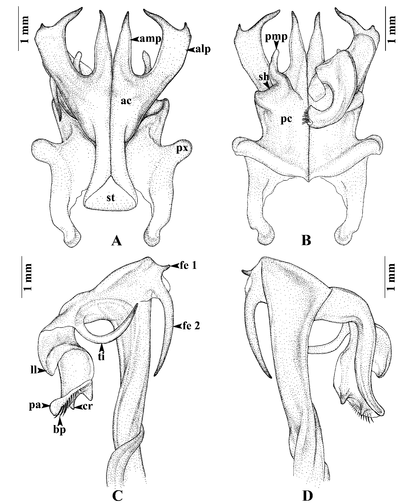
Diagnosis: A species of the bifurcus subgroup. Lateral margin of lateral process of anterior coxal fold (alp) with neither crest nor spine. Similar in this respect to T. casjeekeli and T. enghoffi . Differ from T. enghoffi by having a very short first femoral spine (fe 1), a very long the second femoral spine (fe 2) and a small lamella at both side of base of fe 2. Particularly similar to T. casjeekeli , differing from it by having a double femoral spine (fe) and by the absence of a small spine (ss) at base of apical part, opposite the origin of the tibial spine.
Description: Adult males with 59–64 podous rings, no apodous rings. Length ca. 8–12 cm, width ca. 6.0– 7.1 mm. Adult females with 58–62 podous rings, no apodous rings. Length ca. 9–12 cm, width ca. 6.3–7.7 mm. Overall color of living animal ( Fig. 7 View FIGURE 7. A E) dark brown; legs brownish.
Gonopods ( Figs. 6 View FIGURE 6 A–D): Anterior coxal fold (ac) ( Fig. 6 View FIGURE 6 A): lateral process (alp) flattened, slightly curved, terminating in two cusps, the outer one very short, the inner one very long; mesal process (amp) very long, almost as long as alp, straight, directed distad, pointed. Posterior coxal fold (pc) ( Fig. 6 View FIGURE 6 B) basally with moderately high paracoxites (px), distally truncate, forming shelf (sh) for accommodation of telopodite, mesal process (pmp) slender, directed anteriad. Telopodite ( Figs. 6 View FIGURE 6 C–D) leaving coxite over shelf of posterior coxal fold; double femoral spine (fe), the first fe (fe 1), very short, pointed, situated above fe 2, the second fe (fe 2) very long, as long as ti, slender, curved downward, with a small lamella at both side of base; tibial spine (ti) long, slender, curving in horizontal plane, its tip in situ resting close to base of amp; apical part: lamellar lobe (ll) broad, bent down; palette (pa) simple, gutter-like, with a longitudinal rounded crest (cr) near tip; distally with about nine brownish blepharochaetae (bp).
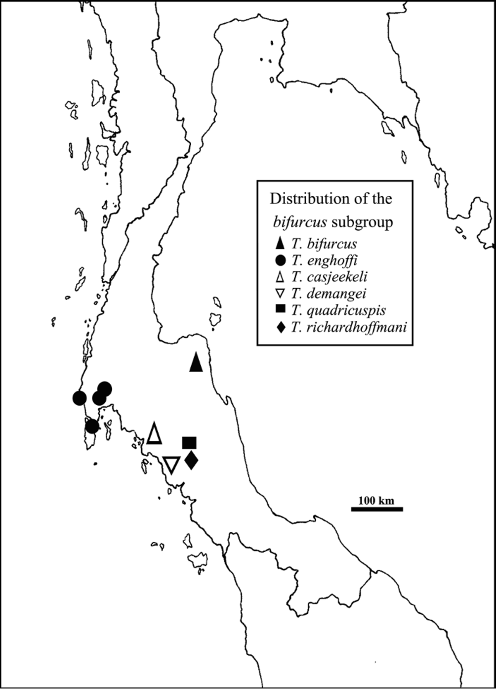
Distribution ( Fig. 8 View FIGURE 8 ): Known only from the type locality.

No known copyright restrictions apply. See Agosti, D., Egloff, W., 2009. Taxonomic information exchange and copyright: the Plazi approach. BMC Research Notes 2009, 2:53 for further explanation.