
Bengalia inermis Malloch, 1927
|
publication ID |
https://doi.org/ 10.11646/zootaxa.2251.1.1 |
|
persistent identifier |
https://treatment.plazi.org/id/B74687E8-851B-0865-4396-FAF3A217571D |
|
treatment provided by |
Felipe |
|
scientific name |
Bengalia inermis Malloch, 1927 |
| status |
|
5. Bengalia inermis Malloch, 1927 View in CoL
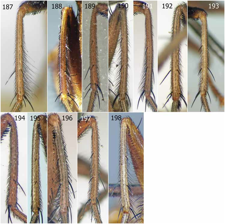
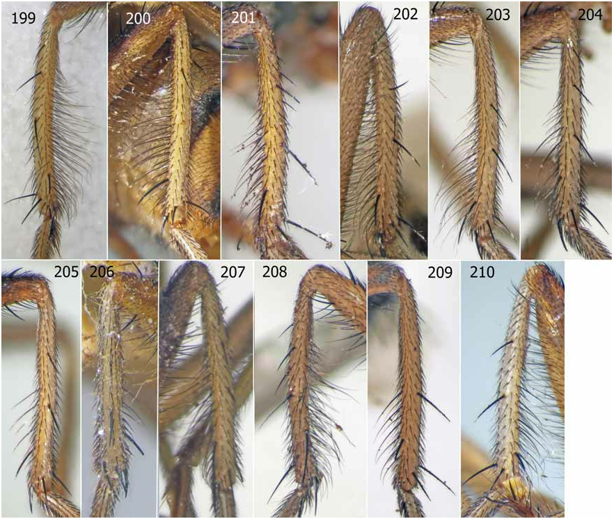
Figs. 64–82 View FIGURES 64–73 View FIGURES 74–82 , 177 View FIGURES 173–186 , 190 View FIGURES 187–198 , 202 View FIGURES 199–210 .
Holotype male, Philippines ( Luzon , Mt. Makiling) ( BMNH), by original designation. For details see Type material below.
“7. Bengalia sp. incerta ♂.”: Bezzi, 1913: 78 (as “sp. incerta Nr. 2.” in key p. 74). “Ein Männchen aus Los Banos, Philippinen, in meiner Sammlung, von Prof. Baker erhalten …“. Examined.
Note. This refers to a male specimen in MSNM which subsequently served as the holotype of Afridigalia laguna Lehrer. As detailed below, it carries a label in Bezzi’s hand reading “ Bengalia inermis n. sp. ” Bengalia inermis was described by Malloch in 1927 from a single “Type” from Mt. Maquiling, Philippines, thus a holotype was designated. The laguna holotype is therefore not a syntype of inermis (even though it carries a printed label reading “inermis SYNTYPUS ”). Malloch (1927: 414) mentions it and thinks that this particular specimen in coll. Bezzi might belong to the same taxon as his own inermis (“[T]his may be the species “sp. incerta No. 2” of Bezzi’s paper on the genus…”). He may have corresponded about it with Bezzi, and Bezzi as a result possibly wrote the name on a label in his own hand and put it on the specimen in anticipation of Malloch’s paper (Bezzi died in 1927 and is mentioned as “the late Dr. M. Bezzi” in Malloch’s paper, p. 412). This might explain the presence of a n. sp. label in Bezzi’s hand on a specimen of a species Bezzi did not describe himself.
Bengalia inermis Malloch, 1927: 400 View in CoL (key), 413 (main entry). Holotype male, by original designation, Philippines ( Mt. Makiling ) (BMNH). Examined.
Bengalia inermis: James, 1977: 529 View in CoL . Catalogue entry. Examined. For details of the specimen, see below. See also “7. Bengalia sp. incerta ♂ ” entry above. Discussed and put into synonymy by Rognes (2006: 464).
Afridigalia nusantara Lehrer, 2005: 58 . Holotype male, by original designation, Philippines (Luzon, Mt. Makiling) (BPBM). Not examined. Discussed and put into synonymy by Rognes (2006: 464).
Afridigalia pinatuba Lehrer, 2005: 63 . Holotype male, by original designation, Philippines (BMNH). Not examined. Syn. nov.
Note. The anepimeron is described as having only yellow setulae. There is nothing in Lehrer’s description and figures of pinatuba that indicate that it differs from inermis View in CoL .
Ashokiana ramsdalei Lehrer, 2005: 78 . Holotype male, by original designation, Philippines (Luzon, Camarines Sur, Mt. Iriga ) (BPBM). Examined. Syn. nov. For details of the specimen, see below.
Bengalia inermis: Verves, 2005: 239 View in CoL . Catalogue entry.
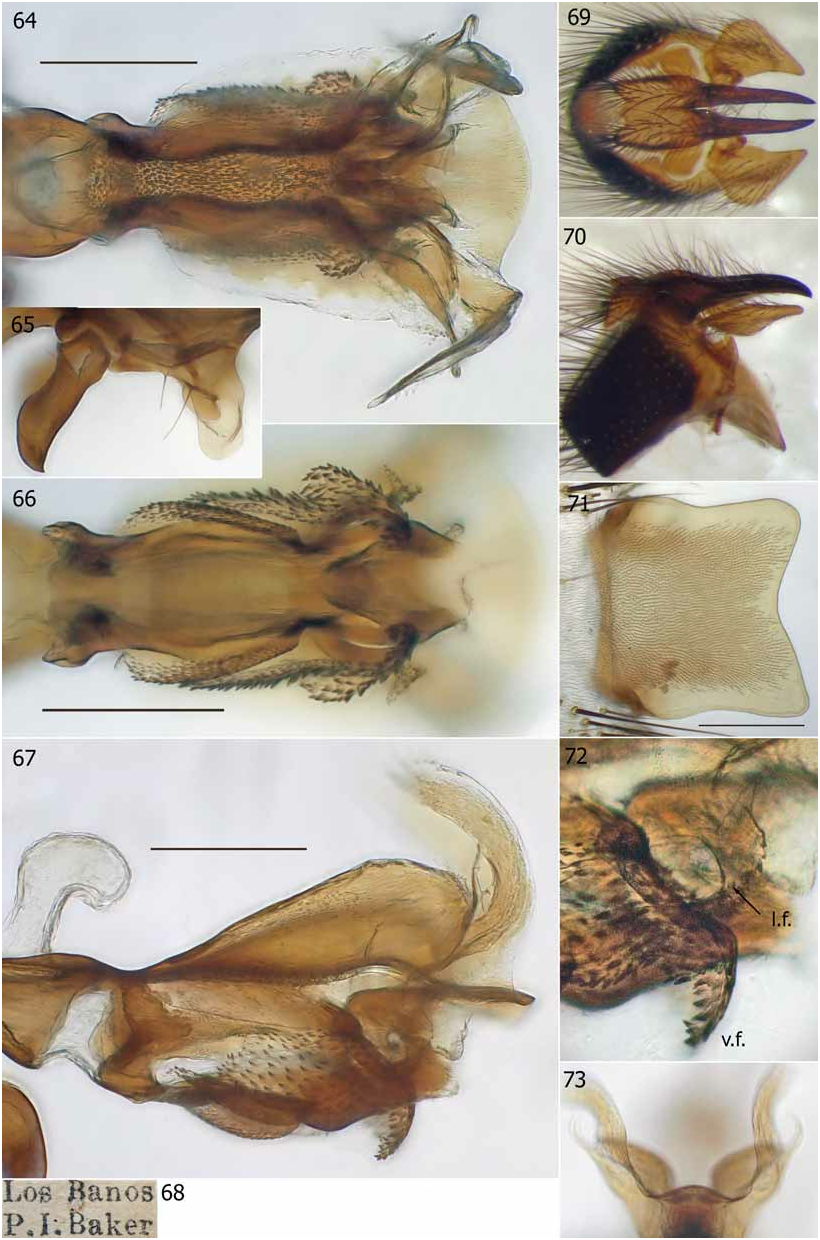
Diagnosis. A bright yellow species. Male. Length: 13mm. Frons at vertex / head width ratio: 0.283 –0.292 (mean 0.285, n=4). Lunula bare. Fronto-orbital plate without proclinate orbital setae. Anepimeron with yellow setulae only. Legs yellow. Fore tibia without spine-like setae on ventral side ( Fig. 177 View FIGURES 173–186 ). Mid tibia with a fringe of long thin pv setae in distal half, longest setae 1.5–2.0x tibial diameter ( Fig. 190 View FIGURES 187–198 ). Hind tibia with a dense fringe of long av, v and pv setae on distal two-thirds ( Fig. 202 View FIGURES 199–210 ). Abdomen almost all yellow with narrow (1/6) black marginal bands. ST5 flap ( Figs. 71 View FIGURES 64–73 , 78 View FIGURES 74–82 ) almost square, with the hind edge straight or slightly concave.
Cerci slightly curved distally. Surstylus without vestiture below. The triangular projection of the bacilliform sclerite short and blunt.
Distiphallus with prominent dorsolateral wings and broad, backwardly curved antlers. Basal tooth strong. Tip of antlers with 2–5 tines. Upper lip projecting beyond base of antlers, distal edge convex in dorsal view, underside concave as seen from in front ( Fig. 73 View FIGURES 64–73 ). Lateral finger small ( Figs. 66, 72 View FIGURES 64–73 ). Ventral finger strongly projecting below midventral wall in profile view, its anterior edge smooth, without denticles ( Figs. 67, 72 View FIGURES 64–73 ). Internal hypophallic lobes only slightly converging in ventral view, distal part of outer hypophallic lobe moderately folded with a distinct shelf ( Fig. 66 View FIGURES 64–73 ).
Female. Unknown to me.
Distribution. Philippines.
Material examined. Type material. Bengalia inermis Malloch, 1927 . Holotype male, in BMNH, labelled (1) “Holo- / type” [printed on circular label with broad red margin]; (2) “ Mt. Makiling / Luzon, Baker” [printed]; (3) “Brit. Mus. / 1923 – 423” [first line printed, second handwritten]; (4) “ Bengalia / inermis / Type / Det. / J R Malloch” [first three lines handwritten in Malloch’s hand, last two lines printed]; (5) “See slide / collection.” [printed]. Label on slide, in BMNH, reads: “ Bengalia / inermis Mall / Holotype / BM: 1923: 423 / [black line across label] / mounted / 25 – 7 – 38 J. SMART” [label handwritten, except “ Holotype ” which is printed on circular white label with broad red margin glued to main label, and “J SMART” which is printed; also black printed line all around label].
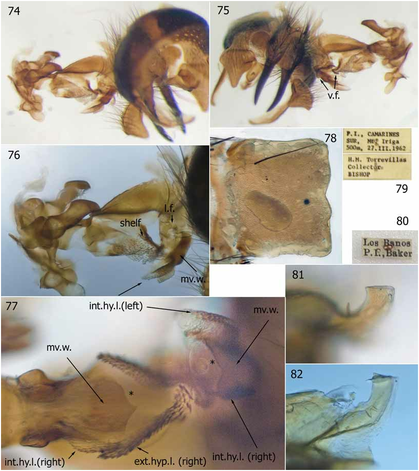
Afridigalia laguna Lehrer, 2005 . Holotype male, in MSNM, labelled (1) “Los Banos / P. I. Baker” [printed; note two pinholes ( Fig. 80 View FIGURES 74–82 )]; (2) “76” [handwritten number]; (3) “ Bengalia sp. nov. / peut être ♂ de / B. javana Macq.” [black-rimmed label with three thin ruled black lines in Surcouf’s handwriting]; (4) “ Bengalia / inermis / n. sp.” [black ink in Bezzi’s handwriting]; (5) “inermis / SYNTYPUS ” [printed museum label]; (6) “ Bengalia ♂ / laguna Lehrer sp.n. / Det. Dr. A. Z. LEHRER / XII.2005 ” [printed]; (7) “ Bengalia ♂ / laguna Lehrer sp.n. / Det. Dr. A. Z. LEHRER / XII.2005 ” [printed]; (8) “ HOLOTYPUS ” [black print on white label, latter glued to larger red label]; (9) “Published as / Afridigalia laguna / Lehrer 2005: 48 / K. Rognes 23.vi.2008 ” [printed]; (10) My determination label ( inermis Malloch ).
Note. The specimen was badly damaged in the mail at reception, but the genitalia, which were kept in a big plastic vial, arrived safely. I have glued head, wings, abdomen, one fore femur, one mid leg and two hind legs to a card on a separate pin labelled “From holotype of / Afridigalia laguna Lehrer, 2005 / Specimen crushed in accident / in mail”. Genital parts in glycerol have been transferred from original big plastic vial to glass microvial on pin where it is placed between labels (9) and (10). The ST5 flap is partly destroyed (as shown in Lehrer’s fig. 19A). In the aedeagus both antlers are broken off halfway ( Figs 81, 82 View FIGURES 74–82 ), basal tooth finger. broken at base on right side, left basal tooth intact. The ventral finger is bent stronger than usual towards base of aedeagus. Upper lip is intact. In his key to species Lehrer (2005: 23) defines the species on the assumption that the “Apophyses latérales postérieures [= antlers] sont large, courtes, ayant le bout taillé en biais ... ”, whereas they are clearly broken, a fact also suggested by Lehrer’s fig. 19C. Afridigalia laguna is therefore clearly based on an artifact. Lehrer seems not to have understood that the antlers are not in their pristine state. Without ever mentioning B. inermis as a valid species in his book, Lehrer nevertheless notes that the “… ptéropleures [= anepimeron] sont brunes avec de poils jaunes…” and he is the only witness to the fact that the fore tibiae are “... exceptionnellement, sans ctenidium proximo-ventral” [fore tibiae both destroyed in mail], exactly the reason why Malloch named his species inermis (= latin for unarmed).
Ashokiana ramsdalei Lehrer, 2005 . Holotype male, in BPBM, labelled: (1) “ H. M. Torrevillas / Collector / BISHOP” [printed]; (2) “P. I. CAMARINES / SUR, Mt. Iriga (500m, 27.III.1962 ” [printed]; (3) “ HOLOTYPE ” [printed white label glued to larger red label] / “17040” [pencil writing on red part of label]; (4) “ Ashokiana ♂ / ramsdalei Lehrer n. sp. / Det. Dr. A.Z. Lehrer / 2004” [printed]; (5) “ Ashokiana ♂ / ramsdalei Lehrer n. sp. / Det. Dr. A.Z. Lehrer / 2004” [printed]; (6) My determination label ( inermis Malloch ). Dissected by Lehrer.
Note. The abdomen is intact except for the genitalia and ST5 flap which have been removed, probably by Lehrer. The epandrial complex (bacilliform sclerite present on one side only, other side lost), aedeagus with pre- and postgonites plus the basal part of the phallapodeme (i.e., the long-legged intermedium piece), and the ST5 flap were stored in a plastic vial at reception of the holotype in the mail. I have transferred the genitalia to glycerol in a glass microvial pinned below the specimen.
The holotype has a rather peculiar aedeagus according to the detailed figure by Lehrer (2005: 78, fig. 34C). Rather than reflecting a separate species and genus, however, I have to conclude that the peculiar structure is simply an artifact resulting from an effort (most likely on the part of Lehrer himself) to pry loose the aedeagus from the epandrium to which it has apparently been glued by accident. This effort has partly destroyed the distiphallus by rupturing its midventral wall. The explanation, as I see it, follows below.
When received by me the aedeagus was glued with its apex to the side of the epandrium near the anal membrane, apparently because of some accident from treatment with glycerol jelly or some other substance (no details are given by Lehrer) ( Figs. 74, 75 View FIGURES 74–82 ). It was impossible to disconnect the aedeagus from the epandrium by heating the joined complex carefully in a water bath at near boiling point. Nevertheless, it was perfectly possible to study most of the aedeagus. The upper lip could not be observed, but the antlers were clearly visible.
Seen from the left side ( Fig 76 View FIGURES 74–82 ), the distiphallus matches perfectly the drawing by Lehrer (2005: 78, fig. 34C). Other angles of view revealed that the peculiar structure of the distiphallus, which led Lehrer to establish the nominal genus Ashokiana ( Lehrer, 2005: 22, 78), is simply an artifact resulting from a failed effort to pry loose the aedeagus from the epandrial complex. Evidently pressure has been applied to the basal part of the aedeagus with the attached pre- and postgonites in the hope that the aedeagus might come loose. In the process the ventral wall of the distiphallus has simply ruptured in the middle (between * * in Fig. 77 View FIGURES 74–82 ). The basal third of the mid-ventral wall (mv. w.) has remained unharmed but displays a distal termination edge which is angular in ventral view. The peculiar appendix described by Lehrer in the key to Ashokiana on p. 22 [“[d]istiphallus a une structure particulière dans sa partie antéro-inférieure, formée d’une portion supérieure membraneuse et pourvue d’épines récurrentes et d’une portion inférieure plus ou moins sclérifiée et sous forme d’auge”] is the combined distal two-thirds of the midventral wall (mv. w.) plus parts of the hypophallic lobes that have been torn away from and forming an angle with the remainder of the distiphallus as seen in lateral view. Seen from the ventral side the free edge of the projecting appendix has an angular excavation of exactly the same shape as the distal edge of the basal part of the ventral wall, together revealing exactly where the rupture has occurred (between * * in Fig 77 View FIGURES 74–82 ). The “portion supérieure membraneuse et pourvue d’épines récurrentes” is simply the torn away internal hypophallic lobes (int. hy. l.), and the “portion inférieure plus ou moins sclérifiée et sous forme d’auge” is the distal two-thirds of the midventral wall (mv. w.). Comparing fig. 19C ( inermis Malloch , as laguna Lehrer ) and fig. 34C ( inermis Malloch , as ramsdalei Lehrer ) in Lehrer (2005) one can immediately convince oneself of this fact. Close inspection reveals that the membranous part is a bilateral structure as expected, and on the right side it is broken closer to the “auge” than on the other, so that a considerable part of the internal hypophallic lobe remains in situ on the right side (int. hy. l. (right) ) to the lower left in Fig. 77 View FIGURES 74–82 ). On the left side the whole internal hypophallic lobe (int. hy. l. (left) ) is torn away from its natural position. Similarly, a close look at the distal part of the ventral wall [“… sclérifiée et sous forme d’auge”] reveals that it is quite similar to the one in a normal inermis distiphallus in ventral view. The pressure applied to the distiphallus has also caused a vertical rupture in the middle of the external hypophallic lobe on the right side, resulting in a posterior displacement of the lateral finger and shelf area away from the proximal parts of the lobe ( Fig. 75 View FIGURES 74–82 ). There has also been a rupture in the lateral wall on the left side resulting in an artificially large distance between the shelf area (shelf) of the external hypophallic lobe and the lateral finger (l. f.) ( Fig. 76 View FIGURES 74–82 ). This is represented by a large clear area in Lehrer’s fig. 34C ( Lehrer 2005: 78). Finally, the removal of the distal parts of the mid-ventral wall from its normal position has displaced the distal parts of the remaining external hypophallic lobes, including the ventral finger (v. f.), towards the midline so as to fill the gap ( Figs. 75, 77 View FIGURES 74–82 ).
In view of Lehrer’s careful reproduction of the Ashokiana distiphallus it is surprising that he was not aware of the fact that it had already been partly destroyed when he made his drawing. It is equally surprising that Lehrer did not report on the condition of the genitalia of his type specimen (fusion of the epandrial complex with the tip of aedeagus, the rupture of the mid-ventral wall of latter, etc.) and that he failed to understand the true reason behind the peculiar shape of the aedeagus of his ramsdalei holotype.
The holotype of ramsdalei can safely be assigned to inermis on account of the shape of the ventral finger (long, curved and denticulate on posterior side only), small curved lateral finger, broad antlers terminating in several small points and with a strong and long basal tooth and broad dorsolateral wings. The shape of the ST5 flap, the chaetotaxy of the legs, only yellow setulae on anepimeron, bright yellow body colour and narrow marginal bands on abdominal tergites support this assignment.
Other material. SDEI: 1 male labelled (1) “Los Banos / P. I. Baker ” [printed; note one pinhole only]; (2) My determination label ( inermis Malloch ). Dissected by K. R. Abdominal T1–5 glued to card on pin, genitalia in glycerol in vial on pin. Genitalia shown in Figs. 64–73 View FIGURES 64–73 .

| BPBM |
Bishop Museum |
No known copyright restrictions apply. See Agosti, D., Egloff, W., 2009. Taxonomic information exchange and copyright: the Plazi approach. BMC Research Notes 2009, 2:53 for further explanation.
|
Kingdom |
|
|
Phylum |
|
|
Class |
|
|
Order |
|
|
Family |
|
|
Genus |
Bengalia inermis Malloch, 1927
| Rognes, Knut 2009 |
Afridigalia nusantara
| Rognes, K. 2006: 464 |
| Lehrer, A. Z. 2005: 58 |
Afridigalia pinatuba
| Lehrer, A. Z. 2005: 63 |
Ashokiana ramsdalei
| Lehrer, A. Z. 2005: 78 |
Bengalia inermis:
| Verves, Yu. G. 2005: 239 |
Bengalia inermis: James, 1977: 529
| Rognes, K. 2006: 464 |
| James, M. T. 1977: 529 |
Bengalia inermis
| Malloch, J. R. 1927: 400 |