
Scapheremaeus rodickae, Norton, Roy A. & Franklin, Elizabeth, 2010
|
publication ID |
https://doi.org/ 10.5281/zenodo.193893 |
|
DOI |
https://doi.org/10.5281/zenodo.6206662 |
|
persistent identifier |
https://treatment.plazi.org/id/B75AE82C-1404-940F-79C1-F93F82B89153 |
|
treatment provided by |
Plazi |
|
scientific name |
Scapheremaeus rodickae |
| status |
sp. nov. |
Scapheremaeus rodickae View in CoL sp. nov.
( Figs. 1 View FIGURE 1 –4, 5B–C, 6A)
Diagnosis. Uniformly reddish-brown. Notogaster without caudal emargination or humeral process; with 10 pairs of setae. Notogastral microsculpture uniformly reticulate-areolate except ventral to circumferential scissure; circumdorsal scissure U-shaped, absent posteriorly and weakly defined anteriorly and laterally; central plate with vaguely defined longitudinal ridges (3 anteriorly, 1 posteriorly). Prodorsum with simple, nearly parallel costulae that lack distal cusps; with ridges forming flattened X-shaped figure between bothridia; sculpturing reticulate medial to costulae, tuberculate lateral to tutorial ridge.
Etymology. We take pleasure in naming this species after Ms. Christine Rodick, River Keeper and Guardian of the Rocks and Shoals Natural Area, in recognition of her valiant efforts in environmental preservation.
FIGURE 4. A–I Scapheremaeus rodickae n. sp.: A) bothridial seta, lateral aspect; B) genu I, abaxial aspect, showing seta l’; C) apobasic notogastral seta p2, arrow on deep tubular invagination containing basal half of seta; D, E) notogastral microsculpture from humeral region and region of longitudinal ridge, respectively, showing thin layer of fine adherent birefringent particles; F) lateral view of ventral plate posteroventral to leg IV (trochanter and part of femur visible), showing tuberculate microsculpture; G) subcapitulum, ventral aspect; H) distal region of tarsus I, arrow on famulus; I) region of palp insertion on subcapitulum, dorsal aspect, arrow on postpalpal seta; J) Scapheremaeus palustris (Sellnick) , prodorsum and anterior region of notogaster of specimen from Austria, showing broken microsculpture pattern between costulae. All light micrographs with differential interference contrast illumination, layered from 2–10 sequentially focused images. Scale bars: 10 μm.
Description of Adult. Dimensions. Medium-sized species, body length (n=10) 403–450 (mean 430) µm, and width 220–240 (mean 193) µm. Sexual species, with males common (sex ratio not determined).
Integument. Reddish brown, well sclerotized and with microsculpture throughout body and legs. Microsculpturing mostly areolate, varying from reticulate to tuberculate, as noted below. Patterns emphasized by dark, relatively thick cerotegument that mostly mimics underlying cuticle; surface of cerotegument finely granulate over tuberculate or elongate microsculpture, smooth over areolate microsculpture. Thin, usually inconspicuous layer of adherent debris present, composed mostly of minute birefringent particles (probably minerals; Figs. 4D–E).
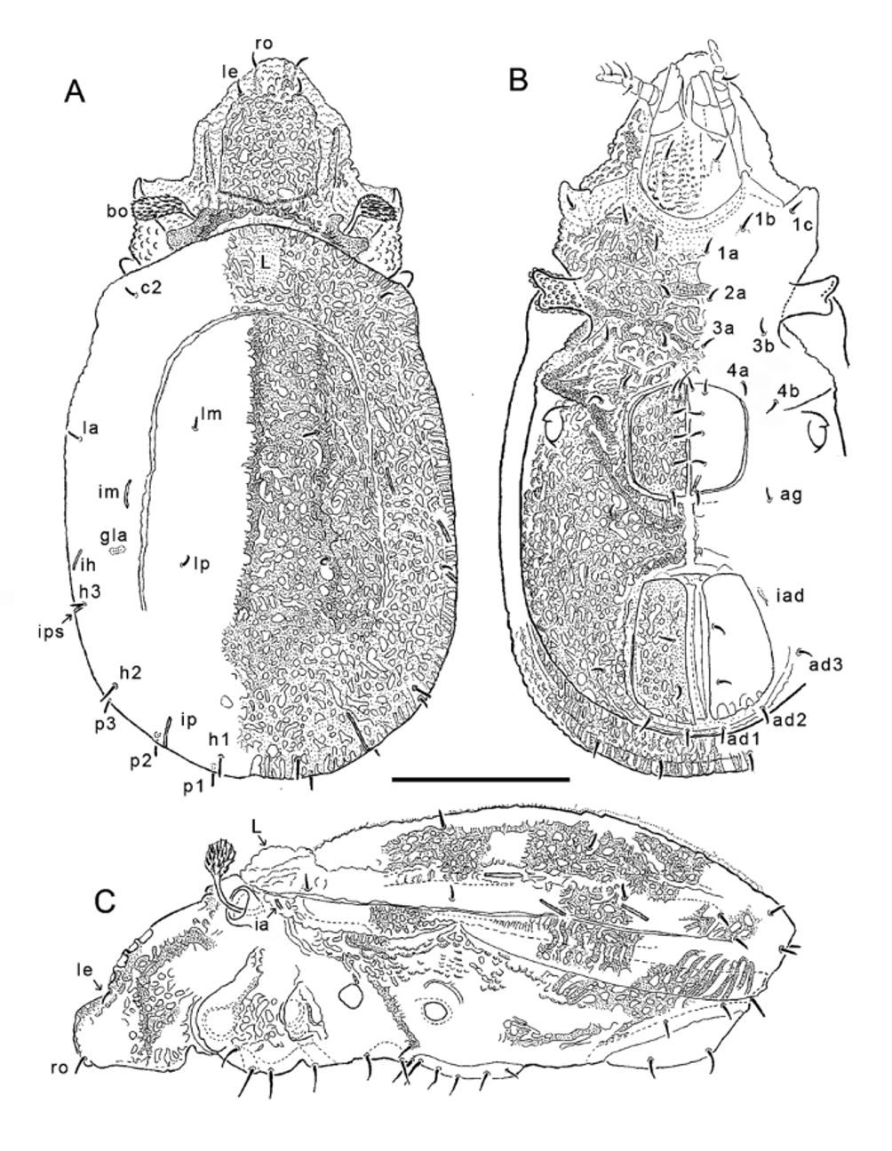
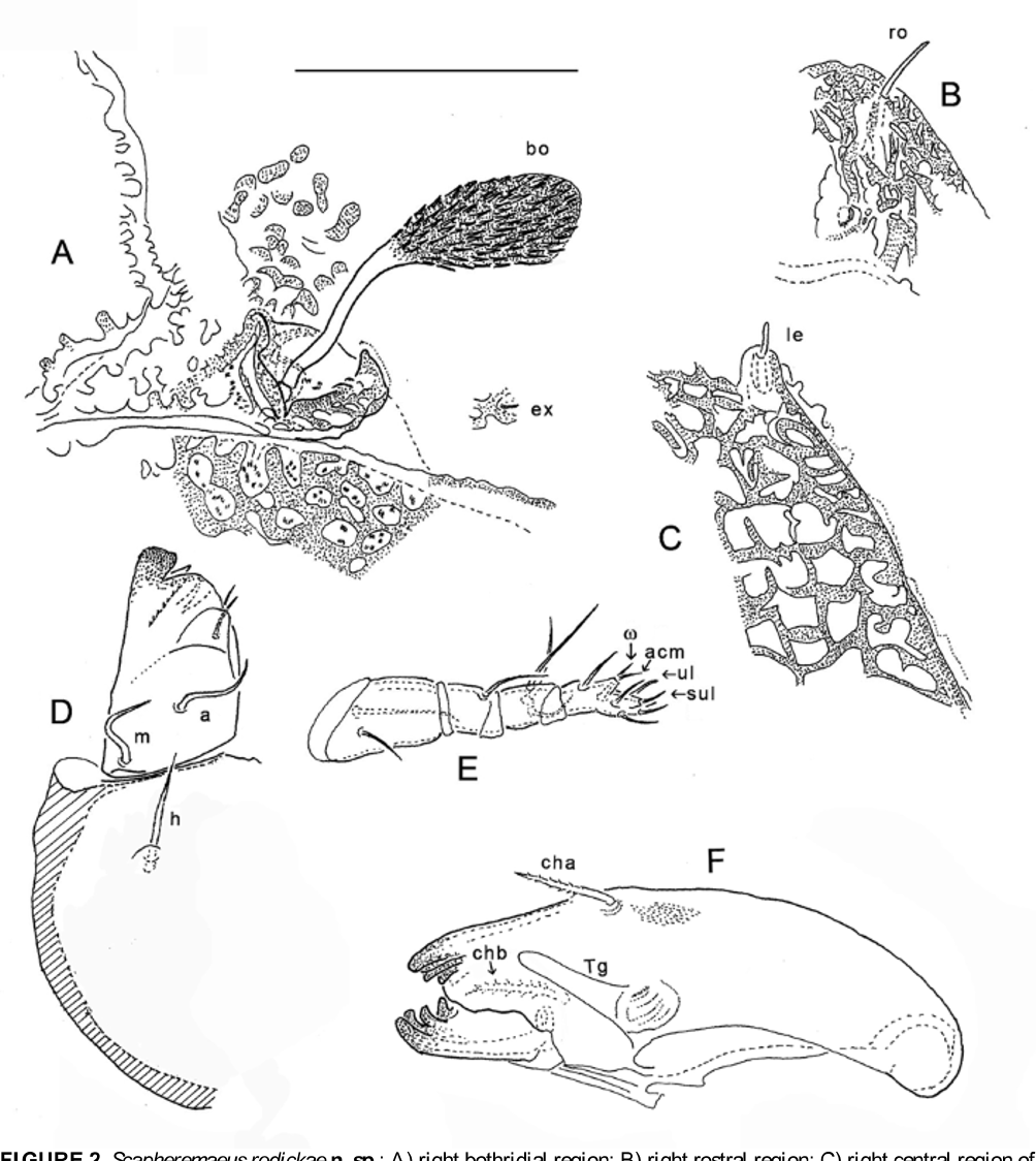
Prodorsum. Prodorsum occupies about one-quarter body length; with rough form of equilateral triangle in dorsal aspect ( Figs. 1 View FIGURE 1 A, 6A); rostrum narrowly rounded, strongly tapering from level of acetabula I. Lateral outline irregular ( Fig. 1 View FIGURE 1 C); transversely concave in anterior quarter, rostrum blunt. With nearly parallel, simple costulae each terminated by small tubercle bearing lamellar seta; with no transverse ridge connecting costulae distally. Posteriorly with irregular ridges forming flattened X-shaped figure between bothridia. Microsculpture reticulate medial to costulae, tuberculate lateral to well-formed lateral carina, almost absent in leg-accommodating groove between costula and carina. Prodorsal setae short, setiform and smooth. Lamellar (le) seta ~6 μm, bacilliform ( Fig. 2 View FIGURE 2 C), mutual distance about 30 μm. Rostral (ro) seta ~10 μm, acuminate, mutual distance 20 μm ( Fig. 2 View FIGURE 2 B), pair usually slightly diverging; exobothridial seta (ex) minute ( Fig. 2 View FIGURE 2 A), apparent only with highest oil immersion lens. Interlamellar seta absent. Bothridial seta (sensillus, bo) curved dorsad and anterolaterad, ~50 μm long; head pigmented, about equal in length to narrow stalk, but otherwise atypical for genus: fan-like, strongly flattened and expanded, with dense longitudinal ridges terminating in minute barbs ( Figs. 2 View FIGURE 2 A, 4A).
Notogaster. Notogaster slightly ovoid, about 1.4 times longer than broad ( Figs. 1 View FIGURE 1 A, 6A); without caudal emargination or humeral process; anterior margin not evenly arched, slightly parabolic. Microsculpture of ventral circumferential plate (below circumferential scissure, not visible from above) with vaguely parallel, radially oriented ridges; all dorsally visible cuticle of notogaster irregularly reticulate-areolate, with relatively small areoles (mostly 5–7 μm diameter) separated by darker reticulation of similar width. Circumdorsal scissure inversely U-shaped, absent posteriorly, weakly defined anteriorly and laterally; circumferential scissure also incomplete posteriorly ( Fig. 1 View FIGURE 1 C). Centrodorsal region with three vaguely defined longitudinal ridges, one medial ridge and lateral pair; one medial ridge present in pygidial region. Lenticulus relatively small, equidistant from circumdorsal scissure and anterior margin of notogaster. With 10 pairs of setae, smooth, sub-bacilliform, tapering slightly to rounded tip; pairs p1, p2, p3 conspicuously project beyond margin in dorsoventral aspect. All setae apobasic, with emergent part 6–7 μm plus similar length in tubular invaginated socket (Fig. 4C). Setal pairs lm and lp well medial to circumdorsal scissure. Lyrifissures long slits (~15–17 μm) but inconspicuous due to microsculpture and cerotegument; five typical pairs present: three pairs close to circumferential scissure, ia ventral to it at level of lenticulus ( Fig. 1 View FIGURE 1 C), ih and ips dorsal to it and lateral to setae lp and h3 respectively; im at midlength of notogaster, slightly lateral to circumdorsal scissure; ip midway between setae h1 and h2. Opisthonotal gland opening (gla) posterior to im and medial to ih.
Ventral Region ( Fig. 1 View FIGURE 1 B). Microsculpture and cerotegument irregular on most of coxisternum, tuberculate anteriorly on mentotectum; areolate posterior to epimere IV, similar to that of notogaster except with wellspaced elongated tubercles dorsal to leg acetabula and posterior to acebabulum IV (Fig. 4F). Epimeral setation 3-1-2-2; setae apobasic, most acuminate with emergent part 6–8 μm, 1b longer, (15 μm) attenuate. Pedotectum II weakly bifurcated in dorsoventral aspect, anterior and poster faces slightly concave. Genital and anal plates isolated from rest of ventral plate by closely surrounding grooves, with medial connecting groove; microsculpture tuberculate in grooves. Genital aperture subquadrate, slightly broader anteriorly; five genital setae aligned on medial margin, sixth closely lateral to most anterior seta; all apobasic, attenuate, emergent part ~8 μm long except anterior seta on medial margin much longer (25–30 μm). Anal 50 % longer than genital aperture, separated from it by about two-thirds length of latter. Anal valves slightly wider posteriorly; with deep longitudinal groove along each medial margin that blends with connecting groove when valves closed; two anal setae sub-bacilliform, apobasic (emergent part 8–9 μm). Aggenital seta similar to shorter genital setae, adanal setae similar to anal setae; ad1 and ad2 inserted parallel to posterior margin of anal valves, close to edge of separating groove; ad3 more removed from anal valves, at their mid-level. Lyrifissure iad near anterolateral corner of anal plate, within surrounding groove, oriented parallel to plate margin.
Gnathosoma . Subcapitulum normal for genus ( Fig. 2 View FIGURE 2 D, 4G), mentum with microstructure of rounded to elongated tubercles; cerotegument thin, with clear microgranules. Setae of gena and mentum apobasic, emergent part ~20 μm, attenuate, smooth to very weakly barbed; postpalpal seta short (6 μm), tapered to blunt tip, smooth, with normal insertion (not apobasic) in relatively thin, soft cuticle (Fig. 4I); two adoral setae with similar form. Palp setal formula (trochanter to tarsus) 0-2-1-3-9, plus solenidion ω; eupathidion acm on tubercle, free from solenidion ( Fig. 2 View FIGURE 2 E). Chelicera of typical robust form ( Fig. 2 View FIGURE 2 F); setae cha and chb strong and barbed.
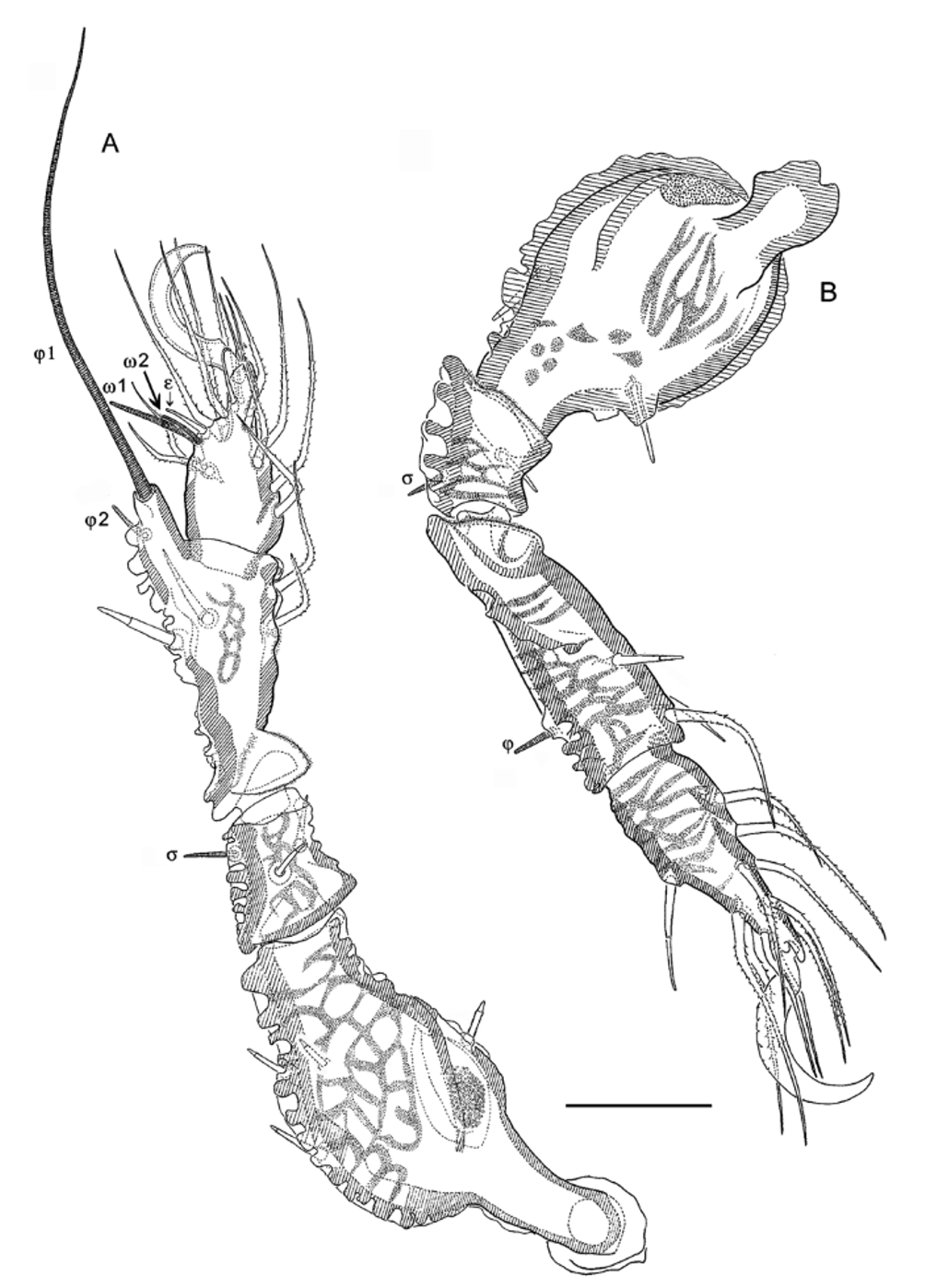
Legs ( Figs. 3 View FIGURE 3 A–B). Well sclerotized, heavily ornamented with microsculpture, including thick cerotegument; pattern in form of irregular ridges on most segments, femora mostly tuberculate adaxially, ridged or areolate abaxially. Paraxial porose areas on trochanters III, IV small but distinct; those of femora I, II large, conspicuous; porose areas of femora III and IV in form of small inconspicuous band on proximal face of femoral bulb. Genua and tibiae with retrotecta (articulate with “sockets”); trochanters III and IV with large ventrodistal protectum. All pretarsi monodactylous; claw robust, dorsally with paired rows of inconspicuous teeth. Setal formulas (legs I-IV) of trochanters 0-0-1-0; femora 4-4-2-2; genua 2-2-1-1; tibiae 4-3-3-3; tarsi (including famulus) 15-13-12-11. Homologies of setae given in Table 1. Setae with three general forms: 1) setae bv ” of femur II and ev’ of femora III-IV spiniform, apobasic, similar to notogastral setae; 2) seta bv ” of femur I, lateral and dorsal setae of trochanter III and all genua and tibiae, and ft” of tarsus IV straight, acute, with thin sleeve of brown cerotegument extending from one-quarter to three-quarters setal length (Fig. 4B); 3) ventral setae of tibiae and all setae of tarsi (except ft” IV) attenuate, clearly barbed, light brown but with smooth, hyaline base. Famulus relatively large (10 μm), with broad base and very slightly expanded tip (Fig. 4H). Solenidial number, form and distribution normal for genus (Table 1).
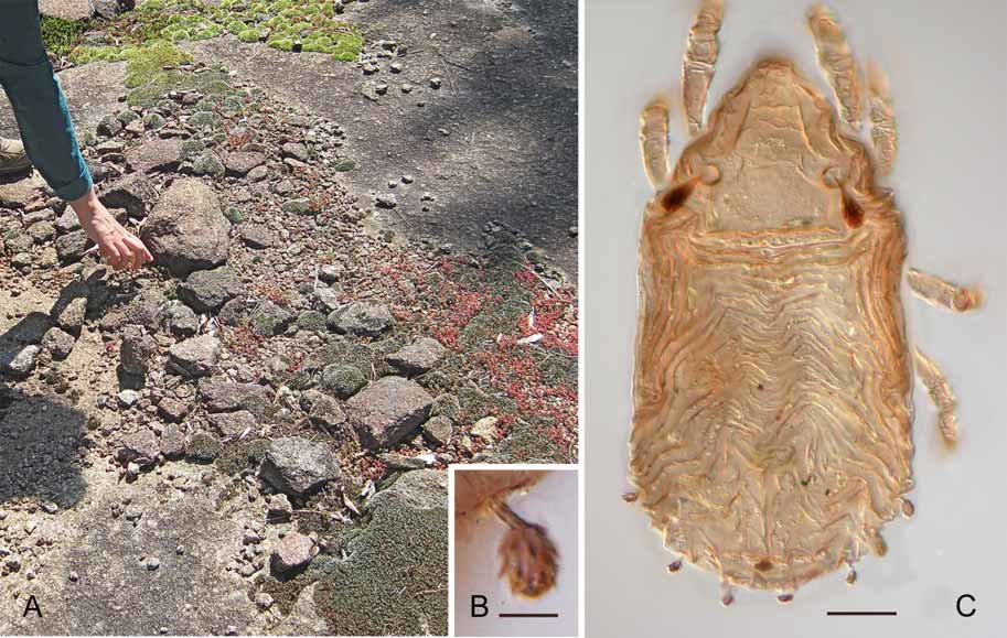
Immatures. Immatures were collected along with the adults, but were not studied in detail. Nymphs have the plicate form typical of Scapheremaeus species (see Travé & Fernandez 1986). The gastronotic setae of nymphs are mostly inconspicuous, but four posteromarginal pairs (lp, h1-3) have a heavily barbed, cupped head attached to a narrow stalk ( Fig. 5 View FIGURE 5. A B–C); with cerotegument included, the head is reminiscent of half the shell of a thorny oyster ( Spondylus ).
Material Examined. Holotype: collected from Rocks and Shoals Natural Area, Clarke County, Georgia, USA (N 33°53’15.5”, W 83°20’0.60”; elevation 230 m. a.s.l.); col. 11-X-2005 by D. A. Crossley and L. Dame, ex: Black Rock Moss, Grimmia laevigata (Bird.) , growing at margin of small, rain-collecting depressions on granite outcroppings ( Fig. 5A View FIGURE 5. A ). Paratypes: 40 with same data as holotype. The holotype and five paratypes are deposited in the Georgia Museum of Natural History (Natural History Building, University of Georgia, Athens, GA 30602); five paratypes are in the Acarology Laboratory of the Ohio State University; five paratypes are in the Canadian National Collection, Ottawa; and the remaining paratypes and immatures are in the personal collection of R.A. Norton. All types are preserved in ethanol, except eight in the latter collection which were dissected and slide-mounted.
Comments. Colloff (2009) made the first attempt to group species within the large genus Scapheremaeus by recognizing 13 species groups. Scapheremaeus rodickae does not have the complete set of attributes of any group, but best seems to fit the Emarginatus group. It differs from the description of this group mainly by having a circumdorsal scissure (“circumnotogastric” was a lapsus in the character list; M. J. Colloff, personal communication 2009), but one that is inconspicuous and incomplete.
Scapheremaeus View in CoL species are commonly associated with moss and lichens growing on tree bark or on otherwise barren rocks. The moss ( Grimmia laevigata View in CoL ) from which S. rodickae View in CoL was collected grows on the edges of temporary rock pools, or lithotelmata, on exposed granite outcrops ( Keever 1957). We recently described Zygoribatula colemani Franklin et al. (in an unrelated superfamily, Oripodoidea), which is abundant both in these mosses and—unlike S. rodickae View in CoL —in the central sediment of the depressions, where the only vegetation is the sedum Diamorpha smalli Britt. ( Franklin et al. 2008) . While some Scapheremaeus View in CoL species have internalized respiratory organs on their legs, S. rodickae View in CoL does not; femora I–IV, as well as trochanters III and IV, have only the normal surface porose areas.
No known copyright restrictions apply. See Agosti, D., Egloff, W., 2009. Taxonomic information exchange and copyright: the Plazi approach. BMC Research Notes 2009, 2:53 for further explanation.
|
Kingdom |
|
|
Phylum |
|
|
Class |
|
|
Order |
|
|
Family |
|
|
Genus |
Scapheremaeus rodickae
| Norton, Roy A. & Franklin, Elizabeth 2010 |
Diamorpha smalli Britt. ( Franklin et al. 2008 )
| Britt. (Franklin et al. 2008 |