
Lithonecrus Nieves-Aldrey & Butterill
|
publication ID |
https://doi.org/ 10.11646/zootaxa.3846.2.3 |
|
publication LSID |
lsid:zoobank.org:pub:B31B3F36-052A-477D-882B-8E555F74832A |
|
DOI |
https://doi.org/10.5281/zenodo.6143081 |
|
persistent identifier |
https://treatment.plazi.org/id/B776D130-5E1C-496C-FF43-FA90FD0FFBC7 |
|
treatment provided by |
Plazi |
|
scientific name |
Lithonecrus Nieves-Aldrey & Butterill |
| status |
|
Lithonecrus Nieves-Aldrey & Butterill , gen. n.
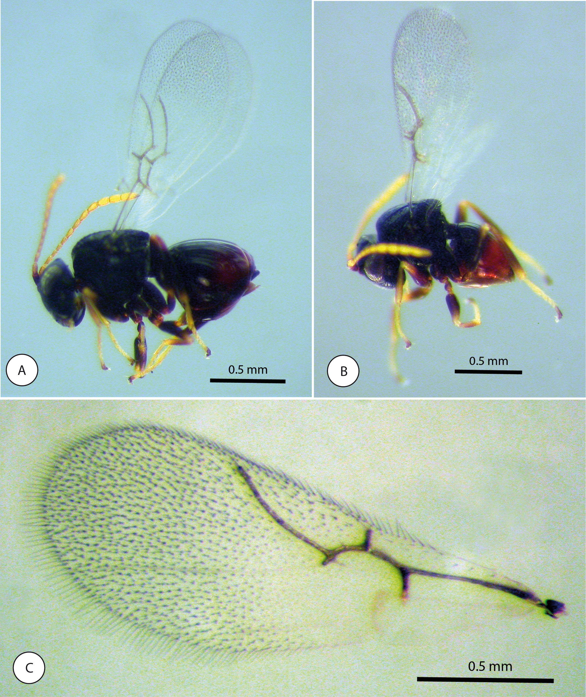
( Figs 1–5 View FIGURE 1 View FIGURE 2 View FIGURE 3 View FIGURE 4 View FIGURE 5 )
Type species. Lithonecrus papuanus Nieves-Aldrey & Butterill , sp. n., by present designation and monotype.
Etymology. A combination of two words: “ Litho” from the host tree species Lithocarpus and “ necrus” from the end word of the related genus Saphonecrus .
Description
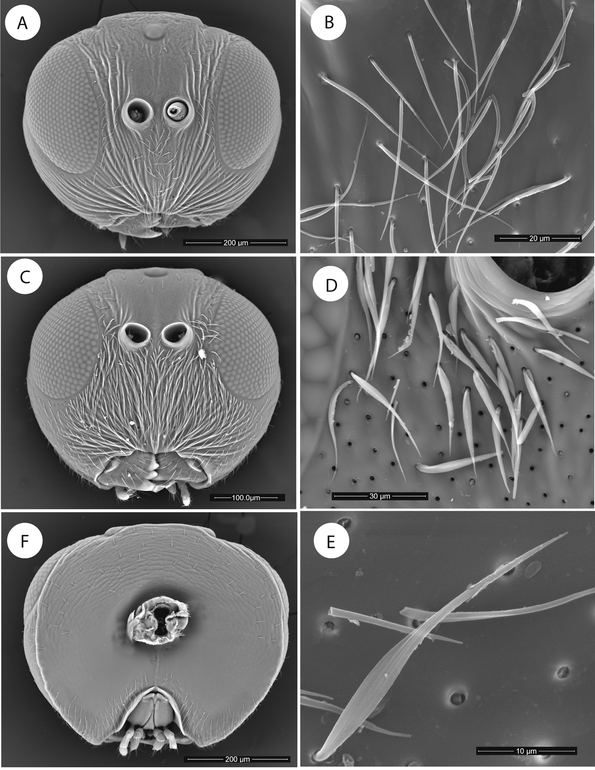
Head. Rounded in females ( Fig. 1 View FIGURE 1 A), oval in males ( Fig. 1 View FIGURE 1 C). Slightly pubescent in females, more heavily in males; scattered long setae on the face medially, the lateral areas, and upper part of frons; in males the setation is dense and whitish, the setae being differentiated, longer and broader basally ( Figs 1 View FIGURE 1 C, 1D, 1E). Gena slightly expanded behind compound eyes. Clypeus indistinct, ventral margin sinuate, not projecting over mandibles. Anterior tentorial pits visible; epistomal sulcus and clypeo-pleurostomal lines indistinct. Face with wide, strong and blunt irradiating striae from clypeus, almost reaching ventral margin of eye and ventral margin of toruli; The facial striae extend dorsally branched in several rows on the lateral area of the frons, reaching the lateral ocelli, but absent medially ( Fig. 1 View FIGURE 1 A). Vertex abruptly limited at the occiput by an occipital carina situated just behind the lateral ocelli ( Fig. 3 View FIGURE 3 A); the occipital carina is well defined and complete, reaching ventral margin of face ( Fig. 1 View FIGURE 1 F). Occiput coriaceous dorsally. Gula relatively long; distance between occipital foramen and oral foramen clearly longer than the height of the occipital foramen. Hypostomal sulci well visible ( Fig. 1 View FIGURE 1 F).
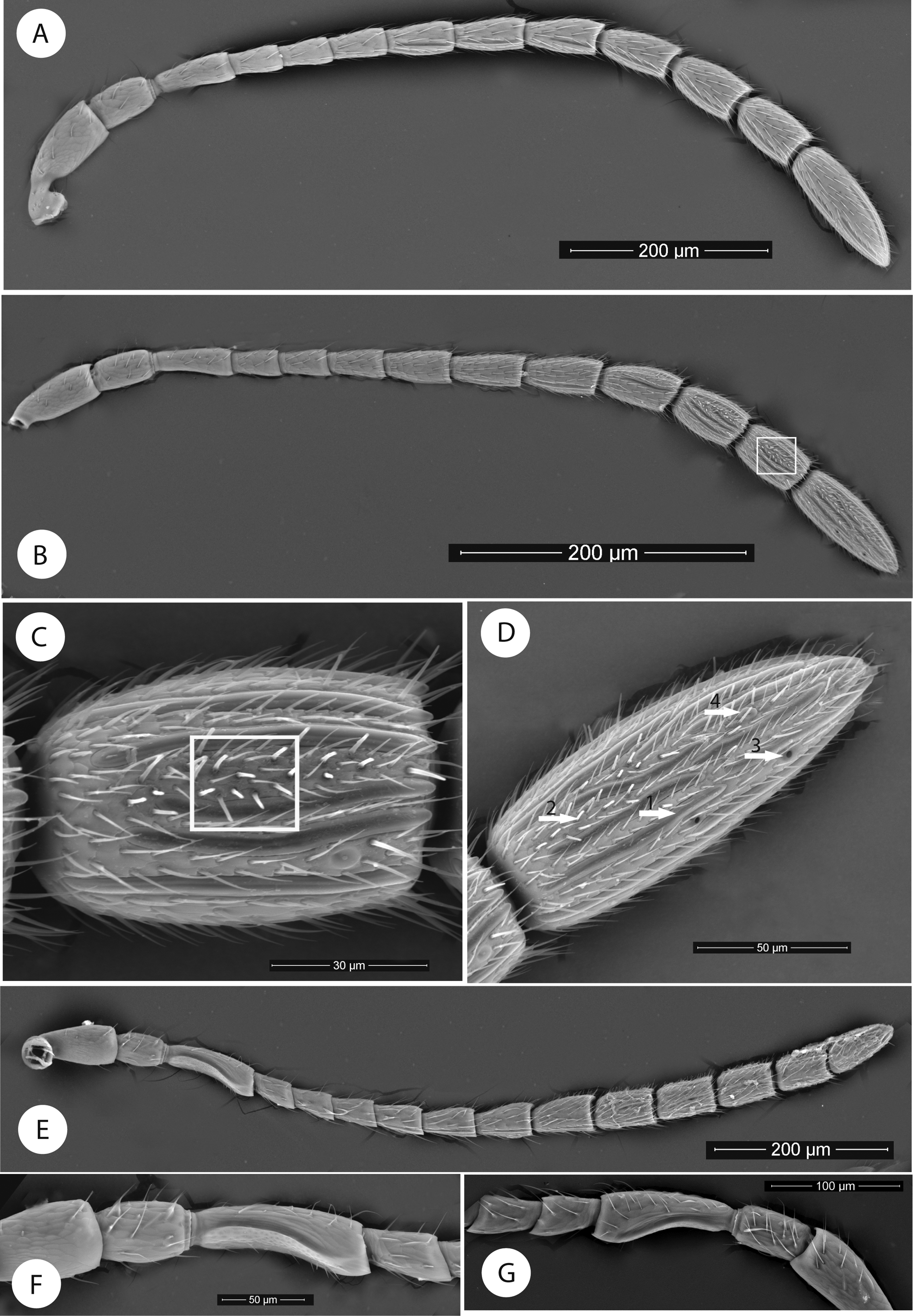
Female antenna 13-segmented ( Figs. 2 View FIGURE 2 A, 2B); flagellum broadened towards apex; with relatively long, erect setae and placodeal sensilla visible on flagellar segments F6–F11. Last three flagellar segments presenting wide areas with basiconic setae ( Figs 2 View FIGURE 2 B, 2C). Pedicel 1.8 as long as wide, broader but shorter (0.7) than F1; F1 1.7 as long as F2 ( Fig. 2 View FIGURE 2 A). Male antenna 15-segmented, not broadened towards apex ( Fig. 2 View FIGURE 2 E). F1 dorsally curved, excavated in the middle and expanded apically ( Figs 2 View FIGURE 2 F, 2G). F1 more than 2x F2.
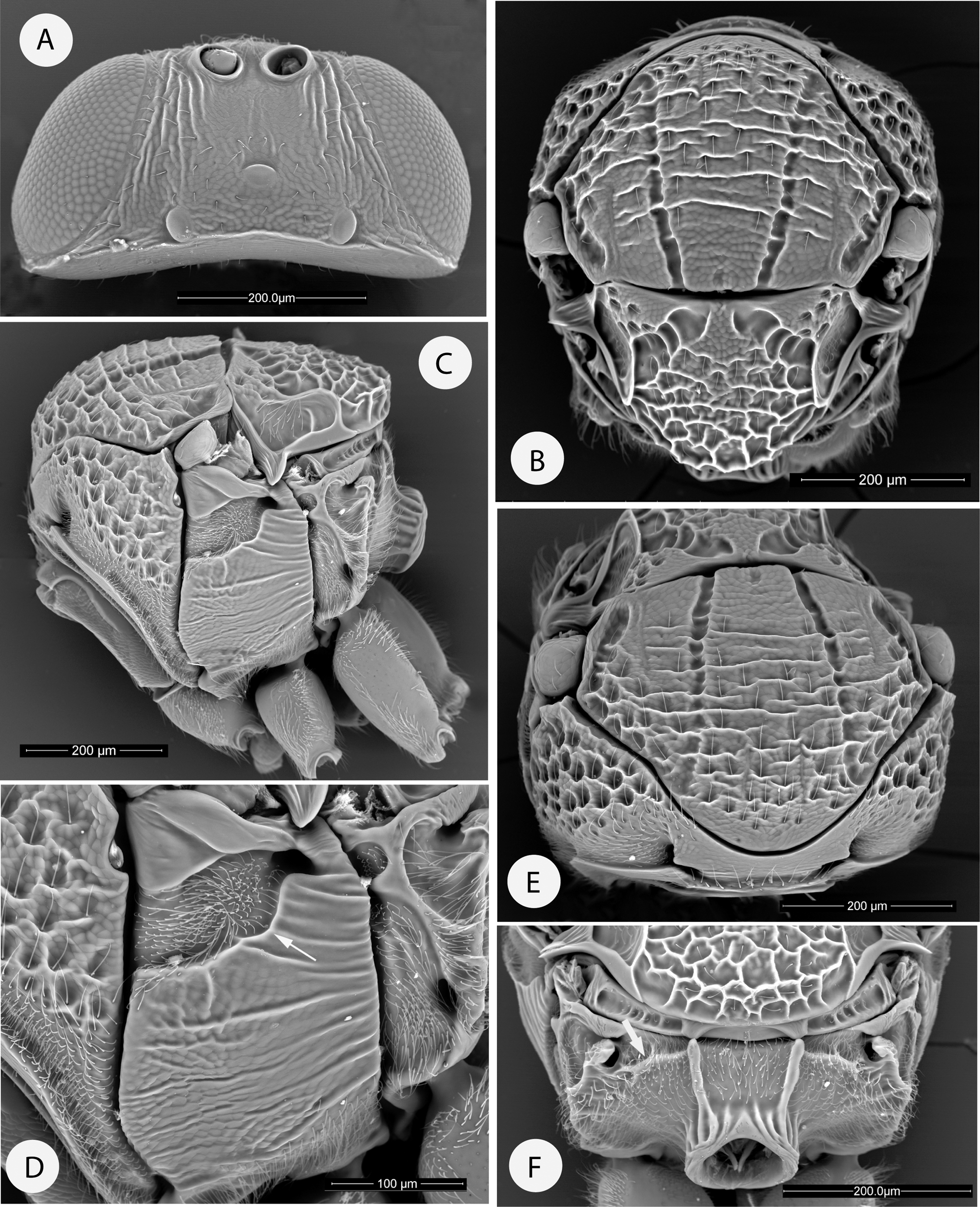
Mesosoma. Pronotum relatively broad medially, measuring nearly 1/5 of the length of the outer lateral margin. Pronotal plate ( Fig. 3 View FIGURE 3 E) relatively well developed, with lateral margins present posteriorly and only faint anteriorly; posterior part of pronotal plate with antero-lateral margins quite well extended to the sides of pronotum. Lateral margins of pronotum rounded, without lateral pronotal carinae ( Fig. 3 View FIGURE 3 C). In lateral view, pronotum very short. Mesoscutum with notauli broad, percurrent ( Fig. 3 View FIGURE 3 B). Median mesoscutal impression invisible. Anteroadmedian signae visible. Parapsidal signa deep, reaching posterior margin of pronotum. Transscutal fissure narrow. Scutellar foveae large, obliquely separated, with smooth sculpture but crossed by some strong carinae ( Fig. 2 View FIGURE 2 B). Mesopleural triangle subrectangular, ventral margin not straight and angulated in the posterior third ( Figs 3 View FIGURE 3 C, 3D). Mesopleuron with longitudinal striae and coriaceous sculpture between the striae ( Figs 3 View FIGURE 3 C, 3D).
Metapectal-propodeal complex. Metapleural sulcus meeting posterior margin of mesopectus at about the height of posterior subalar pit. Lateral propodeal carinae distinct, broad, slightly convergent posteriorly ( Fig. 3 View FIGURE 3 F). Median propodeal areas smooth and pubescent. Dorsally, on the lateral propodeal areas, a deep groove is present, extending from the propodeal spiracles to the dorsal part of the lateral propodeal carinae, the groove being limited posteriorly by a crest ( Fig. 3 View FIGURE 3 F). Nucha dorsally strongly sulcate longitudinally.
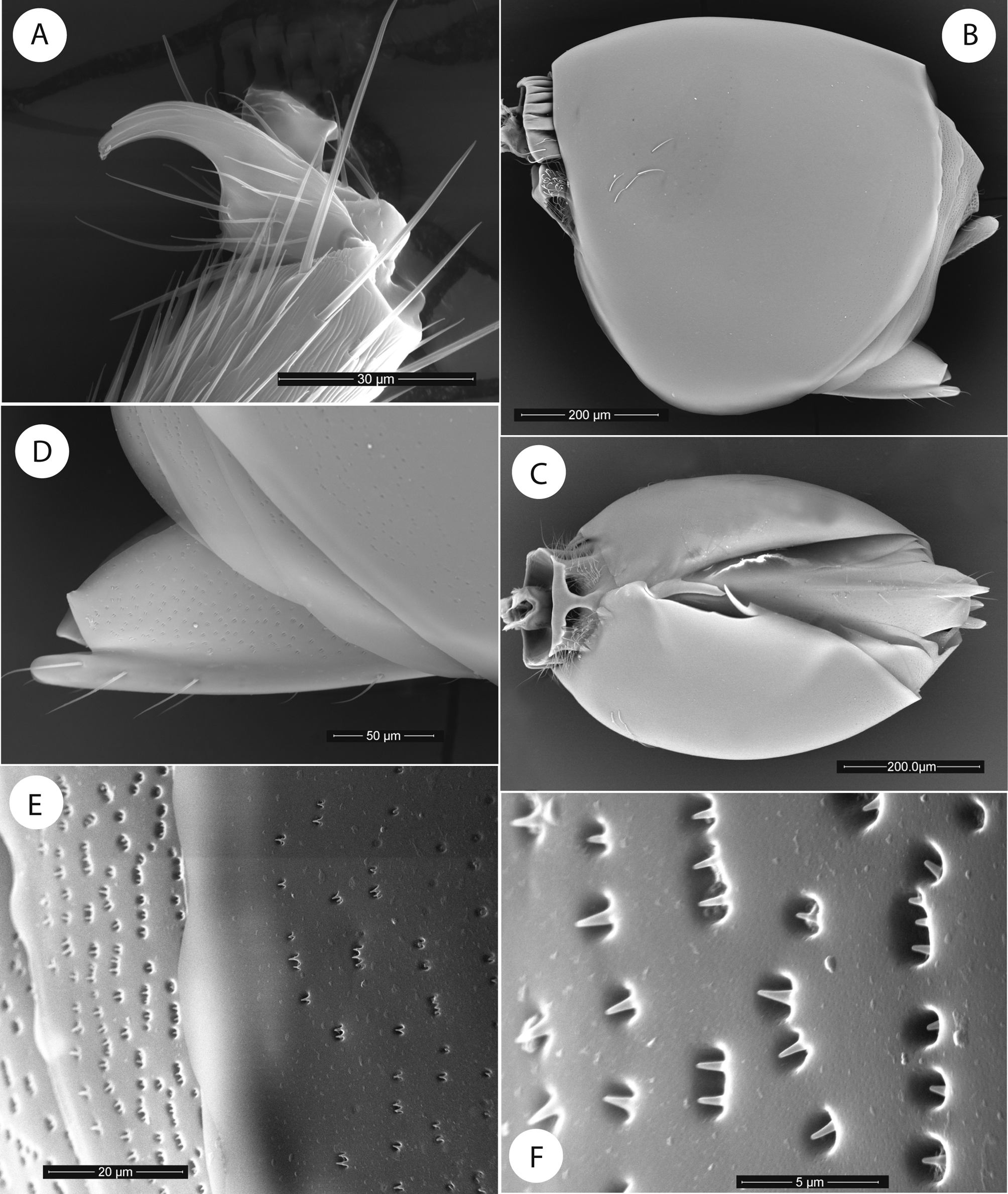
Legs. Metatarsal claw simple, without basal acute lobe or tooth ( Fig. 4 View FIGURE 4 A).
Forewing. Radial cell open along anterior margin; R1 and Rs stopping close to the anterior margin of wing ( Fig. 5 View FIGURE 5 C); R1 forming roughly a 90o angle with anterior margin of wing and R1+Sc; Medial and cubital veins virtually invisible; areolet not present. When visible, the M+Cu1 vein is situated rather higher than apical part of cubital vein (Cu1a). Apical margin of wing with a fringe of long setae ( Fig. 5 View FIGURE 5 C).
Metasoma ( Fig. 4 View FIGURE 4 B). T1 only half ring shaped dorsally; strongly sulcate. T2+3 covering almost the entire metasoma; smooth and shining, except for a narrow posterior band with micropunctures present ( Figs 4 View FIGURE 4 E, 4F). Projecting part of hypopygial spine short, 1.5 times as long as high in profile ( Figs 4 View FIGURE 4 C, 4D); hypopygial spine ventrally with two widely spaced rows of long setae.
No known copyright restrictions apply. See Agosti, D., Egloff, W., 2009. Taxonomic information exchange and copyright: the Plazi approach. BMC Research Notes 2009, 2:53 for further explanation.