
Armascirus gimplei Smiley, 1992
|
publication ID |
https://doi.org/ 10.11646/zootaxa.3194.1.1 |
|
DOI |
https://doi.org/10.5281/zenodo.4901705 |
|
persistent identifier |
https://treatment.plazi.org/id/B818C041-FD4A-F31A-EEC0-FA78FA1AFDCD |
|
treatment provided by |
Plazi |
|
scientific name |
Armascirus gimplei Smiley, 1992 |
| status |
|
Armascirus gimplei Smiley, 1992
( Figs. 11–13 View FIGURE 11 )
Armascirus gimplei Smiley, 1992: 139 , fig. 70A, B; Kalúz 2009: 37.
Diagnosis. Armascirus gimplei most closely resembles A. ozarkensis and A. cerris in that it has a small hysterosomal (median) shield that is not complemented with dorsal setae and has lateral platelets. It can be differentiated from A. cerris because it has 6 setae after coxae II (not including coxal, genital and anal setae) instead of 7. It can be differentiated from A. ozarkensis based on the lateral platelets, which are conspicuous and as long as the median shield in A. ozarkensis and inconspicuous and only as long as or slightly longer than c 2 in A. gimplei .
Remarks. After examining both the holotype and the newly collected specimen, differences between the original description and the specimens were found. Smiley (1992) states that the lateral hysterosomal platelets of A. gimplei are small and that c2 is located on the platelets. In reality the platelets are small and inconspicuous, but occur on the integument between d1 and c2 much as they do in other Armascirus . The structures previously reported as the lateral platelets are the same tiny platelets that occur at the base of all dorsal setae that are situated in the integument away from larger plates and shields.
The integument around the setae laterad of coxae III appears to be more sclerotized than the surrounding cuticle. This area does not bear the reticulated pattern of the coxal or dorsal plates. The structure is not visible in the holotype so we were unable to determine if it is present across the species or an anomaly of the specimen examined.
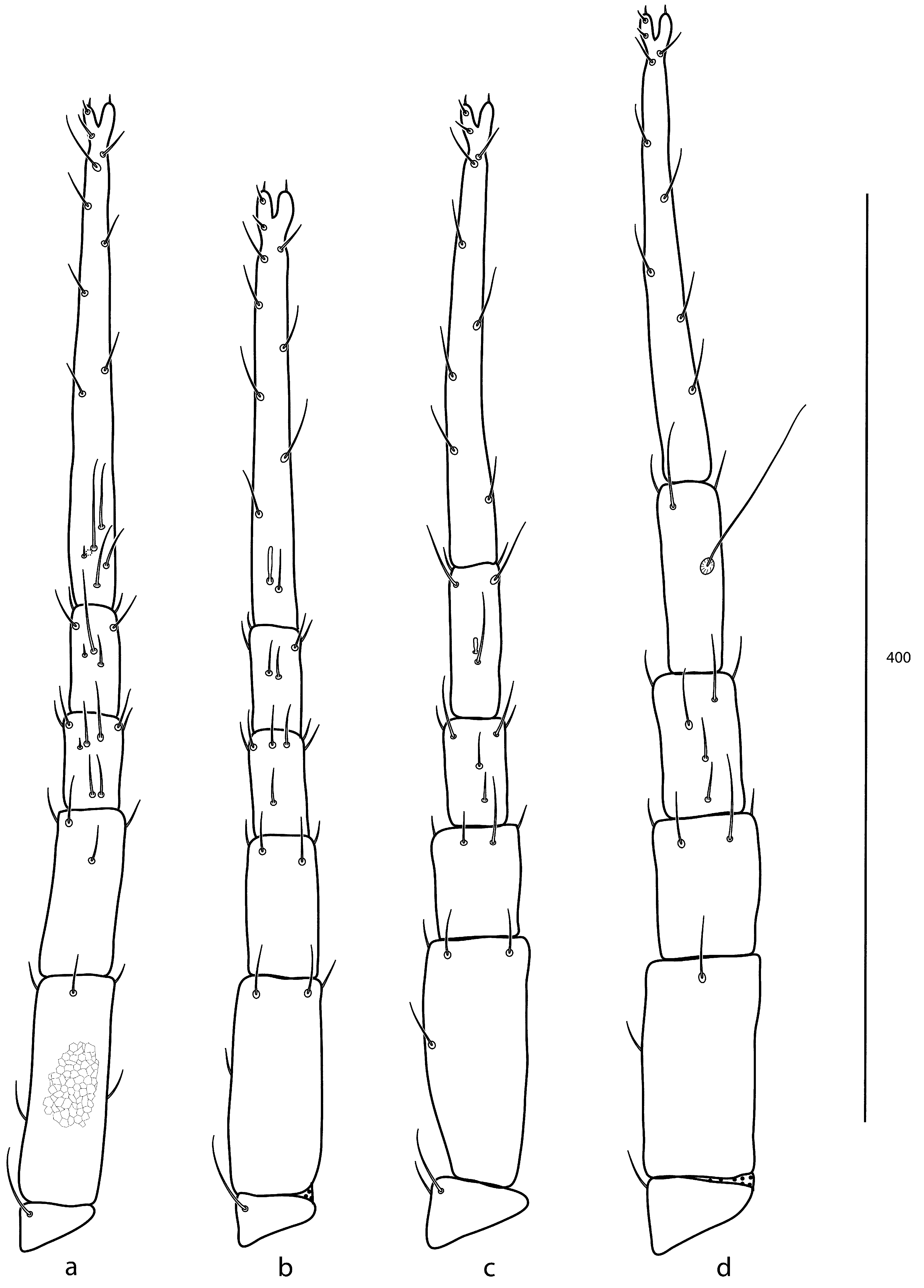
In addition, differences between the new specimen and the published leg setal formulae were found as follows: tibiae I with 2 asl, 1 mst, 4 sts; tibiae III with 1 bsl, 5 sts. Unfortunately the holotype is in a poor condition that does not allow these leg segments to be viewed, and therefore these differences cannot be corroborated with the type. Subcapitulum ( Fig. 11 View FIGURE 11 a), palp ( Fig. 11 View FIGURE 11 b), chelicera ( Fig. 11 View FIGURE 11 c), The idiosoma (12a, b) and legs ( Fig. 13 View FIGURE 13 a–d) have been illustrated based on the Ozark specimen to aid in identification.
Material examined (2 individuals on slides). Female holotype, ex. Tillandsia sp., Mexico, Vera Cruz. 6 April 1966, coll. J. T. Watt. ● 1 female ( APGD 10-0730-005), ex. mixed cedar and deciduous litter, USA, Arkansas, Newton Co., Buffalo National River, Steel Creek (36° 01.924 N, 093° 20.040 W), 30 July 2010, by M. J. Skvarla.

No known copyright restrictions apply. See Agosti, D., Egloff, W., 2009. Taxonomic information exchange and copyright: the Plazi approach. BMC Research Notes 2009, 2:53 for further explanation.
|
Kingdom |
|
|
Phylum |
|
|
Class |
|
|
Order |
|
|
SubOrder |
Prostigmata |
|
Family |
|
|
Genus |
Armascirus gimplei Smiley, 1992
| Skvarla, Michael J. & Dowling, Ashley P. G. 2012 |
Armascirus gimplei
| Kaluz 2009: 37 |
| Smiley 1992: 139 |