
Hydrodynastes bicinctus ( Hermann, 1804 )
|
publication ID |
https://doi.org/ 10.11646/zootaxa.4007.1.4 |
|
publication LSID |
lsid:zoobank.org:pub:9BF26A2B-3D17-4F35-9690-A2B2874579BD |
|
DOI |
https://doi.org/10.5281/zenodo.6122761 |
|
persistent identifier |
https://treatment.plazi.org/id/B82CC024-B910-B858-FF59-FB6AC3514315 |
|
treatment provided by |
Plazi |
|
scientific name |
Hydrodynastes bicinctus ( Hermann, 1804 ) |
| status |
|
Hydrodynastes bicinctus ( Hermann, 1804)
Figs. 3–5 View FIGURE 3 View FIGURE 4 View FIGURE 5
Coluber bicinctus Hermann 1804 . Observationes Zoologicae:276.
Elaps schrankii Wagler in Spix 1824. Serpentum brasiliensium species novae:1. Xenodon bicinctus — Schlegel 1837. Essai sur la physionomie des serpens:95. Hydrodynastes schrankii — Fitzinger 1843. Systema Reptilium:25.
Liophis bi-cinctus — Duméril, Bibron and Duméril 1854. Erpétologie générale vol. 7: 716. Lejosophis bicinctus — Jan 1863. Archives of Zoology, Anatomy and Fisiology:324. Cyclagras bicinctus — Cope 1885. Proceeding of the American Philosophical Society:185. Urotheca bicincta — Boulenger 1894. Catalologue of the snakes in the British Museum vol. 2: 184 Dugandia bicincta — Dunn 1944. Caldasia:70.
Hydrodynastes bicinctus — Hoge 1958. Papéis Avulsos de Zoologia:222.
Hydrodynastes bicinctus bicinctus — Hoge 1966. Ciência e Cultura:143.
Hydrodynastes bicinctus schultzi — Hoge 1966. Ciência e Cultura: 143. New synonymy.
Holotype. Not designated in the original description.
Neotype ( Fig. 4 View FIGURE 4 ). Adult male, MPEG 24628, collected on 24 November 2005 by M.S. Hoogmoed, M.A. Ribeiro Jr., and C. Oliveira Araújo, in the municipality of Novo Progresso (07o02’25’’S, 55o24’55’’W, about 240m above sea level), state of Pará, Brazil.
Diagnosis. Hydrodynastes bicinctus is distinguished from its congeners by the following combination of states of characters: dorsal scales usually in 19/19/15 rows; ventral scales 164–180 in females and 154–179 in males; subcaudals 60–85 in females and 63–93 in males; prediastemal teeth 11–13; no apical pits; dorsum of body brown with darker saddle-shaped blotches; venter checkered with black and cream; postocular stripe “C” shaped, reaching gular region.
Comparisons. Hydrodynastes bicinctus is distinguished from H. gigas and H. melanogigas by having 11–13 prediastemal maxillary teeth (vs. 14–17 in H. gigas and H. melanogigas ) and lacking apical pits (vs. apical pits present in the other two species). Additionally, H. bicinctus has a "C" shaped postocular stripe reaching gular region (vs. postocular stripe longitudinally extended but not reaching gular region in H. gigas and no distinct postocular stripe in H. melanogigas ); venter with checkered pattern (vs. venter composed by black dots distributed in two longitudinal lines along lateral regions in H. gigas and H. melanogigas ).
Description of the neotype ( Fig. 4 View FIGURE 4 ). Adult male, SVL 1280 mm; TL 355 mm (27.7% SVL); head length 42.8 mm (3.3% SVL) from tip of snout to quadrate articulation; broadest head width 28.8 mm; interocular distance 13.9 mm; snout-orbit distance 10.6 mm (0.76 times interocular distance); rostral visible from above; two internasals 0.95 times as broad as high; right prefrontal 0.98 and left prefrontal 0.92 times as long as broad; frontal 1.19 times as long as broad, pentagonal-shaped in dorsal view; each parietal 1.2 times as long as wide; nasal divided; loreal pentagonal-shaped, 1.4 times as long as high; eye diameter 4.2 mm; pupil rounded; three suboculars, third larger than others; preocular single, two postoculars, and supraocular single; temporal 2+2+3/2+2+3; eight supralabials, none contacting orbit; ten infralabials, first to sixth contacting chin shields, except for the fifth scale on right side; three pairs of chin shields, anterior pair smaller than others; gular scale rows six between first ventral and infralabials; thirteen prediastemal maxillary teeth on right side and eleven on left side; two enlarged postdiastemal teeth on each side; dorsal scales 19/19/15, smooth, with no apical pits; ventrals 171; cloacal shield single; subcaudals 85, paired and unkeeled.
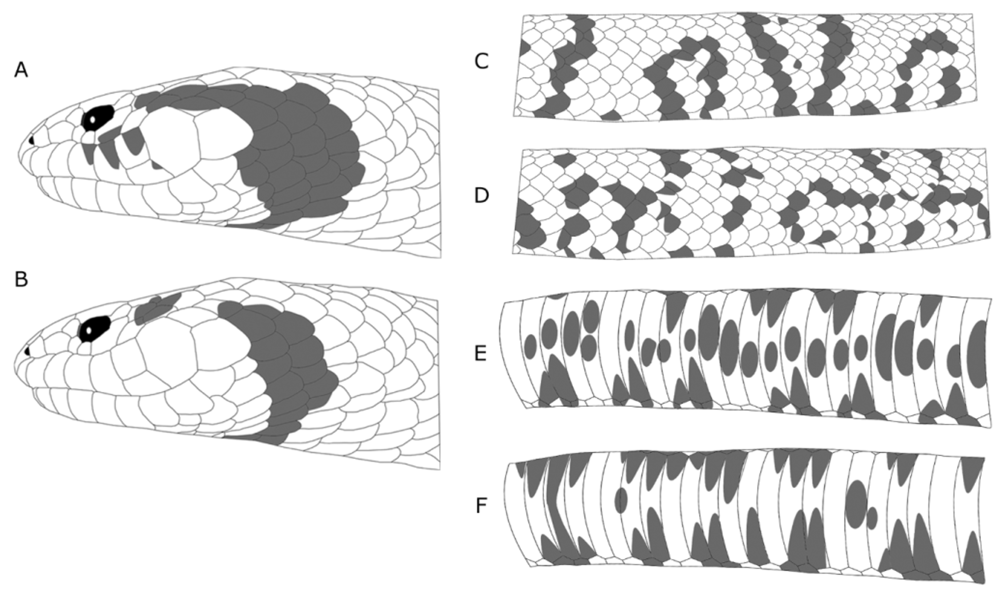
Color of the neotype in preservation (ethanol 70%) ( Fig. 4 View FIGURE 4 ). Head dark brown; two small black spots in anteromedial portion of parietals; two black spots (one on each side) on the level of third scale after parietals; black “C” shaped postocular stripe reaching gular region; supralabials brown; infralabials and chin shields creamishwhite; ventral surface of head with two black blotches (one on each side), on the level of second scale before first ventral scale; dorsum of body brown with 30 dark brown, saddle-shaped blotches reaching the venter, alternating with well-defined dark brown rounded lateral blotches, also reaching venter; ventrals creamish-white with black spots irregularly distributed in a checkered pattern.
Color pattern variation of adults in preservation (ethanol 70%) ( Fig. 1 View FIGURE 1 ). Two basic patterns of dorsal coloration were observed, the “Cerrado pattern” and the “Amazon pattern”. In some individuals the postocular stripe is divided, with a small black postocular dot not connected to the “C” shaped stripe ( Fig. 1 View FIGURE 1 B). Venter often with dots reaching the lateral portion of the body ( Fig. 1 View FIGURE 1 F).
Juvenile color pattern in preservation ( Fig. 5 View FIGURE 5 ). Juveniles with dorsum of body brown, dark brown saddleshaped dorsal blotches bordered by pale lines (these lines are usually vestigial or absent in adults). Rounded lateral blotches usually well-defined in all individuals, regardless of geographic origin. Postocular stripe frequently divided into a small black postocular dot not connected to the “C” shaped stripe.
Morphometric and meristic variation. Largest female SVL 1960 mm and TL 575 mm (IBSP 13824; TL/ SVL = 0.29); largest male SVL 1930 mm and TL 530 mm (MNRJ 4741; TL/SVL = 0.27); TL/SVL 23–38% for females and 23–39% for males; dorsal scales row formula 17/17/15 (n = 1), 19/15/13 (n = 1), 19/15/14 (n = 1), 19/ 15/15 (n = 10), 19/16/15 (n = 1), 19/17/13 (n = 5), 19/17/14 (n = 2), 19/17/15 (n = 35), 19/18/13 (n = 1), 19/18/15 (n = 4), 19/19/13 (n = 1), 19/19/15 (n = 59), 21/17/15 (n = 2), 21/18/15 (n = 1), or 21/19/15 (n = 8); ventral scales in females 164–180 (mean = 172. 2; s = 4.2; n = 48), males 154–179 (mean = 170.1; s = 2.7; n = 81); subcaudals in females 60–85 (mean = 74.1; s = 5.5; n = 38), males 63–93 (mean = 80.9; s = 6.3; n = 70); usually single preocular, three suboculars, and two postocular scales (n = 78); temporal scale formula extremely variable, with 51 distinct combinations; supralabials 7 (n = 1), 8 (n = 111), 9 (n = 24) or 10 (n = 1), usually with different numbers on each side; infralabials 9 (n = 3), 10 (n = 62), 11 (n = 62) or 12 (n = 3), generally with different numbers on each side; maxillary teeth 13–15.
Hemipenis (n = 2, Fig. 6 View FIGURE 6 ). Organ deeply bilobed, lobes occupying about ¼ of total length of hemipenis. Sulcus spermaticus divides near the base of organ, adopting a centrolineal position until proximal region of lobes, where branches diverge to a centrifugal position, ending on lateral tip of lobes. Organ semicapitate and semicalyculate. Capitulum formed by papillate calyces on the sulcate and papillate calyces and body calyces on the asulcate side. Hemipenis with large, calcified lateral spine rows, beginning at the level of bifurcation of sulcus spermaticus and extending along apical portion of hemipenial body, immediately proximal to the beginning of lobes, lateral to asulcate side. Hemipenial body covered with spinules on both sides.
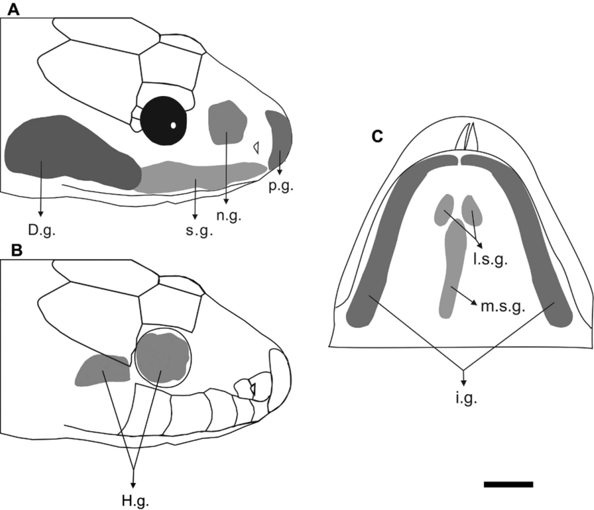
Cephalic glands (n = 2, Fig. 7 View FIGURE 7 ). Premaxillary gland: triangular, contacting anterior region of supralabial glands. Nasal glands: pentagonal, limited by prefrontal bone posteriorly and by nasal capsule anteriorly; the dorsal limit of the nasal glands goes beyond the nostril and dorsal margin of orbit. Supralabial glands: slender glands contacting premaxillary gland anteriorly and rear end overlapped by Duvernoy’s gland. Harderian glands: orbital and postorbital lobes about same size; orbital lobes rounded, completely filling orbit space; postorbital lobes with irregular rectangular shape, showing a constriction on medial portion, limited by musculus adductor mandibulae externus medialis (amem) posteriorly and overlapped by Duvernoy’s gland ventrally. Duvernoy’s glands: large, elliptical, overlapping posterior region of supralabial glands anteriorly, and limited posteriorly by musculus adductor mandibulae externuns superficilais (ames). Rictal glands: absent or indistinct. Infralabial glands: cover lateral portions of ventral surface of head, reaching the level of amem; mesoposterior portion enlarged; anterior portion of infralabials glands touch each other in the region adjacent to symphysial. Sublingual glands: medial sublingual gland elongated, positioned between lateral sublingual glands, and not extending beyond posterior limit of infralabial glands; lateral sublingual glands ellipsoidal, not in contact. Supralingual gland: absent or indistinct.
Skull (n = 2, Figs. 8–10 View FIGURE 8 View FIGURE 9 View FIGURE 10 ). SNOUT: Premaxilla ( Figs. 8–9 View FIGURE 8 View FIGURE 9 ): triangular in frontal view with slender transverse process slightly oblique ventrally, not reaching anterior portion of maxilla; basal portion of ascending process large and apical portion straight, slightly narrower than base; ascending process not touching nasals; vomerine processes divergent in ventral view overlapping vomer. Septomaxillae ( Fig. 9 View FIGURE 9 ): positioned ventral to nasals, dorsal to vomers and posterior to premaxilla; anterior edge simple embedded in the angle formed by ascending and vomerine processes of premaxilla; along with vomer, septomaxilla forms capsule of vomeronasal organ; anterolateral process oblique dorsally, with rounded edge, not overlapping anterior portion of maxilla or transverse process of premaxilla; septomaxilla contacts nasals laterally. Vomers ( Figs. 8–9 View FIGURE 8 View FIGURE 9 ): positioned anteroventrally in midline of skull, posteriorly to premaxilla; anterior process overlapped by vomerine process of premaxilla; posterior process of vomer with large foramen in ventral view and with vertical portion perpendicular to horizontal portion. Nasals ( Figs. 8–9 View FIGURE 8 View FIGURE 9 ): joined medially and with dorsal position in skull, between premaxilla and frontals; lateral edges more slender than medial edges, with pair of nasals forming a diamond shape; anterior and posterolateral margins slightly concave; nasal with elongated posteroventral process in lateral view (the frontal process of nasal) which contacts vertical lamina of frontals; anteriorly, nasals do not contact ascending process of premaxilla.
BRAINCASE: Frontals ( Figs. 8–9 View FIGURE 8 View FIGURE 9 ): joined medially, positioned dorsally between nasals and parietal; pair of frontals with trapezoidal shape in dorsal view and slightly longer than wide; anterolateral portion with prefrontal process, anterior margin wider than posterior margin; frontals, along with parietal, constitutes internal wall and dorsal border of orbit in lateral view; orbital foramen inserted in region of contact between frontal and parietal; most of the foramen inserted into parietal, occupying less than half of height of orbital internal wall in lateral view. Prefrontals ( Figs. 8–9 View FIGURE 8 View FIGURE 9 ): with irregular shape, contacts frontal dorsally and maxilla ventrally, overlapping palatine process of maxilla, and delimiting orbit anteriorly; on its anterior portion, the bone has an acuminate projection; in frontal view, lacrimal foramen crosses the bone as far as its posterior portion; dorsal to this foramen, a lacrimal process is present. Parietal ( Fig. 8 View FIGURE 8 ): with subtriangular aspect in dorsal view; parietal contacts frontals anteriorly, postorbitals (by mean of a conspicuous postorbital process) anterolaterally, supraoccipital posteriorly, and prootics posterolaterally, where parietal is overlapped by supratemporals; two convergent well-developed dorsolateral crests emerge at the level of postorbital process up to region of contact with supraoccipital; dorsolateral crests not in contact. Postorbitals ( Figs. 8–9 View FIGURE 8 View FIGURE 9 ): with tapered and curved shape; delimits orbital cavity posteriorly with ventral edge not reaching maxilla; extends parallel to anterolateral portion of parietal, not reaching frontals; anterior face concave and posterior convex laterally. Supraoccipital ( Fig. 8 View FIGURE 8 ): bone with irregular shape, projecting toward supratemporals and prootics laterally, and exoccipitals posteriorly; concave edge contacts parietal anteriorly; mesodorsal crest crosses supraoccipital longitudinally and its lateroposterior portion has two transverse crests. Exoccipitals ( Fig. 8 View FIGURE 8 ): bones with irregular shape, positioned at posterior portion of braincase, with anterior limit on supraoccipital and posterior limit adjacent to first vertebra (atlas), comprising dorsolateral edge of foramen magnum; exoccipitals contacting prootics anteriorly and basioccipital ventrally; transverse crests of supraoccipitals extending through exoccipitals, and laterally over the level of fenestra ovalis. Prootics ( Fig. 10 View FIGURE 10 ): contacting parietal anteriorly, exoccipitals posteriorly, and dorsal portion of supraoccipital and ventral portion of basioccipital posterolaterally; supratemporals overlap most of dorsal portion of prootics; lateral ridge divides prootic into dorsal and ventral portions; two foramina allow passage to maxillary and mandibular branches of trigeminal nerve lateroventrally; ventral to these foramina, next to region of contact with parabasisphenoid, there are multiple smaller foramina. Columella auris: paired small bones located on lateroposterior region of braincase, articulating with exoccipitals and prootics through fenestra ovalis; each columella consists of a small discoid footplate inserted in a fenestra ovalis and has an elongated shaft extending toward quadrate. Basioccipital ( Fig. 8 View FIGURE 8 ): bone with subtrapezoidal shape, located on ventral surface of skull and contacting parabasisphenoid anteriorly and atlas posteriorly; anterior margin wider than posterior margin, which has a slight bifurcation; anterior portion with inconspicuous dentigerous process, adjacent to suture with parabasisphenoid; basioccipital with slight longitudinal mesial crest. Parabasisphenoid complex ( Fig. 8 View FIGURE 8 ): composed of parasphenoid and basisphenoid, both fused without visible suture; spear shaped, positioned at mesoventral surface of braincase, with posterior portion broader and anterior portion tapering approximately on the level of posterior portion of palatines; the most tapered portion corresponds to anterior end of parasphenoid; posterior opening of Vidian canal situated posterolaterally, in the suture with parietal, next to prootics.
PALATOMAXILLARY APPARATUS: Maxillae ( Figs. 8–9 View FIGURE 8 View FIGURE 9 ): located on anterolateral portion of skull, not contacting premaxilla; maxilla with elongated and arched shape; lateral portion convex and medial portion concave, extending to the level of postorbitals; palatine process completely overlapped by prefrontals, with broad base, extending from sixth up to ninth teeth, with medial edge narrowing at the level of eighth tooth; posterior edge enlarged in the region of contact with ectopterygoids; each maxilla bears twelve or thirteen curved prediastemal and two enlarged and ungrooved postdiastemal teeth (aglyphous); diastema equals size of one alveolus; anterior maxillary teeth smaller. Palatines ( Figs. 8–9 View FIGURE 8 View FIGURE 9 ): elongated and narrow, located on the mesoventral portion of skull, contacting vomer-septomaxilla complex anteriorly, maxillae laterally, and pterygoids posteriorly; lateral maxillary process overlapped by prefrontal, slightly anterior to choanal process, extending from fourth to sixth teeth; maxillary process with wide base and straight edge; choanal process extending from sixth to eighth teeth, close to parabasisphenoid, but not reaching it; posterior portion with small bifurcation where palatine articulates with anterior portion of pterygoid; each palatine bears ten or eleven curved teeth of nearly equal size. Pterygoids ( Fig. 10 View FIGURE 10 ): bones located medially on ventroposterior region of skull, elongated, tapered anteriorly, with length more than 50% of skull length; pterygoid contacts palatine anteriorly, ectopterygoid mesolaterally, and quadrate-compound bone joint posteriorly; anterolateral portion of pterygoid articulates with ectopterygoid at the level of seventh to fifteenth teeth; pterygoid becomes broader anteroposteriorly, up to the end of the row of teeth, where the bone tapers, directing laterally on the posterior portion; mesoposterior portion with dorsolateral longitudinal crest emerging just after joint with ectopterygoid and extending to its posterior edge; each pterygoid has fifteen to eighteen curved teeth of nearly equal size. Ectopterygoids ( Fig. 10 View FIGURE 10 ): located on the mesolateral portion of skull, elongated, with anterior edge bifurcated contacting posterior portion of maxilla and posterior portion contacting pterygoid, which has approximately two times the length of ectopterygoid.
SUSPENSORIUM AND MANDIBLE: Supratemporals ( Fig. 10 View FIGURE 10 ): elongated, located on the posterodorsal portion of skull with anterior portion slightly wider than posterior portion; contacts parietal and prootic anteriorly, overlapping much of dorsal portion of prootic; posterior end extends beyond braincase, reaching the level of rear end of atlas. Quadrates ( Fig. 10 View FIGURE 10 ): well-developed bones located on the posterolateral portion of skull, lateral to supratemporals; articulates with supratemporal dorsally and glenoid cavity of compound bone ventrally, representing quadrate-articular joint; quadrate has about same length of supratemporal, elongated shape, and dorsal portion wider than ventral portion; anterolateral crest toward anterior portion of skull present; longitudinal and oblique rod-shaped process on posterior edge. Dentaries ( Fig. 10 View FIGURE 10 ): elongated, located on anterior edges of mandibles; posteroventral portion contacts dorsal surface of splenial and anterodorsal edge of angular in medial view; anterior portion medially curved toward opposite hemimandible; posterior edge bifurcated in lateral view, forming dorsal and ventral processes of dentary, between which the anterior edge of compound bone fits; dorsal process slightly longer than ventral; dentary forms—along with splenial—the Meckel groove; fifteen to seventeen curved teeth with nearly equal size distributed from anterior edge to the end of dorsal process; lateral face with a foramen on the level of seventh tooth. Splenials ( Fig. 10 View FIGURE 10 ): triangular with anterior edge tapered and posterior end vertical; in medial view, contacts posteroventral portion of dentary; mylohyoid anterior foramen close to posterior joint with angular. Angulars ( Fig. 10 View FIGURE 10 ): triangular, located on medial face, with anterior edge vertical and posterior end tapered, exceeding the level of posterior edge of dorsal process of dentary; anterior portion of angulars with mylohyoid posterior foramen; anterodorsal portion contacts posterior portion of dentary; angular contacts posterior portion of splenial anteriorly, and it is completely overlapped by anteroventral portion of compound bone dorsally; joint with splenial visible in medial and ventral views. Compound Bones ( Fig. 10 View FIGURE 10 ): corresponding to the largest bone of the mandible; contacts dentary, splenial, and angular anteriorly, and rear end articulates with quadrate; elongated, with narrow anterior portion fitting between dorsal and ventral process of dentary; posterior portion with glenoid cavity; after glenoid cavity, rear end of compound bone with narrow projection; articular and surangular crests well developed, first crest higher than second, lying anterior to glenoid cavity; posterior orifice of inferior dental canal located on anterior portion of a large cavity between articular and surangular crests; small crest on the posterior portion of compound bone in lateral view.
Geographic distribution ( Fig. 2 View FIGURE 2 ). Hydrodynastes bicinctus occurs in the Amazon, Tocantins, Paraná and Atlantic, stretch North/Northeast, hydrographic basins, at Amazon rainforest and Cerrado open formations, through Colombia (Vaupés, Dunn 1944; Orinoco and Amazonas Rivers, Pérez-Santos and Moreno 1988), Guyana (no specific locality, Boulenger 1894; Onora Falls, Cole et al. 2013), Suriname (no specific locality, Duméril et al. 1854; Kwamalasemoetoe, Abuys 2003; Sipaliwini, Avila-Pires et al. 2010), Venezuela (Caracas, Jan 1863; between Guaramaco and San Fernando, and San Carlos, Negro River, Roze 1966), French Guiana (Amazon basin and Guianas Shield, Chippaux 1987), and Brazil (in the states of Amazonas, Amapá, Goiás, Maranhão, Mato Grosso do Sul, Mato Grosso, Minas Gerais, Pará, Rondônia, São Paulo, and Tocantins).


| MPEG |
Museu Paraense Emilio Goeldi |
No known copyright restrictions apply. See Agosti, D., Egloff, W., 2009. Taxonomic information exchange and copyright: the Plazi approach. BMC Research Notes 2009, 2:53 for further explanation.
|
Kingdom |
|
|
Phylum |
|
|
Class |
|
|
Order |
|
|
Family |
|
|
Genus |
Hydrodynastes bicinctus ( Hermann, 1804 )
| Murta-Fonseca, Roberta A., Franco, Francisco L. & Fernandes, Daniel Silva 2015 |
Coluber bicinctus
| Hermann 1804 |