
Melledobythus, Hlaváč, Peter, Nakládal, Oto & Jalžić, Branko, 2014
|
publication ID |
https://doi.org/ 10.11646/zootaxa.3835.4.7 |
|
publication LSID |
lsid:zoobank.org:pub:2913D41F-E4A7-4550-A908-5699DA7E481B |
|
DOI |
https://doi.org/10.5281/zenodo.6138911 |
|
persistent identifier |
https://treatment.plazi.org/id/B93A87DF-E65B-0C76-FF69-76CDFD4AF801 |
|
treatment provided by |
Plazi |
|
scientific name |
Melledobythus |
| status |
gen. nov. |
Melledobythus View in CoL gen. nov.
Type species. Melledobythus bilandzijae sp. n. Gender masculine.
Etymology. The name is the combination of Melleda, ancient name of the Island Mljet and “bythus” reflecting the affiliation of the genus to the tribe Bythinini .
Diagnosis. Small, anophthalmous Bythinini with eleven-segmented antennae, scape long and slender, antennomere III smaller than pedicel, palpomere IV slightly longer than wide, pronotum with two lateral foveae connected by antebasal sulcus, elytra lacking sutural striae.
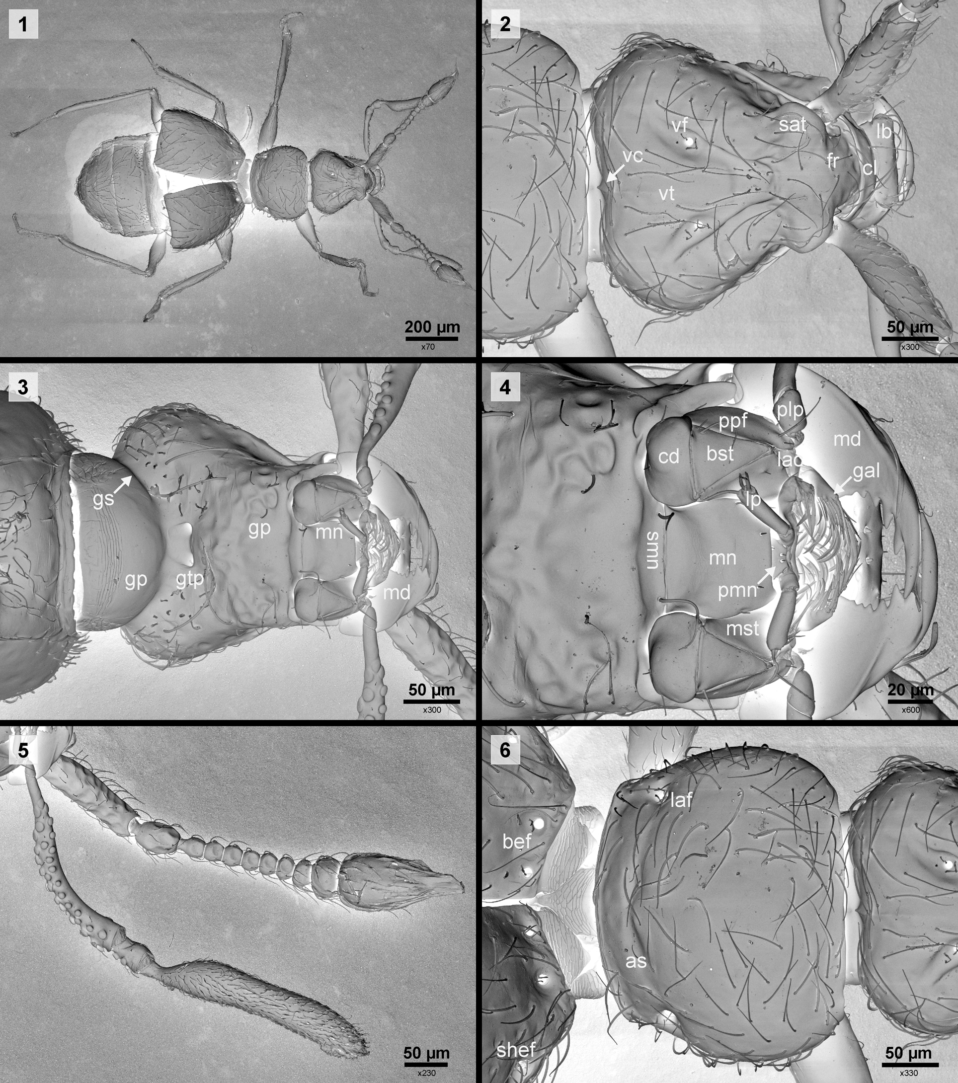
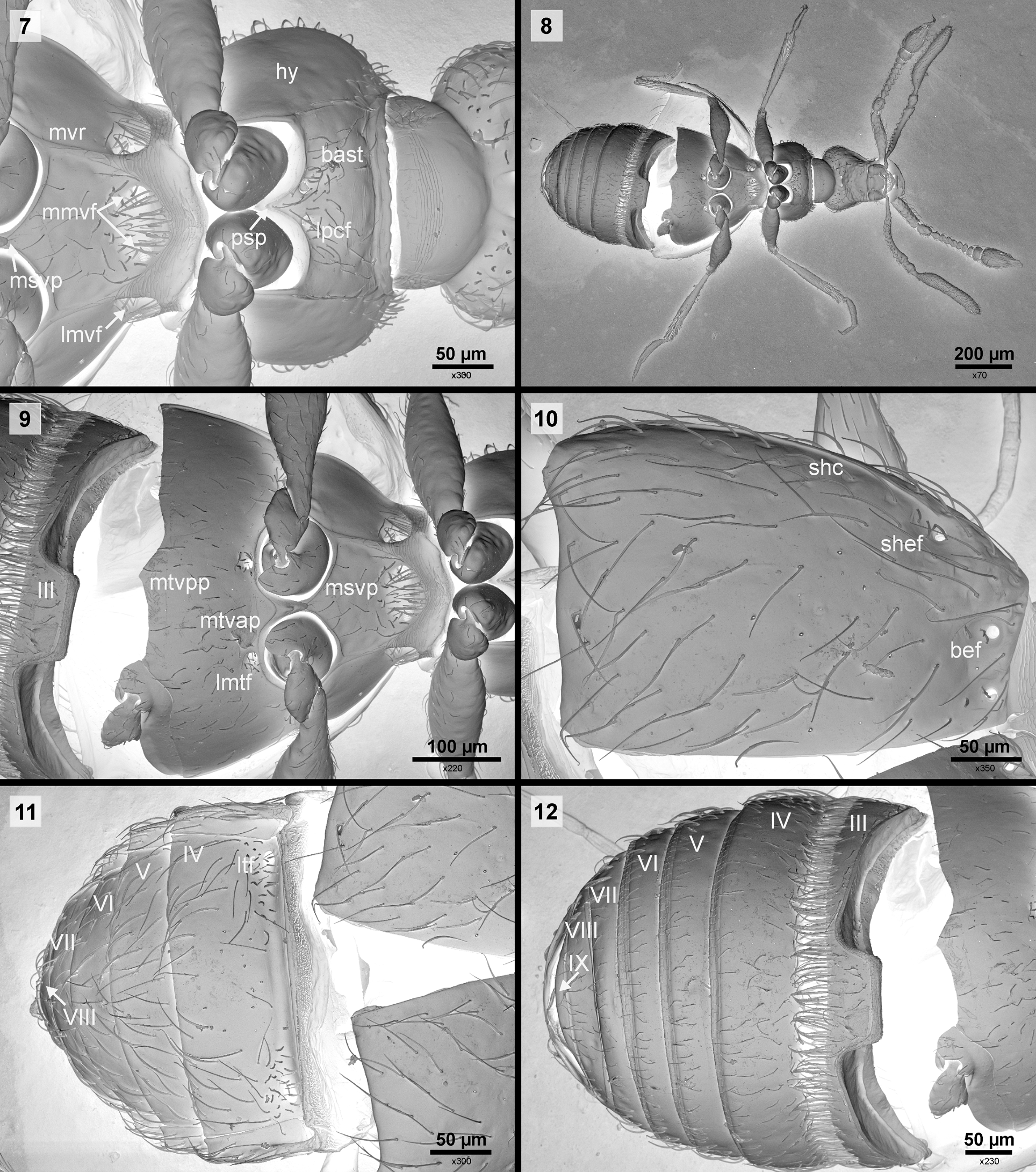
Description. Body ( Fig. 1 View FIGURES 1 – 6 ) shiny, light reddish-brown, with long, sparse setation on whole body. Head triangular, about as long as wide, eyes completely atrophied, supra-antennal tubercles ( Fig. 2 View FIGURES 1 – 6 , sat) prominent, frontal fovea absent, vertexal foveae ( Fig. 2 View FIGURES 1 – 6 , vf) well-defined, vertexal carina ( Fig. 2 View FIGURES 1 – 6 , vc) short, clypeus ( Fig. 2 View FIGURES 1 – 6 , cl) short, anterior margin rounded, with two setae, labrum ( Fig. 2 View FIGURES 1 – 6 , lb) rhombic, expanded anteriorly with four long setae on each side. Mandibles ( Fig. 3, 4 View FIGURES 1 – 6 , md) subtriangular and almost symetrical, with 5–6 teeth and sharply pointed apex. Maxilla with subrectangular cardo and triangular basistipes ( Fig. 4 View FIGURES 1 – 6 , cd, bst), lacinia and galea ( Fig. 4 View FIGURES 1 – 6 , lac, gal) short, palpifer ( Fig. 4 View FIGURES 1 – 6 , ppf) long, about twice as long as palpomere I, with one long and one short apical seta; maxillary palpi ( Fig. 5 View FIGURES 1 – 6 ) four-segmented, palpomere I miniscule ( Fig. 4 View FIGURES 1 – 6 , plp), palpomeres II and III with many distinct granules, II very long, pedunculate at base, widest at apex, III slightly longer than wide, terminal palpomere pedunculate at base, with short dense setation, parallel-sided, tapered at apex. Labium ( Fig. 4 View FIGURES 1 – 6 ) with short and transverse submentum ( Fig. 4 View FIGURES 1 – 6 , smn), mentum ( Fig. 4 View FIGURES 1 – 6 , mn) large, longer than wide, prementum ( Fig. 4 View FIGURES 1 – 6 , pmn) short, labial palpi ( Fig. 4 View FIGURES 1 – 6 , lp) two-segmented, palpomere II more than three times as long as I, with about three apical setae of different length. Gular plate ( Fig. 3 View FIGURES 1 – 6 , gp) large with well-defined gular sutures ( Fig. 3 View FIGURES 1 – 6 , gs) demarcating ventrally 'neck region' from anterior part of head, two gular tentorial pits ( Fig. 3 View FIGURES 1 – 6 , gtp) fused. Antennae ( Fig. 5 View FIGURES 1 – 6 ) short, reaching base of elytra, antennal club 3-segmented, scape about three times as long as wide and more than twice as long as pedicel, antennomere III much smaller than pedicel, III–VII subequal in size, VIII slightly more transverse, IX strongly transverse, terminal antenomere about as long as five preceding. Pronotum ( Fig. 6 View FIGURES 1 – 6 ) wider than long, lateral antebasal foveae ( Fig. 6 View FIGURES 1 – 6 , laf) well-defined and connected by shallow but well-defined antebasal sulcus ( Fig. 6 View FIGURES 1 – 6 as). Prosternum ( Fig. 7 View FIGURES 7 – 12 ) with long basisternal part ( Fig. 7 View FIGURES 7 – 12 , bast), prosternal intercoxal process ( Fig. 7 View FIGURES 7 – 12 , psp) long and sharp, median procoxal fovea absent, two, closely located lateral procoxal foveae ( Fig. 7 View FIGURES 7 – 12 , lpcf) well-defined, hypomera ( Fig. 7 View FIGURES 7 – 12 , hy) long, elongate and large, hypomeral ridge absent, replace by line of about 6 setae. Mesoventrite ( Figs. 7, 8 View FIGURES 7 – 12 ) slightly longer than metaventrite, with two median mesoventral foveae ( Fig. 7 View FIGURES 7 – 12 , mmvf) located in large depression and two large lateral mesoventral foveae ( Fig. 7 View FIGURES 7 – 12 , lmvf), mesoventral ridge ( Fig. 7 View FIGURES 7 – 12 , mvr) separating median and lateral parts of mesoventrite, mesocoxae separated by meso and metaventral processes ( Fig. 9 View FIGURES 7 – 12 , mtvpp). Metaventrite ( Fig. 9 View FIGURES 7 – 12 ) strongly transverse, with long median anterior metaventral process ( Fig. 9 View FIGURES 7 – 12 , mtvap) and with two lateral metaventral foveae ( Fig. 9 View FIGURES 7 – 12 , lmtf) located in middle of mesocoxal sockets, median posterior metaventral proces ( Fig. 9 View FIGURES 7 – 12 , mtvpp) wide, moderately concave.
Elytra ( Fig. 10 View FIGURES 7 – 12 ) longer than pronotum, each elytron with two basal ( Fig. 6 View FIGURES 1 – 6 , 10 View FIGURES 7 – 12 , bef), one subhumeral ( Fig. 6 View FIGURES 1 – 6 , 10 View FIGURES 7 – 12 , shef) fovea and subhumeral carina ( Fig. 10 View FIGURES 7 – 12 , shc), lacking sutural stria.
Abdomen ( Fig. 11, 12 View FIGURES 7 – 12 ) narrower than elytra, first visible tergite (IV) slightly less than twice as long as second (V), tergites V–VIII, each shorter than preceeding one, tergite IV with well-defined lateral tergal foveae ( Fig. 11 View FIGURES 7 – 12 , ltf), paratergites IV–VI present. First visible sternite (III) at posterior margin with dense, long setae, second visible sternite (IV) about as long as V and VI combined, VII in middle shorter than VIII.
Legs relatively slender and long, with all tibiae simple, posterior tibiae slightly curved in apical third.
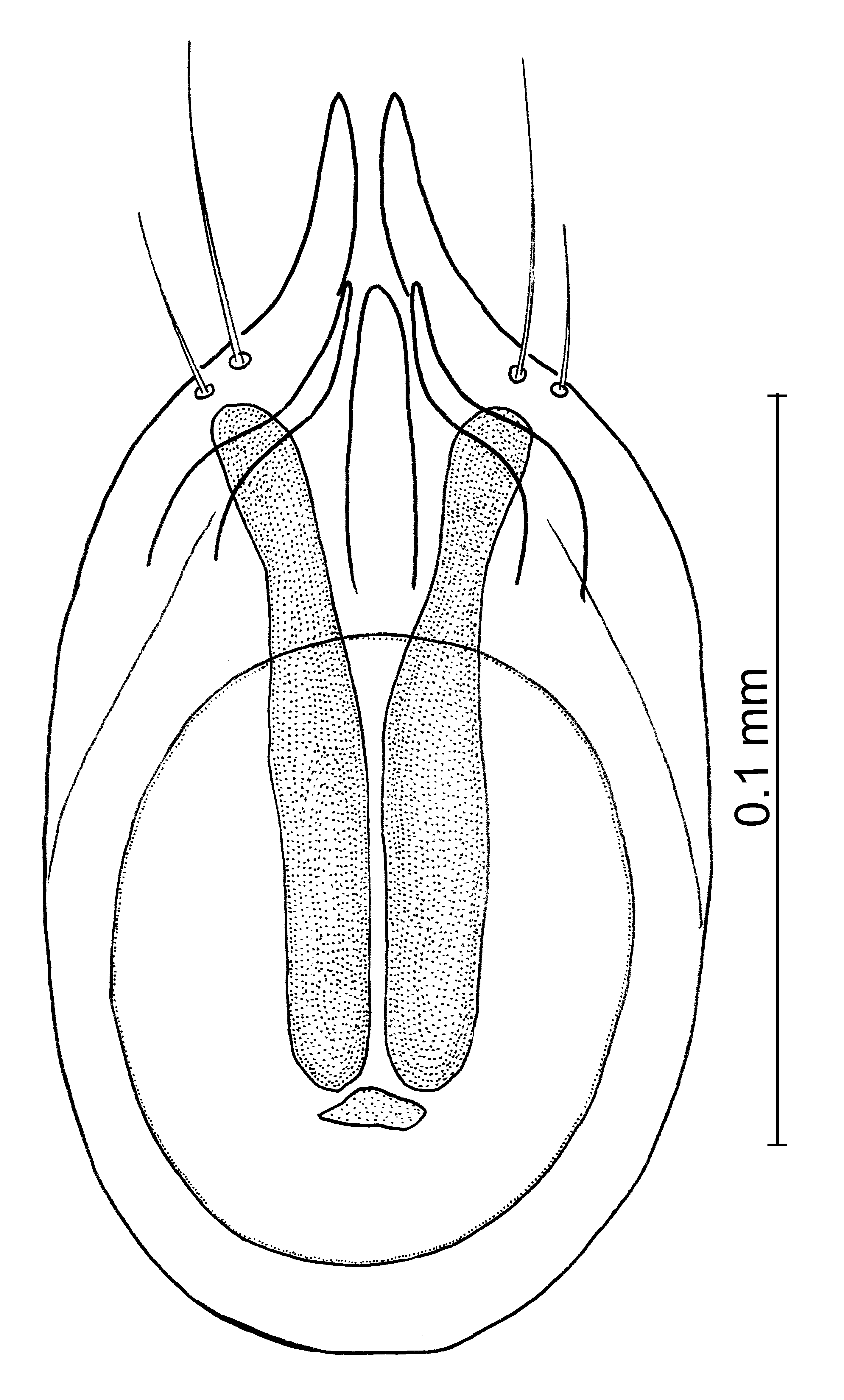
Aedeagus ( Fig. 13 View FIGURES 13 ) elongate, symmetrical in dorso-ventral view, parameres pointed and separated at apex, bearing setae, endophallous with sclerotized corpuscles.
Sexual dimorphism: Not apparent.
Differential diagnosis. At first glance Melledobythus resembles genera Tychobythinus Ganglbauer, 1896 and Gasparobythus Poggi, 1992 . From Tychobythinus it is easily separated by the lack of sutural striae, always welldefined in Tychobythinus . From Gasparobythus it differs by having a well-defined pronotal antebasal sulcus that is absent in Gasparobythus , and by the long terminal maxillary palpomeres that are longer than the head, being always shorter than the head in Gasparobythus .
No known copyright restrictions apply. See Agosti, D., Egloff, W., 2009. Taxonomic information exchange and copyright: the Plazi approach. BMC Research Notes 2009, 2:53 for further explanation.