
Parmulodes verrucosus Wilson, 1944
|
publication ID |
https://doi.org/10.11646/zootaxa.4612.2.7 |
|
publication LSID |
lsid:zoobank.org:pub:F07FA574-675E-4F90-8466-E64E1C29127E |
|
persistent identifier |
https://treatment.plazi.org/id/BB3E87F9-5172-FFD6-FF55-FBCCFC9C5DB1 |
|
treatment provided by |
Plazi |
|
scientific name |
Parmulodes verrucosus Wilson, 1944 |
| status |
|
Parmulodes verrucosus Wilson, 1944
( Figs 4–6 View FIGURE 4 View FIGURE 5 View FIGURE 6 )
Material examined. Holotype ♀ ( USNM 79000 About USNM ), collected in tidal pools of coral reefs, Key Matecumbe , Florida, USA, July, 1925.
Description of female. Body length (excluding caudal setae) 1,124 µm and body width 841 µm. Body with prosomal shield flattened ( Fig. 4A View FIGURE 4 ). Pedigerous somite 1 fused to cephalosome with slightly projected margins. Pedigerous somite 2 narrower than others. Pedigerous somite 3 and 4 fused forming posterior shield recovering urosome. Margins of cephalosome and fused pedigerous somites 3 and 4 with digitiform bands ( Fig. 4B View FIGURE 4 ).
Urosome 4-segmented ( Fig. 4C View FIGURE 4 ). Genital double-somite 150 µm long and maximum width 106 µm, length: width ratio = 1.4:1, anteriorly enlarged, with set of small setules after genital apertures which are located anterolaterally. Two postgenital somites, both wider than long, 56 × 75, 44 × 66 µm, respectively. Anal somite with anal plate on medial-posterior portion. Caudal rami short ( Fig. 4D View FIGURE 4 ), 62 × 47 µm; Length: width ratio 1.3: 1 µm, armed with six setae; seta I absent.
Antennule ( Fig. 4E View FIGURE 4 ) slender, 335 µm long (not including setae), and 17-segmented. Length of segments measured in proximal to distal order: 91, 22, 10, 9, 18, 12, 15, 2, 11, 11, 18, 14, 14, 17, 22, 24 and 49 µm, respectively. Segmental homologies and setation as follows: 1(I) - 2; 2(II - III)-4; 3(VI) - 2; 4(V)-2; 5(VI-VII) - 3; 6(VIII) - 2; 7(IX– XII) - 6; 8(XIII)-1; 9(XIV) - I+1; 10(XV) - 2; 11(XVI) - 2; 12(XVII) - 1; 13(XVIII) - 2; 14(XIX) - 1; 15(XX) - 1; 16(XXI) - 1 +ae; 17(XXII–XXVIII) - 11. All setae smooth. Aesthetasc 47 µm long.
Antenna ( Fig. 5A View FIGURE 5 ) 268 µm long (including distal claw); coxa and basis unarmed. Exopod 1-segmented, 52 µm long, with apical seta and 3 small lateral setules. Endopod 3-segmented; first segment 80 µm long, with distal seta and row of setules on outer margin; second segment 15 µm long with subdistal seta; third segment 11 µm long, ornamented with row of setules along outer margin, and armed with 2 naked and thin setae, located proximally and subdistally, and 1 distal robust terminal seta, close to distally straight claw with curved tip, 54 µm long, with row of spinules and 4 teeth distally.
Oral cone 580 µm long, corresponding to half the length of the body, club-shaped, distal part sharp, reaching insertion between of legs 3 and 4 ( Fig. 4A View FIGURE 4 ). Mandible comprising stylet 319 µm long and slender 2-segmented palp measuring 82 and 27 µm long, respectively ( Fig. 5B View FIGURE 5 ). Stylet slender and serrated, 480 µm long. Palp with second segment armed with 2 apical, plumose setae, one of them almost twice longer the other. Maxillule bilobed ( Fig. 5C View FIGURE 5 ), inner lobe 94 µm long, armed with 1 smooth and 3 unilaterally plumose setae. Outer lobe 29 µm long, armed with 4 naked setae, one of them reduced. Maxilla ( Fig. 5D View FIGURE 5 ) with syncoxa and curved claw measuring 197 and 130 µm long, respectively.
Maxilliped ( Fig. 5E View FIGURE 5 ) 5-segmented, 275 µm long (excluding claw); syncoxa 63 µm long, unarmed; basis 130 µm long, unarmed with row of setules on medial distal inner margin. Endopod 3-segmented, segments measuring 17, 40 and 25 µm long, respectively; first segment with 2 setae and setules on outer margin; second and third segments both with single seta; curved claw ( Fig. 5F View FIGURE 5 ) measuring 57 µm long with subdistal inner tooth, both margins with spinules.
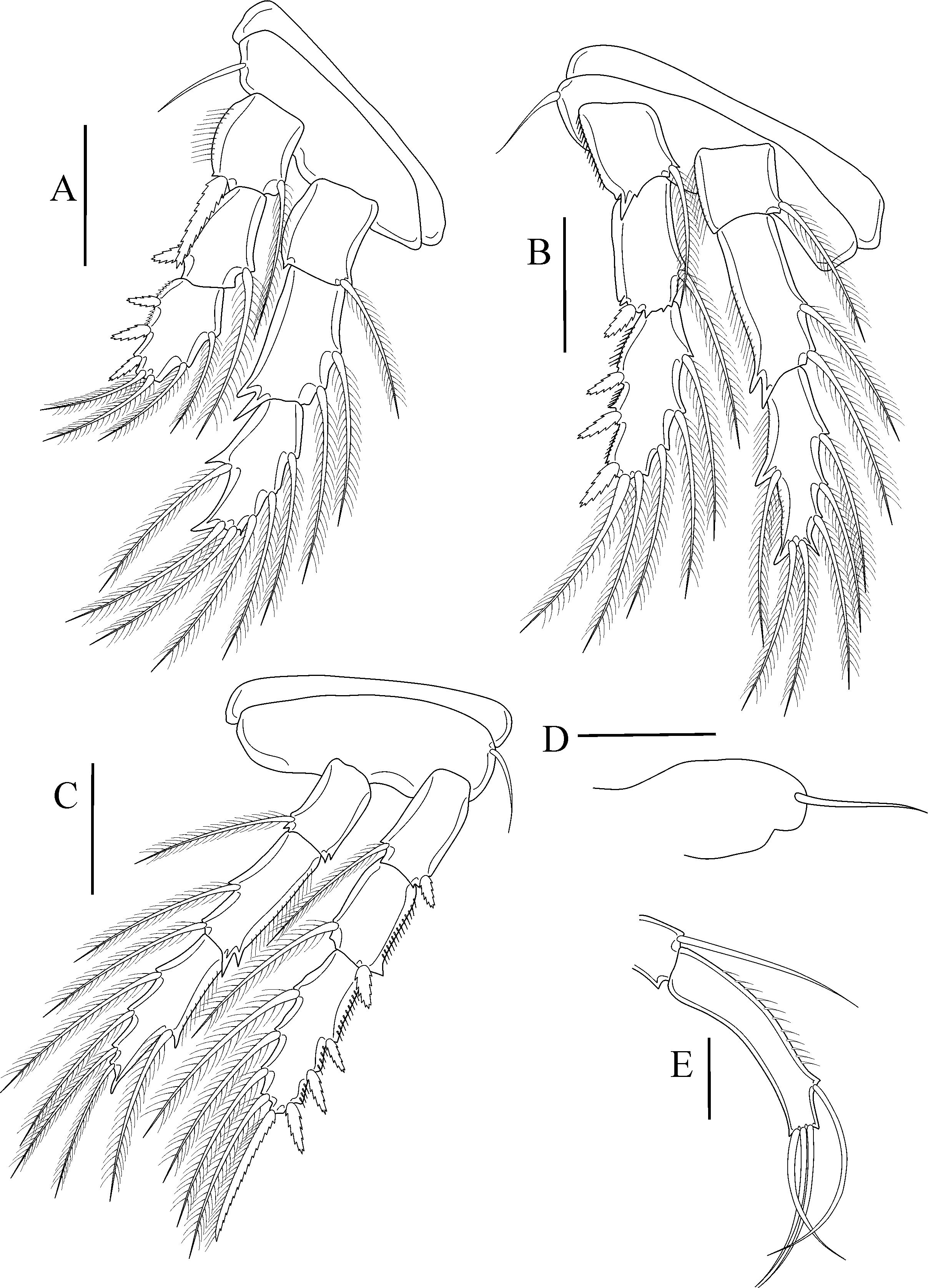
Legs 1–3 ( Figs. 6 View FIGURE 6 A−C) biramous, with 3-segmented rami. Leg 4 ( Fig. 6D View FIGURE 6 ) reduced to a bud bearing a seta. Armature formula of legs 1−3 as follows:
Second endopodal segments of leg 1 to leg 3 with two tooth-like processes on distal outer corner ( Figs. 6 View FIGURE 6 A-C). Setules on outer margin of first exopodal segment of leg 1; and outer margins of most endopodal and exopodal segments of legs 2 and 3.
Leg 5 ( Fig. 6E View FIGURE 6 ) protopodal fused to fifth pedigerous somite, with single outer seta. Free segment with 3 apical and 1 subdistal setae and setules on outer margin.
Male. Material not analyzed.
Remarks. The analysis of the holotype shows many differences regarding the original description. Wilson (1944) describes the pedigerous somites 3 to 5 as fused; but, only the pedigerous somites 3 and 4 are fused. The antennule was described as 18-segmented intead of 17-segmented as observed here due to fusions among basal elements II and III. The illustration provided by Wilson (1944, fig. 153) indicates a possible fusion of these segments once there is a seta located in the division of segments I and II. Consequently, if this seta refers to second segment it would imply in the existence of a fused segment as observed in the present redescription.
In the original description, Wilson (1944, pg 544) states: “The basal segment of the second antenna is stout and longer than the other two segments combined, with a short spine at its inner distal corner” that, together with the illustration ( Wilson 1944, fig. 154) reinforces the conception that the endopod was formed by only 2 segments, the first unarmed and the second with small spine near to terminal claw, and the exopod would be the short spine mentioned. In fact, the endopod is 3-segmented, the first and the second segments with a seta both and the third possessing three setae, one of them robust and spine-like. The short spine of the basis refers in fact to the exopod as presently observed.
The endopod of the maxilliped also differs from Wilson’s original description (1944) once it is 3-segmented with terminal claw instead large claw as illustrated by the author.
The legs illustrated by Wilson (1944; Figs. 159 and 160) also shows many divergences concerning the holotype analyzed. The main dissemblance consists in the statement made by the author that Parmulodes verrucosus has 4 pairs of biramous legs, with 3-segmented rami in each. Leg 4 rises in the adult as a reduced protuberance with a seta on its border as observed in the original slide and also stated by Stock (1992).
Parmulodes possess only one species. The material analyzed in this work as the same described by Wilson (1944) and consist of only a female. Posteriorly, Stock (1992) described the male of P. verrucosus and discovered that the host of this species is the sponge Chondrilla nucula Schmidt. The most of male’s appendices resembles those of the female, as described by Stock (1992), only with a reduction of its size. Among the differences observed in both sexes, include (1) Segmental homologies and setation of male’s antennule: I-2; II-2; III-2; IV-VII-8; VIII- 2; IX-XII-5+ae; XIII-1; XIV-1; XV-XVI-4; XVII-2; XVIII-2; XIX-XX-2; XXI-XXIII-4+ae; XXIV-XXVIII-8+ae reflecting a different fusion pattern in the proximal region of the antennule; and (2) leg 6 represented by two setae.
Despite Parmulodes verrucosus was redescribed by Eiselt (1959) and subsequently by Stock (1992), who made some further comments, the original drawings are too poorly made and a complete redescription was necessary to fulfill gaps concerning details and measures of the appendages.


No known copyright restrictions apply. See Agosti, D., Egloff, W., 2009. Taxonomic information exchange and copyright: the Plazi approach. BMC Research Notes 2009, 2:53 for further explanation.
|
Kingdom |
|
|
Phylum |
|
|
Class |
|
|
Order |
|
|
Family |
|
|
Genus |
Parmulodes verrucosus Wilson, 1944
| Canário, Roberta, Hurbath, Thiego, Da Rocha, Carlos E. F., Neves, Elizabeth G. & Johnsson, Rodrigo 2019 |
Parmulodes
| Wilson 1944 |
P. verrucosus
| Wilson 1944 |
Parmulodes verrucosus
| Wilson 1944 |
Parmulodes verrucosus
| Wilson 1944 |
Chondrilla nucula
| Schmidt. The 1862 |