
Homalinotus depressus (Linnaeus, 1758)
|
publication ID |
https://doi.org/ 10.11646/zootaxa.4311.4.10 |
|
publication LSID |
lsid:zoobank.org:pub:74E6B1Fd-8366-4B29-8Fbb-372C3239A824 |
|
DOI |
https://doi.org/10.5281/zenodo.6023331 |
|
persistent identifier |
https://treatment.plazi.org/id/BC3F7865-104F-7700-FF1E-1F2E27822CFF |
|
treatment provided by |
Plazi |
|
scientific name |
Homalinotus depressus (Linnaeus, 1758) |
| status |
|
Homalinotus depressus (Linnaeus, 1758) View in CoL
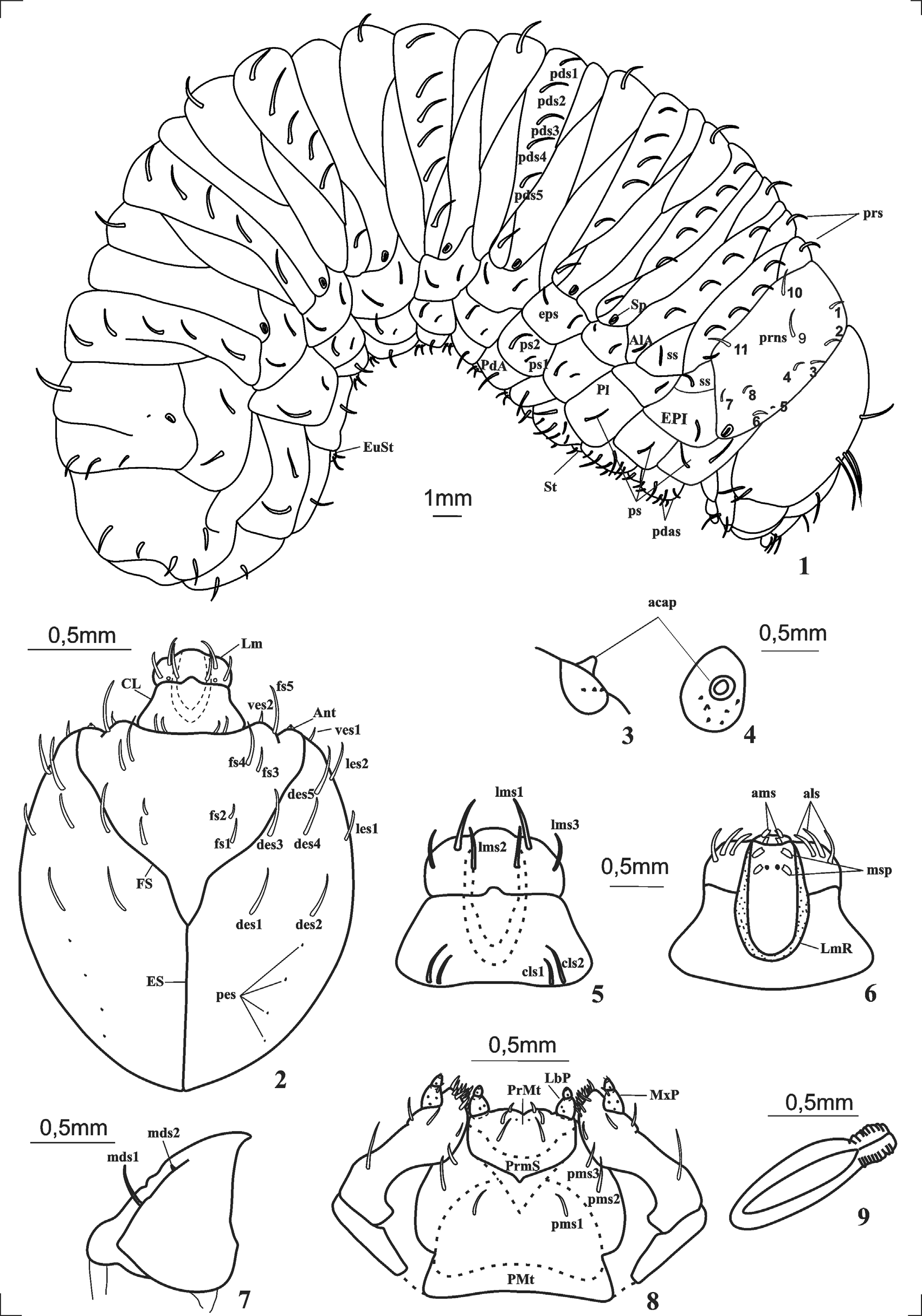
Mature larva description ( Fig. 1 View FIGURES 1 – 9 ). Robust body, sub-circular in cross-section, moderately to strongly dorsoventrally curved; 18–23 mm in length; width of prothorax 0.7–0.9 mm, head capsule width 5–6 mm; yellowish cream color, except for pronotum which is light brown, devoid of roughness.
Head ( Fig. 2 View FIGURES 1 – 9 ). Free, hypognathous, sclerotized, head capsule with greater length than width, sides rounded, light brown, except on the leading edge of the front and mandibles, which are dark brown; clypeus slightly darkened at the base, darkened labrum. Anterior and posterior stemma absent. Antenna (Ant) ( Figs. 3–4 View FIGURES 1 – 9 ) consisting of a membranous article containing an accessory sensory appendage (acap) and six minute processes. Catapophyses distinct and in the same plane as frons. Hypopharyngeal bracon membranous, easily visible in ventral view after removal of mouthparts. Epicranial suture (ES) narrow, extending to half the length of the head capsule. Frontal suture (FS) incomplete, not extending to the mandibles, sinuate and in the form of "V". Median endocarina absent. Epicranium with five pairs of dorsal epicranial setae (des 1–5); two pairs of lateral epicranial setae (les 1, les 2), les 2 located next to the anterior angle of the head capsule; two pairs of ventral epicranial setae (ves 1, ves 2); four pairs of minute posterior epicranial setae (pes), more or less aligned and forming irregular oblique row. Frons containing five pairs of frontal setae (fs), fs 1, fs 2 and fs 3, smaller than fs 4 and fs 5, which are subequal in size. Clypeus (Cl) ( Fig. 5 View FIGURES 1 – 9 ) transverse, wider than long, with two pairs of clypeal setae (cls1 and cls 2), cls 1 smaller than cls 2; sensilla absent. Labrum (Lm) ( Fig. 5 View FIGURES 1 – 9 ) transverse, lateral margin rounded, with three pairs of labral setae, lms 1 larger than lms 2 and lms 3; sensilla absent. Epipharynx (EPX) ( Fig. 6 View FIGURES 1 – 9 ) containing two pairs of anteromedian setae (ams) subequal; three pairs of anterolateral setae (als 1–3); two pairs of median spines (msp); one pair of sensilla epipharyngeal between the median spines, labral rods (LMR) converging and fused posteriorly in a "U" form. Mandibles ( Fig. 7 View FIGURES 1 – 9 ) triangular, symmetric, robust, strongly sclerotized, dorsally bearing two mandibular setae (mds) aligned longitudinally. Maxillary palp (MxP) ( Fig. 8 View FIGURES 1 – 9 ) with two articles, proximal article larger and wider than distal one, with one seta and three sensilla on the ventral surface; distal article with one sensillum on the ventral surface and two setae; stipes elongated with four setae on ventral surface, seta located on the inner margin of the apical third, smaller than the others; mala with eleven setae arranged in a row. Labial palps (LbP) with two articles, proximal article elongated, longer and wider than distal, which is conical and contains one sensilla. Prementum (PrMT) ( Fig. 8 View FIGURES 1 – 9 ) bearing one pair of subequal setae and a pair of sensilla; ligula with two pairs of short setae; premental sclerite (PrmS) slightly sclerotized, with sinuous posterior margin. Postmentum (PMt) with three pairs of postmental setae (pms).
Thorax ( Fig. 1 View FIGURES 1 – 9 ). Pro-, meso- and metathorax transverse. Pronotum with a slightly sclerotized plate, brownish; eleven pronotal setae (prns1–11) on each side, prns1–4 subequal in size and located on anterior margin near head, prns 9 longer than prns 1–4 and situated in the median region of the pronotum, prns 10 and prns 11 in the posterior region and subequal to prns 9, prns 5–8 close to the thoracic spiracles, prns 5 very short. Spiracle (Sp) ( Fig. 9 View FIGURES 1 – 9 ) bicameral, peritreme subeliptic, air tubes annulated, corresponding to one third of the peritreme length. Pro-dorsum of meso and metathorax each with one prodorsal setae (prs) long ( Fig. 1 View FIGURES 1 – 9 ). Postdorsal area of meso- and metathorax with four postdorsal setae (pds) each ( Fig. 1 View FIGURES 1 – 9 ). Alar area (A1A) of meso- and metathorax with one short seta. Spiracular area of meso- and metathorax each bearing one spiracular seta (ss). Epipleurum (EPI) of the mesothorax and metathorax with one epipleural seta (eps) ( Fig. 1 View FIGURES 1 – 9 ). Pleuron (PL) of prothorax with two long pleural setae (ps); mesothorax and metathorax with one long pleural seta each. Pedal area (PdA) of each thoracic segment with seven setae of pedal area (pdas). Sternum (St) of each thoracic segment with one moderately long seta on each side of the mid-ventral line.
Abdomen ( Fig. 1 View FIGURES 1 – 9 ). Eight pairs of bicameral spiracles, subequal, smaller than prothoracic spiracle, but with the same shape and structure. First seven abdominal segments each with three dorsal folds (folds II, III and IV); two dorsal folds on segment VIII; folds not distinct on ninth segment; segment X reduced, circular and ventral. Lateral fold (fold I) developed on segments II–VIII, narrow, without setae, sometimes much reduced and not clearly distinct on eighth segment. Segments I–VII each with the following setae described on one side: one long prodorsal setae (prs); five postdorsal setae (pds); two epipleural setae (eps) on epipleuron (EP1); two pleural setae (ps) in the pleurum (Pl), ps 1 shorter than ps 2; one long setae in the pedal area (PdA); and two moderately long setae in eusternum (EuSt). Sternellum non-distinguishable. Abdominal segment VIII with one very short seta above the spiracle and three dorsal setae, setae 2 and 3 subequal and longer than seta 1; chaetotaxy from other areas as in the segments I–VII. Abdominal segment IX with seven setae on each side, being two longs dorsal setae, two longs lateral setae moderately long, and three short latero-ventral setae. Anus subterminal, coming from four prominent lobes; lateral lobes with four setae each, three longs and one very short.
Material examined. Brazil. Pará: Moju , 30-III-2011, host plant coconut ( Cocus nucifera L.), 12 larvae, two dissected, M. Moreira leg. ( DZUP).
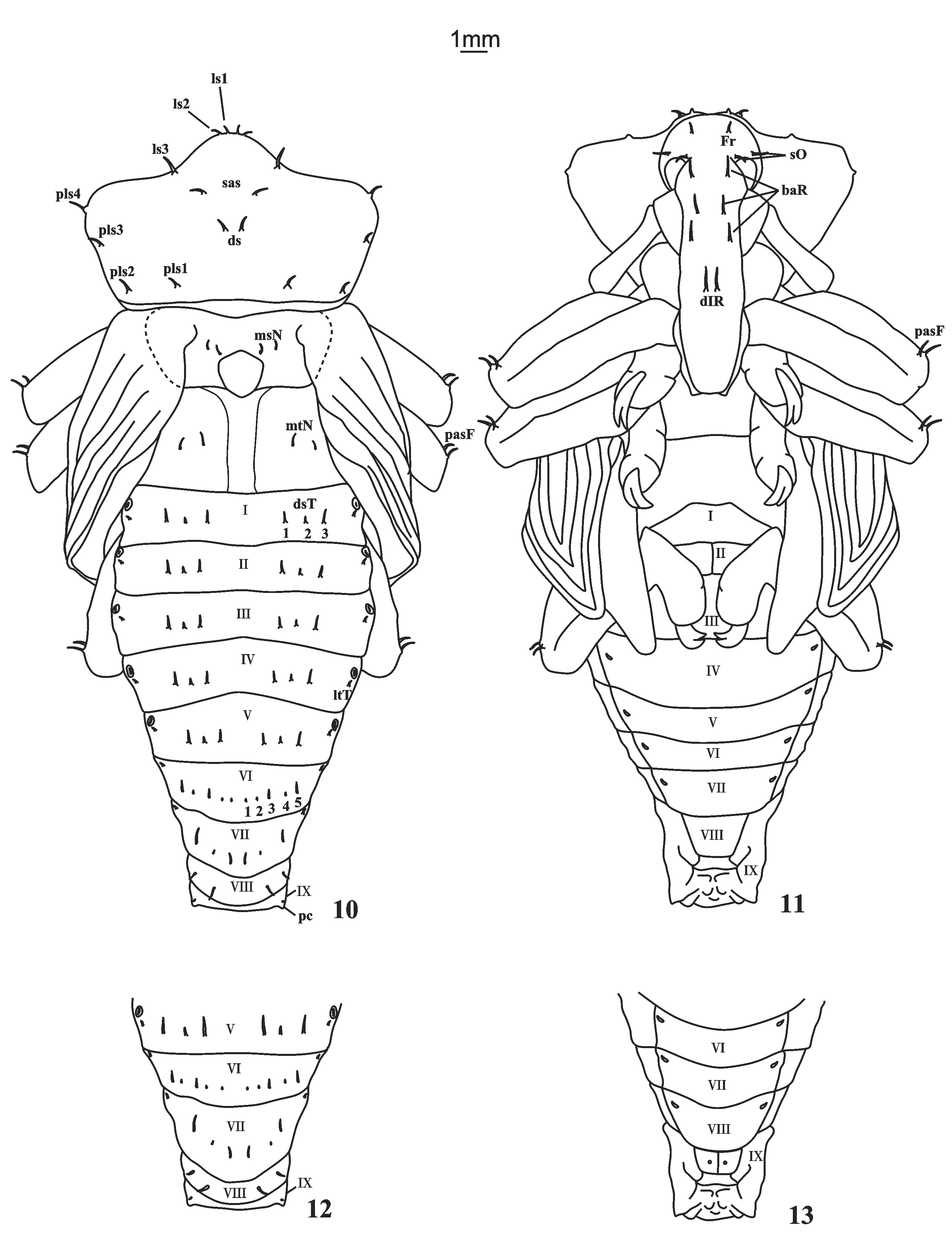
Pupa description ( Figs. 10–13 View FIGURES 10 – 13 ). Elongate body ( Figs. 10–11 View FIGURES 10 – 13 ). Size 23–30 mm. Adecticous and exarate. Color cream white, except for eyes, which are pale to dark; body setae placed on tubercles with different sizes, long and thick distributed on rostrum, head, pro-, meso- and metanotum and femora, smaller and thinner on abdomen.
Head covered by pronotum in dorsal view; a pair of frontal setae (Fr); two pairs of supraorbital setae (sO). Rostrum surpassing mesocoxae; three pairs of basirostral setae (baR); one pair of distirostral setae (diR); epistomal setae absent.
Prothorax trapezoidal ( Fig. 10 View FIGURES 10 – 13 ). Prothoracic depression absent; spiracle well developed and visible laterally; pronotum with one pair of suprapical setae (sas), one pair of discal setae (ds), three pairs of lateral setae (ls) and four pairs of posterolateral setae (pls). Mesothorax ( Fig. 10 View FIGURES 10 – 13 ). Anteronotal setae absent; two pairs of mesonotal setae (msN) similar to those of the prothorax; scutellum subtriangular. Metathorax ( Fig. 10 View FIGURES 10 – 13 ). longitudinally sulcate and divided into two parts, each side with one pair of metanotal setae (mtN) similar to mesothorax, arranged in transverse row and moderately separated; anteronotal setae absent. Legs ( Figs. 10–11 View FIGURES 10 – 13 ). Each femur containing one pair of pre-apical setae (pasF) inserted in a small rounded protuberance.
Abdomen ( Fig. 11 View FIGURES 10 – 13 ). Nine segments visible dorsally, anterotergal setae absent, segments I–V bearing three pairs of discotergal setae (dsT), the central setae minute and sometimes not visible; sixth segment with five pairs of discotergal setae, setae 1, 2 and 4 inconspicuous; the seventh segment with three pairs of discotergal setae, setae 2 minute; segments I–VII bearing one pair of laterotergal setae (ltT) located above and after the spiracles; eighth segment with two pairs of discotergal setae; ninth segment with inconspicuous pseudocerci (pc) and one pair of inconspicuous laterotergal setae; one pair of very small laterosternal setae, sometimes indistinct in some segments.
Sexual dimorphism. Usually the seventh abdominal segment of the male ( Fig. 10 View FIGURES 10 – 13 ) is shorter than on female ( Fig. 12 View FIGURES 10 – 13 ). Female with small round convexity located on each side of sternum on ninth abdominal segment ( Fig. 13 View FIGURES 10 – 13 ).
Material examined. Brazil. Pará: Mojú , 30-III-2011, host plant coconut ( Cocus nucifera L.), eight pupae, six females and two males, M. Moreira leg. ( DZUP).
| DZUP |
Universidade Federal do Parana, Colecao de Entomologia Pe. Jesus Santiago Moure |
No known copyright restrictions apply. See Agosti, D., Egloff, W., 2009. Taxonomic information exchange and copyright: the Plazi approach. BMC Research Notes 2009, 2:53 for further explanation.