
Rhaphidophora shii Lu & Bian, 2022
|
publication ID |
https://doi.org/ 10.11646/zootaxa.5087.1.6 |
|
publication LSID |
lsid:zoobank.org:pub:716394A9-D00F-4154-9952-C1D7B6A911E2 |
|
DOI |
https://doi.org/10.5281/zenodo.5819909 |
|
persistent identifier |
https://treatment.plazi.org/id/BC5787E1-053F-FFFE-1AF0-FED1FA91FCE3 |
|
treatment provided by |
Plazi |
|
scientific name |
Rhaphidophora shii Lu & Bian |
| status |
sp. nov. |
Rhaphidophora shii Lu & Bian View in CoL sp. nov.
石氏Ēȃ
Figs 14–15 View FIGURE 14 View FIGURE 15
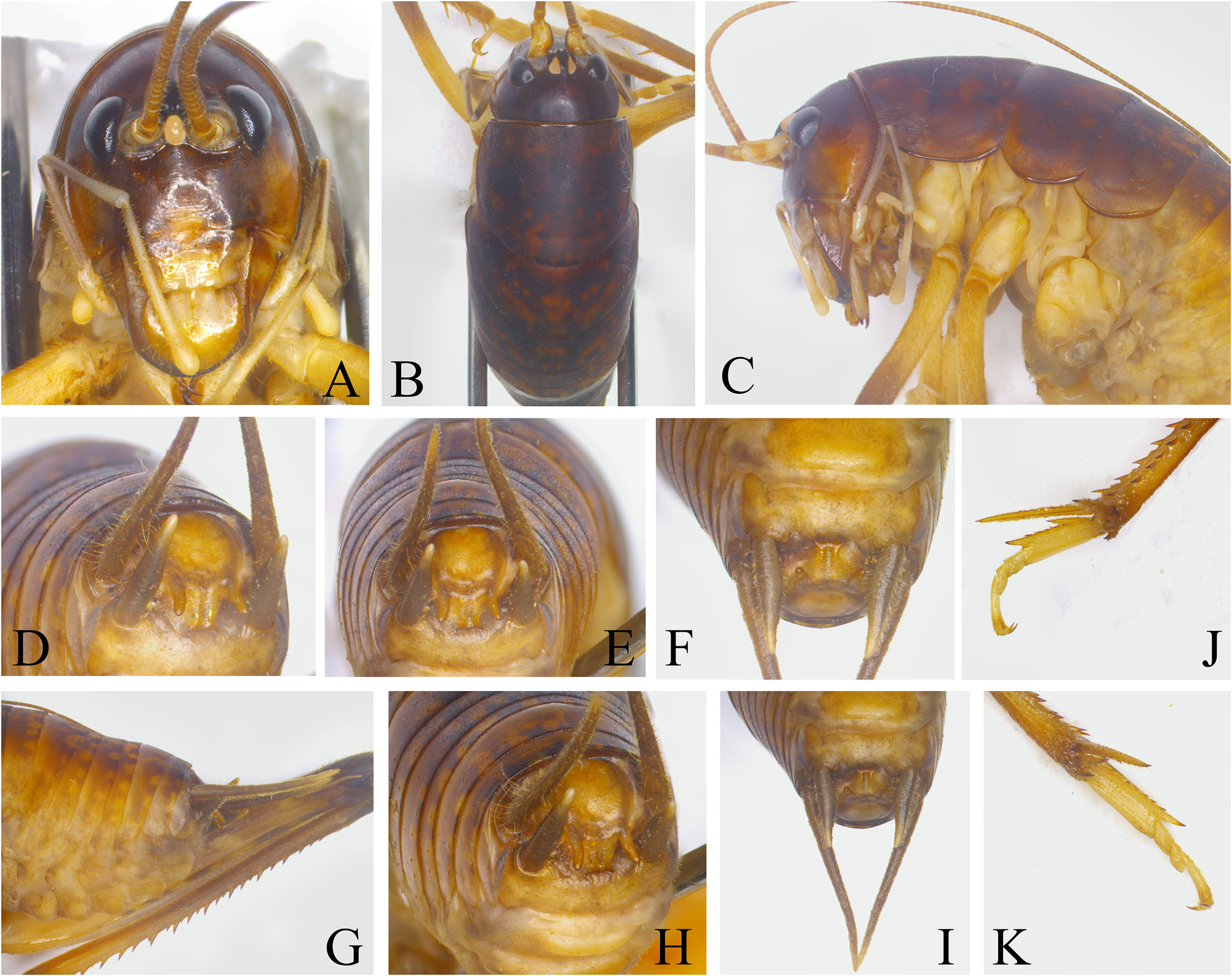
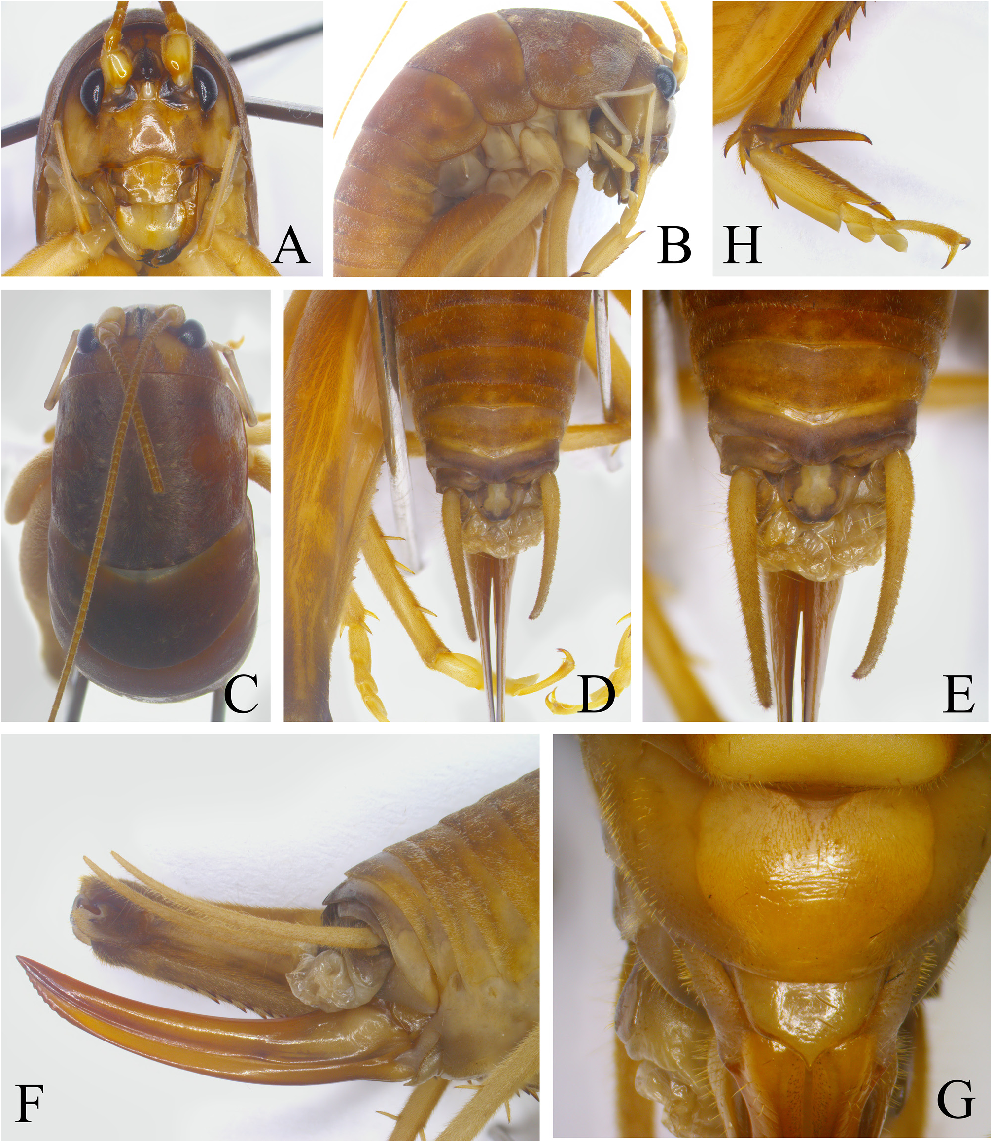
Description. Male. Body medium. Median ocellus as large as lateral ocelli ( Fig. 14A–B View FIGURE 14 ). Apical segment of maxillary palpi longer than subapical segment, apices slightly inflated. Anterior margin of pronotum arched, posterior margin widely rounded ( Fig. 14B View FIGURE 14 ). Fore coxae obviously inflated, with 1 laterally compressed spine; femora with 1 short ventral spine on internal margin; tibiae with 1 internal and 2 external spines ventrally, apices with 1 pair of spines on ventral surface. Middle femora with 1 pair of apical spines on ventral surface; tibiae dorsally with 2 pairs of spines, ventral surface with 1 internal and 3 external spines, apices with 1 pair of spines on dorsal and ventral surfaces separately. Hind tibiae dorsally with 17–19 spines, 1 small subapical spine and 1 apical spine on both sides, ventral surface with 2 pairs of apical spines; basitarsi with 5 spines and 1 apical spine on dorsal surface along the midline ( Fig. 14K View FIGURE 14 ). Abdominal tergites without any processes. Epiproct directing downwards, the basal two thirds nearly rectangular, the lateral margins convex, dorsal surface smooth ( Fig. 14E View FIGURE 14 ); the lateral margin of apical third area with 1 pair of processes, the ventral surface of process slightly expanded, the apex of process obtuse ( Fig. 14H View FIGURE 14 ); apical area inverted trapezoidal, lateral margins of subapical area obviously constricted, apical margin with 1 small lateral spine on each side ( Fig. 14E View FIGURE 14 ). Cerci slender, conical, apices acute. Subgenital plate wider than long, posterior margin slightly arched; styli long, conical, inserted on the posterolateral area of subgenital plate ( Fig. 14F View FIGURE 14 ).
Female. Posterior margin of eighth abdominal tergite slightly projected; ninth abdominal tergite posteriorly obtusely projected, dorsal surface with longitudinal furrow in the middle; tenth abdominal tergite arched concave on posterior margin ( Fig. 15D–E View FIGURE 15 ). Epiproct trapezoidal, apical margin almost straight ( Fig. 15E View FIGURE 15 ). Ovipositor shorter than hind femora, moderately upcurved, dorsal margins smooth, apices acute; apical area of ventral margins with indistinct teeth ( Fig. 15F View FIGURE 15 ). Lateral margins of subgenital plate narrowing, posterior margin with small spine in the middle ( Fig. 15G View FIGURE 15 ).
Coloration. Body brown. Terga with black spots.
Measurement (mm). BL: ♂ 18.2, ♀ 22.1; PL : ♂ 5.5, ♀ 7.5; FFL : ♂ 6.5, ♀ 8.8; MFL : ♂ 6.5, ♀ 8.6; HFL : ♂ 15.0, ♀ 19.2; HBL : ♂ 3.5, ♀ 4.3; OvL : 12.9.
Material examined. Holotype: male, Bubang, Menghai , Yunnan, August 16, 2019, coll. by Minying Guo . Paratype: 1 female, Bubang, Mengla , Yunnan, August 4, 2019, coll. by Minying Guo, Haiqing Huang & Xun Bian. Other specimen : 1 male nymph, Bubang, Mengla , Yunnan, August 16, 2019, coll. by Minying Guo, Haiqing Huang & Xun Bian .
Distribution. Yunnan (Menghai, Mengla).
Discussion. This new species differs from Rhaphidophora spinifera Gorochov, 2013 by: the epiproct of male shorter, apical margin with small spines ( Fig. 14E View FIGURE 14 ); posterior margin of male subgenital plate slightly arched without median concavity, styli longer ( Fig. 14F View FIGURE 14 ); posterior margin of female ninth abdominal tergite with median furrow ( Fig. 15E View FIGURE 15 ), tenth abdominal tergite posteriorly arched, epiproct trapezoidal, posterior margin of female subgenital plate with 1 small median spine ( Fig. 15G View FIGURE 15 ).
Etymology. We wish to thank Professor Fuming Shi for his great contribution to the Chinese Ensifera.
| PL |
Západoceské muzeum v Plzni |
No known copyright restrictions apply. See Agosti, D., Egloff, W., 2009. Taxonomic information exchange and copyright: the Plazi approach. BMC Research Notes 2009, 2:53 for further explanation.
|
Kingdom |
|
|
Phylum |
|
|
Class |
|
|
Order |
|
|
Family |
|
|
Genus |