
Uvarovia longipennis Bolívar, 1930
|
publication ID |
https://doi.org/ 10.11646/zootaxa.5093.5.3 |
|
publication LSID |
lsid:zoobank.org:pub:12334384-266E-4873-9A2C-8E0DF9456DE4 |
|
DOI |
https://doi.org/10.5281/zenodo.6204809 |
|
persistent identifier |
https://treatment.plazi.org/id/BC7DBE62-7375-ED61-50AA-1EFCFB652225 |
|
treatment provided by |
Plazi |
|
scientific name |
Uvarovia longipennis Bolívar, 1930 |
| status |
|
Uvarovia longipennis Bolívar, 1930 View in CoL
( Figs. 1–4 View FIGURE 1 View FIGURE 2 View FIGURE 3 View FIGURE 4 )
Uvarovia longipennis Bolívar, 1930: 202 View in CoL
Mnesicles sp. (misidentification)— Tan, 2012: 12; Tan et al., 2015: 47
Material examined (image). Holotype (female), PENINSULAR MALAYSIA, Isla Dinding [= Manjung District (Perak)], H. N. Ridley leg., det. C. Bolívar, 1922 ( NHMUK012499122 View Materials ).
Material examined. 3 males and 3 females, SINGAPORE : 1 male, Mandai Avenue , felled forest, M. K. Tan & R. W. J. Ngiam leg., 17 March 2011 ( ZRC.ORT.248) ; 1 male, Nee Soon pipeline, edge of freshwater swamp forest, M. K. Tan, R. W. J. Ngiam & W. L. Lim leg., 31 May 2011 ( ZRC.ORT.293) ; 1 female, Belukar Track near Bukit Timah Nature Reserve, forest edge, M. K. Tan & M. R. B. Ismail leg., 30 June 2011 ( ZRC.ORT.313) ; 1 female; Mandai , secondary forest, M. K. Tan leg ; 1 male, Bukit Timah Hindhede Drive , near primary forest, H. K. Lua leg., 27 August 1989 ( ZRC.ORT.563) ; 1 female, Admiralty Park (Nature Park), secondary forest, M. K. Tan leg., 30 September 2021 ( ZRC) .
Type locality. PENINSULAR MALAYSIA: Perak: Manjung District
Distribution. Malay Peninsula (i.e., Peninsular Malaysia and Singapore)
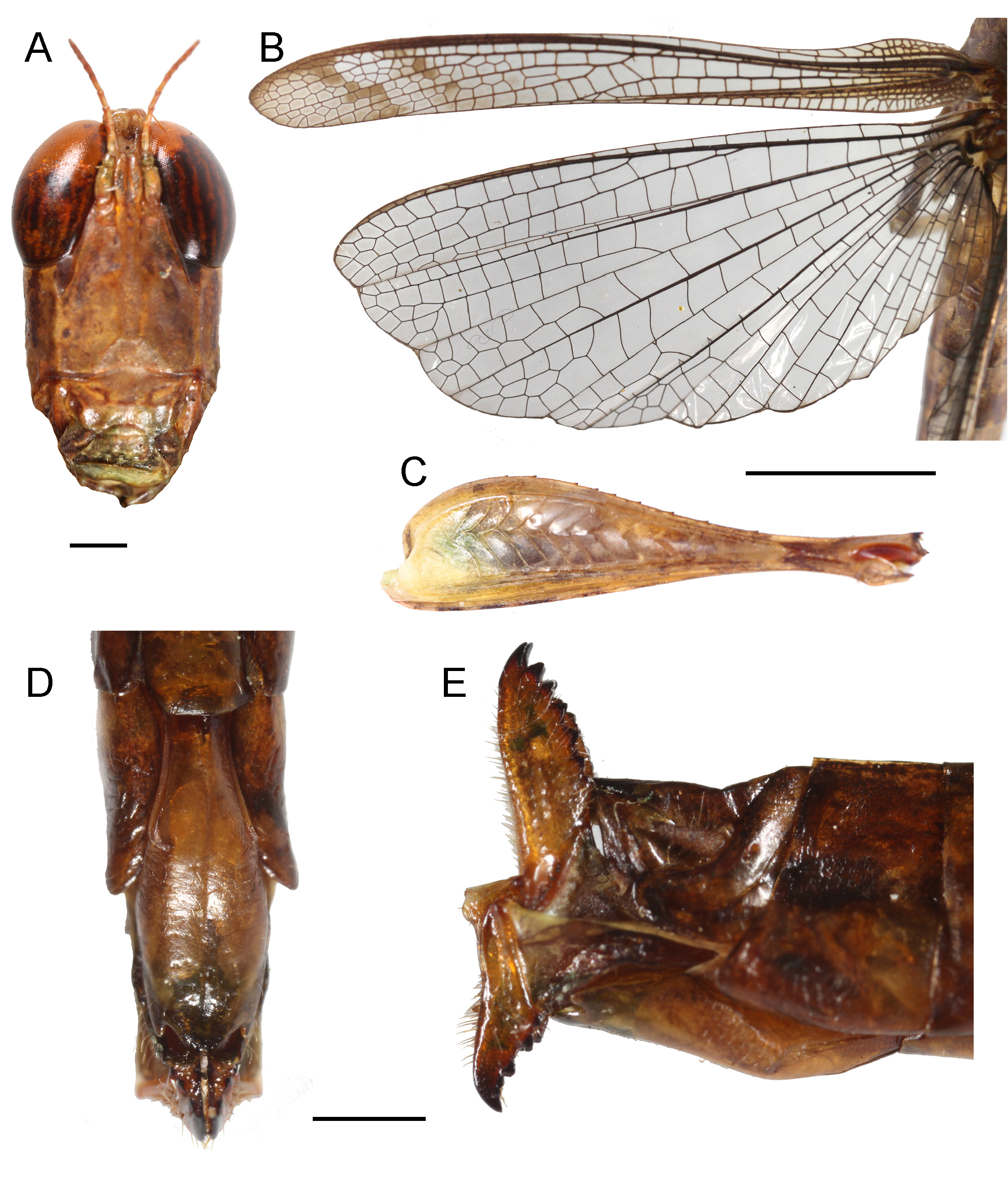
Male description. Habitus as shown in Figs. 2 View FIGURE 2 , 3A View FIGURE 3 . Vertex with faint middle keel ( Fig. 3B View FIGURE 3 ). Dorsally, fastigium narrow, 1.3 times as long as wide, slightly enlarged at the apex; separated from vertex by a transverse furrow ( Fig. 3B View FIGURE 3 ). Frons narrow between antennal scapes with lateral keels sinuously converging dorsally, diverging ventrally (most far apart) between scapes before converging ventrally ( Fig. 3C View FIGURE 3 ). Frontal costa with faint lateral keels ( Fig. 3C View FIGURE 3 ). Scapus elongated, antenna with 11 segments; segment flattened, tend to widen apically ( Fig. 3D View FIGURE 3 ). Pronotal dorsal disc with anterior margin broadly concave and posterior margin obtuse, rounded; with transverse furrow continuing to lateral lobe. Pronotal lateral lobe with anterior margin convex; ventral margin straight in anterior half and oblique in posterior half; ventro-posterior angle 90°; posterior margin straight. Tegmina and hind wings well surpassing beyond apex of hind femora and abdominal apex ( Fig. 3D View FIGURE 3 ). Tegmina with fine veining and delimiting fairly large areoles; slightly curved back at apical part, at the apex subacutely-rounded ( Fig. 3E View FIGURE 3 ). Hind wings 2.0 times as long as wide ( Fig. 3E View FIGURE 3 ). Hind tibia armed with 20–22 external and 14–15 internal spines.
Abdomen enlarged apically ( Fig. 1 View FIGURE 1 ). 8 th tergite normal, with lateral portion extending posteriorly into a narrow lobe with truncated apex ( Fig. 1A View FIGURE 1 ). Central portion of 9 th tergite elongated, narrow slightly after basal third and forming a shaft-like process, apex bilobed with apices of lobes narrow, round and pointing externally ( Fig. 1B View FIGURE 1 ). Lateral portion of 9 th tergite fused to 10+12 th tergites with distinct suture; lateral portion forming three lobes: ventral lobe triangular and pointing posteriorly with subacute apex, middle lobe more narrow and elongated, separated from ventral lobe by deep and wide excision, dorsal projection longest and narrowest, pointing dorsad (slightly curved posteriorly in the middle) with lamellate and truncated apex ( Fig. 1A View FIGURE 1 ). 10 th and 12 th tergites, although united, clearly delimited ( Fig. 1B View FIGURE 1 ). 10 th tergite in dorsal view producing two lateral posterior process with subacute apex, slightly diverging from one another at apex ( Fig. 1B View FIGURE 1 ). 12 th tergite forming tongue-shaped supra-anal plate. Cercus very long and U-bent; basal part slightly less than twice as long as apical part, slightly narrowed towards apex (in dorsal view) ( Fig. 1B View FIGURE 1 ), although in lateral view, it has parallel margins ( Fig. 1A View FIGURE 1 ); the apical part pointing anteriorly (parallel to basal part), narrower than basal part, chitinised, somewhat compressed and very sharp towards the apex ( Fig. 1B View FIGURE 1 ). Subgenital plate with deeply V-excised basal margin; in the middle with narrow furrow along midline; posterior end bilobed, V-excised (not as deep as basal margin) between the lobes. Apical lobe of subgenital plate rectangular in vento-posterior view, slightly longer than on the posterior margin and converging apically, apex truncated.
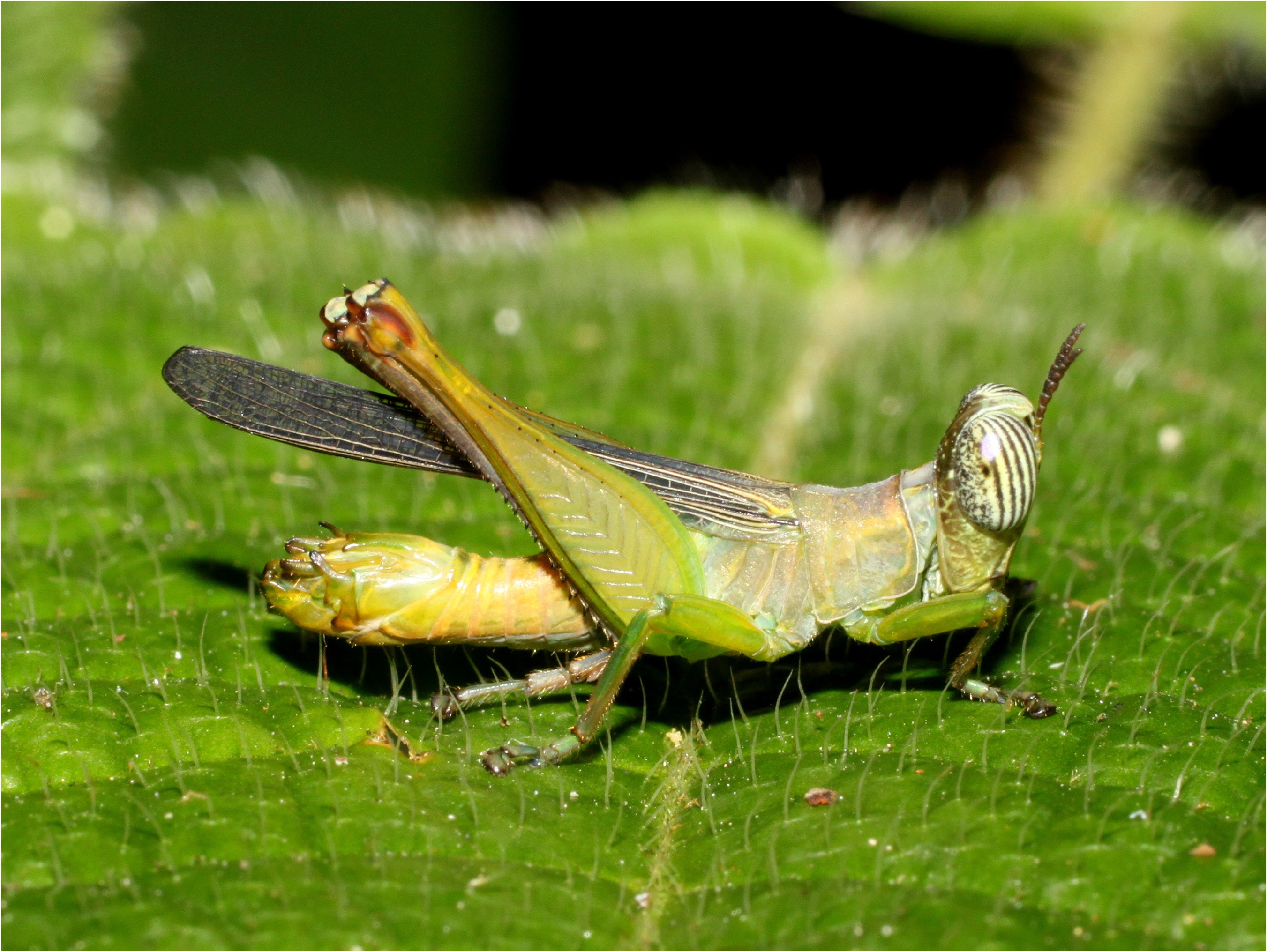
Colouration. Generally yellow with tint of green when alive ( Fig. 2 View FIGURE 2 ), green colouration lost when dry-preserved ( Fig. 3A View FIGURE 3 ). Head with vertex mottled dark and lateral keel yellow; fastigium sometimes lighter in colouration at apex. Eyes white when alive ( Fig. 2 View FIGURE 2 ), brown when dry-preserved, with numerous vertical dark stripes ( Fig. 3D View FIGURE 3 ). Scapus yellow green, antennal segments brown to dark brown ( Figs. 3B, 3D View FIGURE 3 ). Labrum green in dorsal half and mottled dark in ventral half. Maxillary palps grey green with brown rings. Pronotal dorsal disc generally grey (dark when drypreserved) with anterior margin brown; lateral lobe yellow green with tint of blue grey ( Figs. 2 View FIGURE 2 , 3D View FIGURE 3 ). Tegmina with dark brown veins; cells completely hyaline, apex with trace of brown ( Fig. 3E View FIGURE 3 ). Anterior and middle legs generally yellow green to green with brown spines. Hind femora generally yellow green, basal parts more green, apical part more yellow with brown knees ( Figs. 2 View FIGURE 2 , 3A View FIGURE 3 ). Hind tibia and tarsus grey. Thoracic tergites grey yellow, abdominal tergites yellow with tints of green ( Fig. 2 View FIGURE 2 ). Lateral parts of 9 th tergite with apices of processes dark. Cercus apical half brown with black apex ( Fig. 1 View FIGURE 1 ).
Measurements (in mm). BL = 15.6, PL = 2.1, FWL = 12.2, HWL = 11.5, HFL = 9.1, HFW = 2.3, HTL = 8.7, HTaL = 3.9.
Female ( Fig. 4 View FIGURE 4 ). Newly acquired specimens match the original female description: FW with two brown oblique bands, narrowly and imperfectly marked, in its apical third ( Fig. 4B View FIGURE 4 ). Wings being 2.4 times as long as wide ( Fig. 4B View FIGURE 4 ). Abdominal apex with a dorsal tongue-shaped plate with lateral margins emarginated in the middle. Subgenital plate elongated shaft, narrow at the base, widening until basal third, lateral margins parallel; narrowed angularly near the apex into a narrow and truncated apex ( Fig. 4D View FIGURE 4 ). Ovipositor as shown in Fig. 4E View FIGURE 4 .


| ZRC |
Zoological Reference Collection, National University of Singapore |
No known copyright restrictions apply. See Agosti, D., Egloff, W., 2009. Taxonomic information exchange and copyright: the Plazi approach. BMC Research Notes 2009, 2:53 for further explanation.
|
Kingdom |
|
|
Phylum |
|
|
Class |
|
|
Order |
|
|
Family |
|
|
Genus |
Uvarovia longipennis Bolívar, 1930
| Tan, Ming Kai, Japir, Razy & Chung, Arthur Y. C. 2022 |
Mnesicles sp.
| Tan, M. K. & Yeo, H. & Lee, J. X. Q. 2015: 47 |
| Tan, M. K. 2012: 12 |
Uvarovia longipennis Bolívar, 1930: 202
| Bolivar, C. 1930: 202 |