
Microstomum laurae, Atherton & Jondelius, 2018
|
publication ID |
https://doi.org/ 10.5852/ejt.2018.398 |
|
publication LSID |
lsid:zoobank.org:pub:58C075B0-7409-41B7-A6F4-900A5A6BFECE |
|
DOI |
https://doi.org/10.5281/zenodo.5991921 |
|
persistent identifier |
https://treatment.plazi.org/id/49378EBE-7046-49D3-BC3C-DD783580A90E |
|
taxon LSID |
lsid:zoobank.org:act:49378EBE-7046-49D3-BC3C-DD783580A90E |
|
treatment provided by |
Plazi |
|
scientific name |
Microstomum laurae |
| status |
sp. nov. |
Microstomum laurae sp. nov.
urn:lsid:zoobank.org:act:49378EBE-7046-49D3-BC3C-DD783580A90E
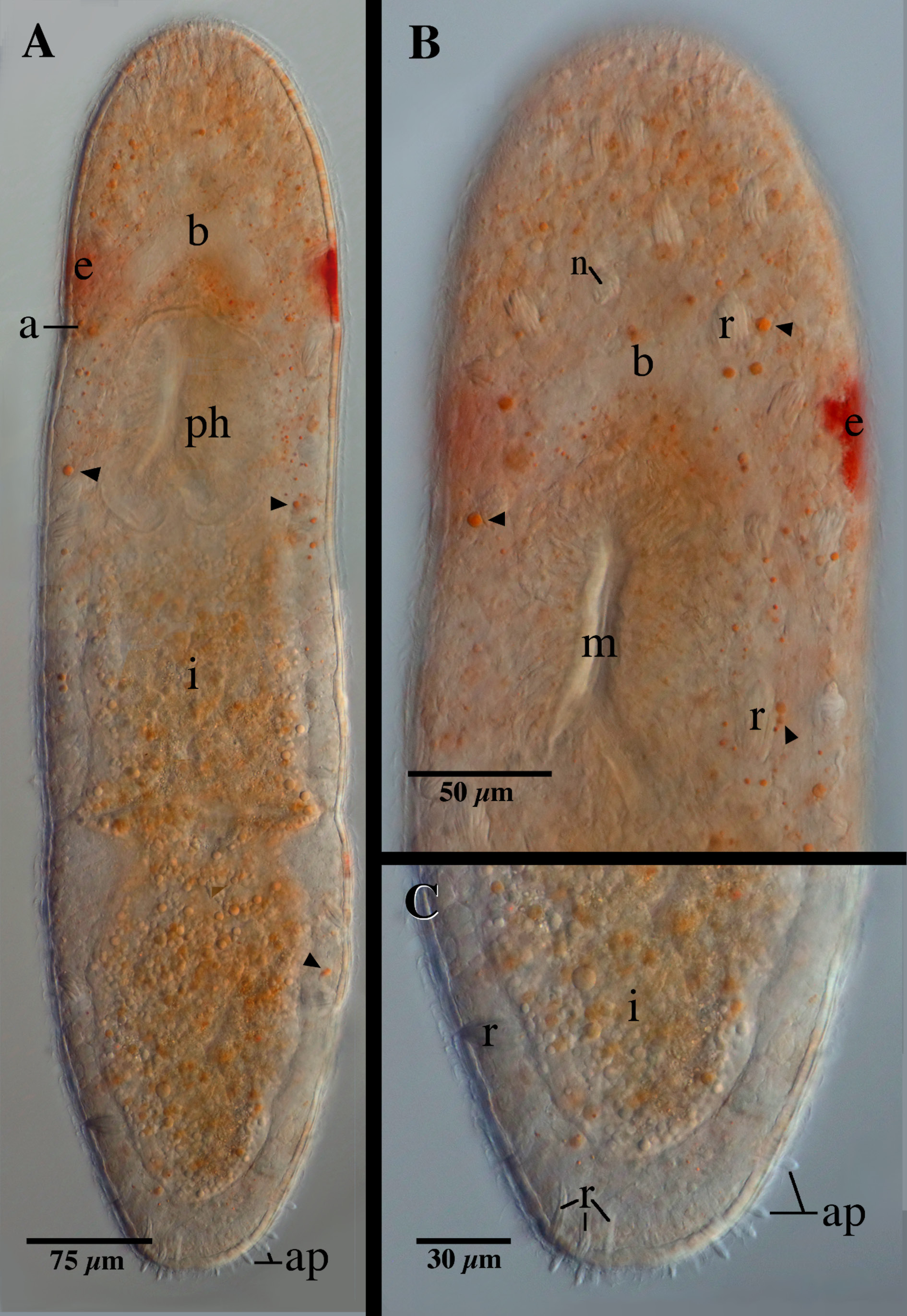
Fig. 1 View Fig. 1
Diagnosis
Strap-shaped Microstomum with body length of 760 µm (two vegetative zooids) and rounded anterior and posterior ends. Posterior rim with approximately twenty adhesive papillae. Paired red eyespots 43 µm long and located in the lateral margins at level of the brain. Rhabdites concentrated in the anterior end above the pharynx. Nematocysts present. Preoral gut extending anterior to brain. Reproductive system unknown. GenBank accession number for partial COI sequence MF185712 View Materials -3.
Etymology
This species is dedicated to Laura R. Atherton, mother of the first author.
Material examined
Holotype
SWEDEN: vegetative specimen, Strömstad , Saltö , 58°52′29″ N, 11°08′41″ E, 10 cm, 17 Jun. 2016, marine, eulittoral sand, S. Atherton leg. ( SMNH-Type-8903 , Genbank accession MF185712 View Materials ). GoogleMaps
Additional material
SWEDEN: vegetative specimen, Strömstad , Saltö , 58°52′41″ N, 11°06′56″ E, 10 cm, 19 Jun. 2016, marine, eulittoral sand, S. Atherton leg. ( SMNH-Type-8904 , Genbank accession MF185713 View Materials ). GoogleMaps
Description
Microstomum with a total body length of 760 µm and two vegetative zooids. Body strap-shaped with very slight constrictions between zooids and at the level of the ciliary pits. Ratio of body width:length 1: 5 in slightly compressed animal. Anterior and posterior ends rounded.
Ciliary pits very small and shallow ( Fig. 1A View Fig. 1 ), below eyespots, 185 µm from anterior. Paired red eyespots approximately 43 µm long and present in the lateral margins of the body level with brain ( Fig. 1A–B View Fig. 1 ).
Many small (max. diameter 10 µm) orange lipid droplets derived from food scattered across body, heavily concentrated around pharynx and anterior end ( Fig. 1A–B View Fig. 1 ). Body otherwise colorless or reflective of intestine.
Epidermis uniformly covered with cilia. Nematocysts present. Rhabdite bundles, 20–30 µm long, scattered about the body, particularly concentrated in the anterior end above the pharynx ( Fig. 1B View Fig. 1 ). Approximately twenty adhesive papillae, 8–10 µm long, on the rim of the posterior end ( Fig. 1C View Fig. 1 ).
Mouth slit-like and 55 µm long. Pharynx encompassing the second fifth of the body. Preoral gut extending well anterior to brain. Intestine evenly filled with orange droplets.
Reproductive system unknown.
Remarks
Eight of the currently recognized Microstomum species ( M. bioculatum Faubel, 1984 , M. gabriellae Marcus, 1950 , M. giganteum Hallez, 1878 , M. groenlandicum Levinsen, 1879 , M. lineare Ørsted, 1843 , M. melanophthalmum Steinbock, 1933 , M. paràdii Graff, 1913 and M. spiriferum Westblad, 1953 ) possess pigmented eyespots. The eyespots of M. melanophthalmum and M. paràdii are black with distinct lenses, while the eyespots of M. laurae sp. nov. are red. The eyespots in M. bioculatum , M. gabriellae , M. giganteum , M. lineare and M. melanophthalmum are situated far in the front of the animal, whereas they are level with the brain in M. laurae sp. nov. Finally, the eyespot of M. groenlandicum is unpaired, red and located medially above the brain.
Microstomum laurae sp. nov. is most similar to M. spiriferum , being alike in general body size and shape as well as their small ciliary pits and large bundles of rhabdites. However, M. spiriferum can be differentiated based on the dorsal richly yellowish pigment cells and absence of adhesive papillae ( Westblad 1953). M. laurae sp. nov., on the other hand, is colorless or sometimes colored by its gut contents; there are no pigment cells apart from the eyespots, and it possesses a distinct line of adhesive tubes along the posterior rim. Though both species are found on the Swedish west coast, M. spiriferum was described from sublittoral habitats between 15 and 60 m depth, while M. laurae sp. nov. was collected from eulittoral sand at around 10 cm depth.
No known copyright restrictions apply. See Agosti, D., Egloff, W., 2009. Taxonomic information exchange and copyright: the Plazi approach. BMC Research Notes 2009, 2:53 for further explanation.
|
Kingdom |
|
|
Phylum |
|
|
Class |
|
|
SuperOrder |
Macrostomorpha |
|
Order |
|
|
Family |
|
|
Genus |