
Neuraphes
|
publication ID |
https://doi.org/10.11646/zootaxa.4018.3.8 |
|
publication LSID |
lsid:zoobank.org:pub:44D5F826-7B66-46C3-90D7-A013B060C30D |
|
DOI |
https://doi.org/10.5281/zenodo.6122286 |
|
persistent identifier |
https://treatment.plazi.org/id/BD76878B-2A15-FF9E-FF04-FC2BFBB642A8 |
|
treatment provided by |
Plazi |
|
scientific name |
Neuraphes |
| status |
|
Neuraphes View in CoL (s. str.) kazakhstanicus sp. nov.
( Figs. 1–14)
Material studied. Holotype, male: KAZAKHSTAN: Almaty reg., 43°16'13"N 77°22'14"E, 1650 m, Talgar distr., Ак Булак [Ak Bulak], 12.–15.v.2014, O. Nakládal & M. Kocian lgt. ( CPH). Paratypes, 6 males, 4 females: the same data as holotype, ( CPH, CPJ, CHM).
Etymology. Locotypic, named after the country name of Kazakhstan, where the new species was collected.
Diagnosis. Medium size, reddish-brown, shiny Neuraphes s.str. with deep and each remote posterior vertexal pits, head with robust posterior lateral vertexal projections.
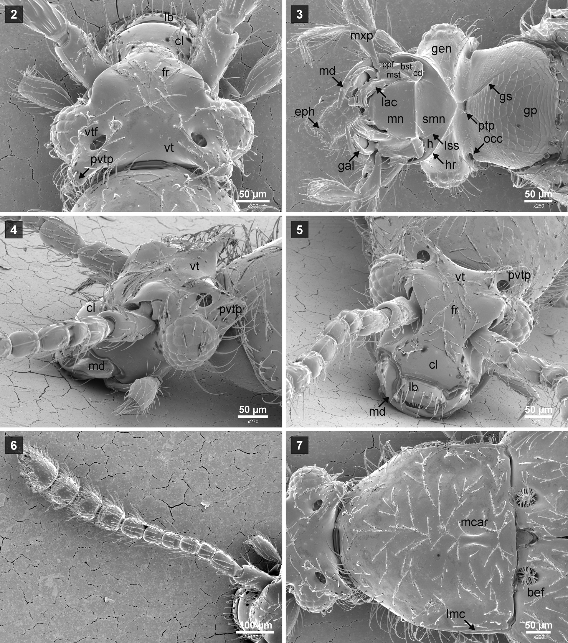
Description. Male: Body 1.4–1.5 mm long, 0.64–0,66 mm wide, colour from light to dark reddish-brown, usually pronotum darker than elytra, maxillary palpi, antennae and legs always lighter, whole body shiny, entirely covered by long, sparse setae. Head transverse, 1.45 times as wide as long, head capsule ( Figs. 2–5 View FIGURES 2 – 7 ) divided by occipital constriction ( Fig. 3 View FIGURES 2 – 7 , occ) into large anterior and smaller posterior part (i.e., 'neck region'), the posterior part retracted into pronotum. Anterior part of head subtriangular, broadest at level of eyes; vertex ( Figs. 2, 4–5 View FIGURES 2 – 7 , vt) transverse, twice as wide as long, temples as long as eyes and each forming long posterior vertexal process ( Figs. 2, 4–5 View FIGURES 2 – 7 , pvtp), anteriorly confluent with frons ( Fig. 2, 5 View FIGURES 2 – 7 , fr); supra-antennal tubercles well developed; genae ( Fig. 3 View FIGURES 2 – 7 ; gen) small. Clypeus ( Figs. 2, 5 View FIGURES 2 – 7 , cl) not separated from frons. Ventral side of head ( Fig. 3 View FIGURES 2 – 7 ) flattened except for strongly convex neck region; gular plate ( Figs. 3 View FIGURES 2 – 7 , gp) subtriangular, large with distinct gular sutures ( Fig. 3 View FIGURES 2 – 7 , gs), feebly demarcated from submentum ( Fig. 3 View FIGURES 2 – 7 , smn) by shallow groove; posterior tentorial pits located between gular plate and submentum. Mouthparts ( Figs. 2–5 View FIGURES 2 – 7 ). Labrum ( Fig. 5 View FIGURES 2 – 7 , lb) transverse, dorsal surface with eight long setae, epipharynx ( Fig. 3 View FIGURES 2 – 7 , eph) with several anterior marginal seta-like sensilla. Mandibles ( Figs. 3–5 View FIGURES 2 – 7 , md) symmetrical, long and slender. Maxillae ( Fig. 3 View FIGURES 2 – 7 ) elongate, with transverse cardo ( Fig. 3 View FIGURES 2 – 7 , cd) bearing long sub-basal seta; basistipes ( Fig. 3 View FIGURES 2 – 7 , bst) triangular, with one long basal seta; mediostipes ( Fig. 3 View FIGURES 2 – 7 , mst) elongate; palpifer ( Fig. 3 View FIGURES 2 – 7 , ppf) elongate, with one long lateral sub-apical seta; galea ( Fig. 3 View FIGURES 2 – 7 , gal) elongate and slender; lacinia ( Figs. 3 View FIGURES 2 – 7 , lac) elongate and wider than galea; maxillary palps ( Fig. 3 View FIGURES 2 – 7 , mxp) long, palpomere I small, about as long as broad, asetose, palpomere II pedunculate, with sparse setae in apical part, palpomere III about as long as II and pedunculate, broadest behind middle, covered with sparse and long setae; palpomere IV small, slender and subconical, with elongate and pointed apical part, with dense and moderately long setae in basal half. Labium ( Fig. 3 View FIGURES 2 – 7 ) with elongate, trapezoidal submentum ( Fig. 3 View FIGURES 2 – 7 , smn) laterally separated from hypostomae ( Fig. 3 View FIGURES 2 – 7 , h) by lateral sutures ( Fig. 3 View FIGURES 2 – 7 , lss) and bearing pair of long latero-anterior setae; mentum ( Fig. 3 View FIGURES 2 – 7 , mn) subtrapezoidal with one pair of long and one short latero-anterior setae. Posteriorly and laterally mouthparts demarcated by hypostomal ridges ( Fig. 3 View FIGURES 2 – 7 , hr) strongly convergent caudad; hypostomae ( Fig. 3 View FIGURES 2 – 7 , h) subtriangular and elongate. Antennae ( Fig. 6 View FIGURES 2 – 7 ) slender, gradually thickening towards apices, with feably defined 4-segmented antennal club, antennomeres moderately compactly assembled, scape cylindrical, wider than and about as long as pedicel, antennomere III smallest, antennomere IV about 1.2 times as long as III, antennomeres IV–VII elongate, VIII–X wider than long, terminal antennomere pointed at apex, shorter than IX–X combined, all antennomeres sparsely covered with suberect to erect setae. Pronotum ( Figs. 7–8 View FIGURES 2 – 7 View FIGURES 8 – 12 ) moderately convex, 1.05 times as wide as long and 1.30 times longer than head, in dorsal view ( Fig. 7 View FIGURES 2 – 7 ) approximately hexagonal with slightly rounded anterior part, lacking foveae, with short median ( Figs. 7–8 View FIGURES 2 – 7 View FIGURES 8 – 12 , mcar) and longer lateral marginal ( Figs. 7–8 View FIGURES 2 – 7 View FIGURES 8 – 12 , lmc) carinae, median carina on both sides flanked by shallow impressions, anterior corners weakly defined, posterior corners of pronotum well defined. Prosternum ( Fig. 9 View FIGURES 8 – 12 ) about 3.5 times as wide as long, considerably shorter than pronotum; procoxal cavities confluent, separated only in anterior part by slender, short prosternal process ( Fig. 9 View FIGURES 8 – 12 , pstp); procoxal sockets closed. Hypomera ( Fig. 9 View FIGURES 8 – 12 , hy) elongate, not demarcated from pronotum by hypomeral ridge, demarcated from prosternum by notosternal sutures ( Fig. 9 View FIGURES 8 – 12 , nss). Mesoventrite ( Figs. 9–10 View FIGURES 8 – 12 ) very short, much broader than long, with short, narrow median intercoxal proces ( Figs. 9–10 View FIGURES 8 – 12 , msvp); mesocoxal cavities separated, posterior margin of mesocoxal cavities with dense setae. Metaventrite ( Figs. 9, 10 View FIGURES 8 – 12 ) much longer than mesoventrite, wider than long, anteriorly separated from mesoventrite, with distinct metaventral anterior process ( Figs. 9–10 View FIGURES 8 – 12 , mvap), lateral margins slightly rounded, at middle posterior margin expanded caudad and forming broad and short sharp metaventral posterior intercoxal process ( Figs. 10, 12 View FIGURES 8 – 12 , mvpp) with distinct median notch. Elytra ( Fig. 11 View FIGURES 8 – 12 ) 1.55 times as long as wide and 1.30 times longer than pronotum, elliptical, with rounded apices; humeri lacking denticle or calli; elytral base with one well-defined, deep circular and setose basal fovea ( Figs. 7 View FIGURES 2 – 7 , 11 View FIGURES 8 – 12 , bef). Legs moderately long and slender, without any special characters. Abdomen ( Fig. 12 View FIGURES 8 – 12 ) elongate, abdominal sternites III–VIII ( Fig. 12 View FIGURES 8 – 12 , st3–st8) gradually narrowing towards abdominal apex, all of about same length. Aedeagus ( Figs. 13–14 View FIGURES 13 – 14 ) small, 0.15 mm in length, broadening from base to apex, almost twice as long as wide, strongly bent (90o) at base in lateral view.
Sexual dimorphism. Not apparent.
Differential diagnosis. The new species is easily recognized from known species of the subgenus Neuraphes (s.str.) by the presence of the long posterior vertexal processes and by the strongly bent base of median lobe.
Biology. All specimens were collected in a narrow, periodically flooded, flat valley along a small stream. The valley was covered by sandy soil with many large boulders. A deep humus layer with leaf litter was only around trees and roots of bushes in elevated places. The habitat was overgrown by dense tree and bush vegetation with dominant Populus sp. and Padus sp. All specimens were collected by sifting leaf litter and wood debris especially at the base of the trunks or tree hollows of old trees.
Distribution. Kazakhstan, Almaty reg. (Talgar, Ile-Alatau National Park).


No known copyright restrictions apply. See Agosti, D., Egloff, W., 2009. Taxonomic information exchange and copyright: the Plazi approach. BMC Research Notes 2009, 2:53 for further explanation.
|
Kingdom |
|
|
Phylum |
|
|
Class |
|
|
Order |
|
|
Family |
|
|
SubFamily |
Scydmaeninae |