
Notoplanella inarmata Bock, 1931
|
publication ID |
https://doi.org/ 10.11646/zootaxa.3780.1.4 |
|
publication LSID |
lsid:zoobank.org:pub:E1E25433-72CD-4592-B9C2-62F4D11F2D82 |
|
DOI |
https://doi.org/10.5281/zenodo.6142162 |
|
persistent identifier |
https://treatment.plazi.org/id/BD77527A-FF91-3A60-1CBD-41DB8DCCFB31 |
|
treatment provided by |
Plazi |
|
scientific name |
Notoplanella inarmata Bock, 1931 |
| status |
|
Notoplanella inarmata Bock, 1931 View in CoL
Figs. 1 View FIGURE 1 (1), 2, 3
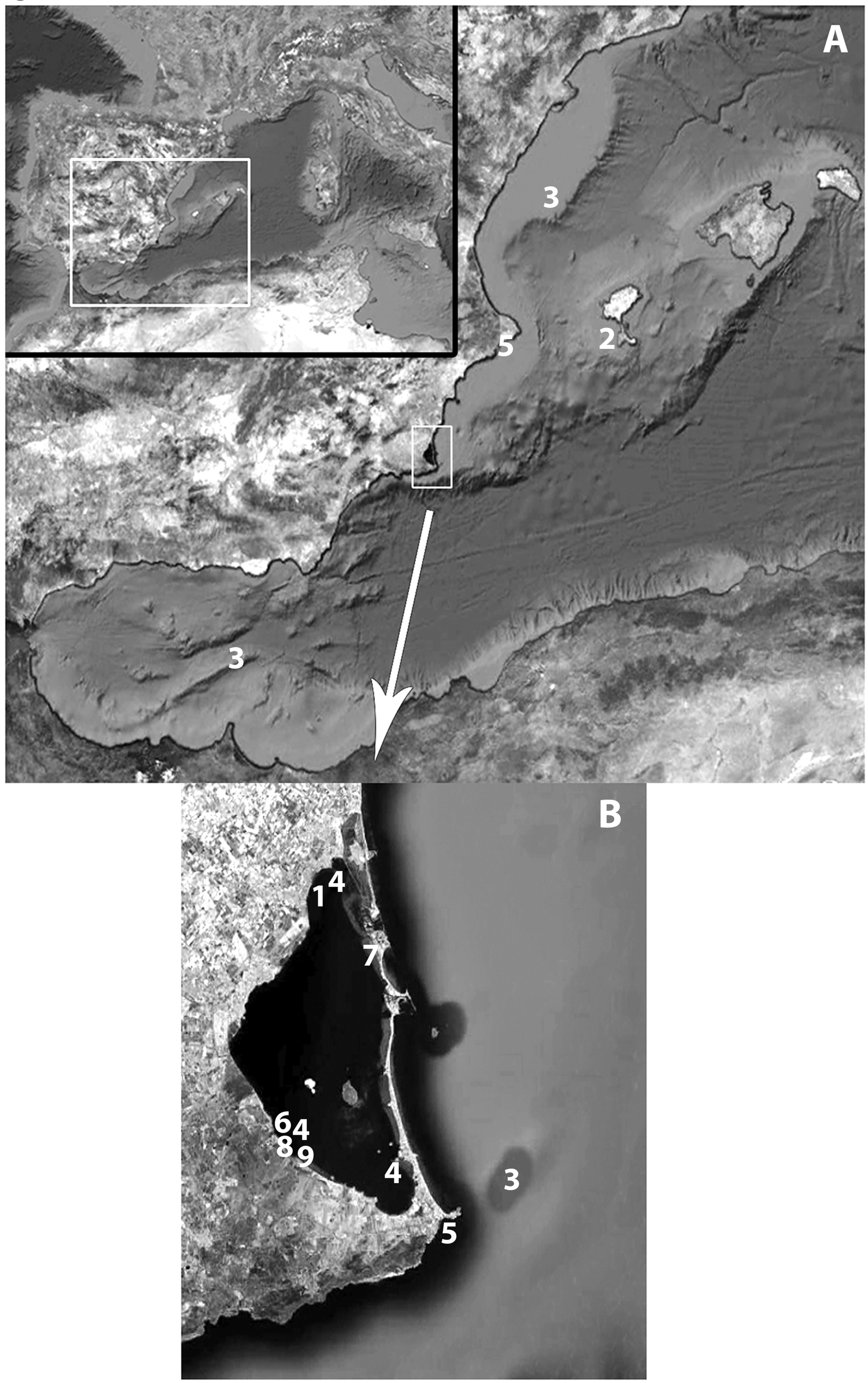
Material examined. One mature specimen sagitally sectioned, collected in Formentera Island from rocky substrate found between 126–134 metres deep.
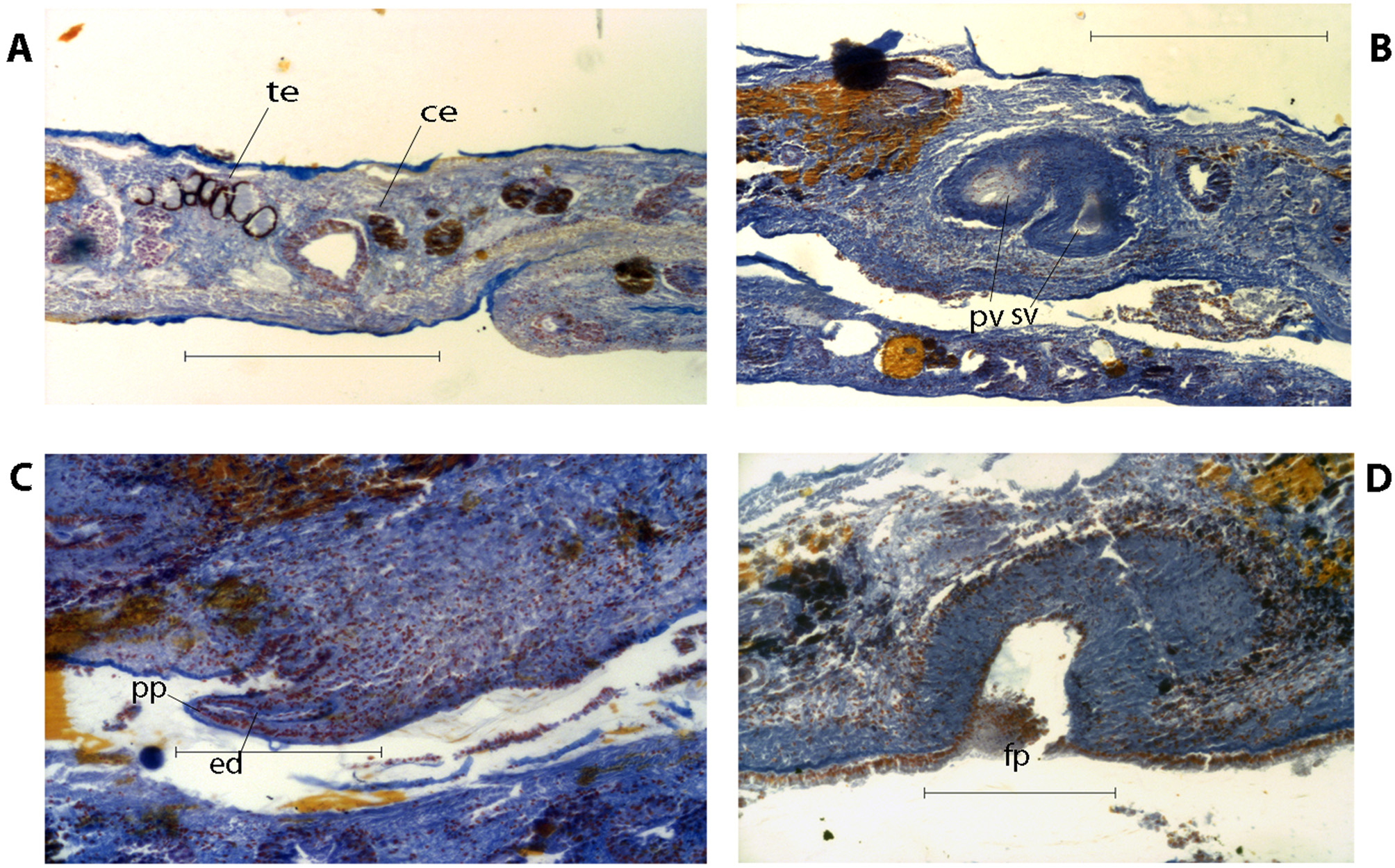
Morphological remarks. This specimen is consistent with Bock’s description of Notoplanella inarmata . Unfortunately, the description was realized only on fixed material, therefore the information about the colour is scarce. The fixed individual presents an oblong-oval body shape, lacking tentacles and whitish-grey background colouration with reddish-brown speckles arranged in lines, diverging from the centre to the margins. Marginal band and pharynx region free of pigment spots ( Fig. 2 View FIGURE 2 A). Paired cerebral and tentacular eye clusters present ( Fig. 2 View FIGURE 2 B, 3A). Pharynx ruffled central, oral pore at the posterior end ( Fig. 2 View FIGURE 2 C).
Within the male reproductive system ( Fig. 2 View FIGURE 2 D), a narrow duct connects the seminal vesicle to the prostatic vesicle ( Fig. 3 View FIGURE 3 B). The prostatic vesicle is interpolated, enlarged, with numerous secretory ducts. Unarmed penis papilla ( Fig. 3 View FIGURE 3 C). The female apparatus consists of a muscular external vagina, with the common oviduct clearly differentiated and without Lang’s vesicle. Unlike to the description of N. inarmata from Cape Town ( Bock 1931), both gonopores, female and male, are well separated ( Fig. 2 View FIGURE 2 D), and the musculature of the body wall surrounding the female genital pore ( Fig. 3 View FIGURE 3 D) is considerably thickened. This feature was also recognised by Prudhoe (1989), when he examined specimen from Cape Province dredged in 19 metres deep, also from Danger Point and Saldanha Bay.
Distribution. South Africa South Africa (Simon’s Bay, Bock 1931; False Bay, Day et al. 1970; Cape Province, Danger Point, Saldanha Bay, Prudhoe 1989). This is the first record for the Mediterranean Sea.

No known copyright restrictions apply. See Agosti, D., Egloff, W., 2009. Taxonomic information exchange and copyright: the Plazi approach. BMC Research Notes 2009, 2:53 for further explanation.