
Planocera graffi Lang, 1879
|
publication ID |
https://doi.org/ 10.11646/zootaxa.3780.1.4 |
|
publication LSID |
lsid:zoobank.org:pub:E1E25433-72CD-4592-B9C2-62F4D11F2D82 |
|
DOI |
https://doi.org/10.5281/zenodo.6142172 |
|
persistent identifier |
https://treatment.plazi.org/id/BD77527A-FF99-3A6E-1CBD-45178A89FE12 |
|
treatment provided by |
Plazi |
|
scientific name |
Planocera graffi Lang, 1879 |
| status |
|
Planocera graffi Lang, 1879 View in CoL
Figs. 1 View FIGURE 1 (5), 9, 10
Type locality. Gulf of Naples, associated with other invertebrates such as Balanus, Halichondria, Penares and Lithodomus ( Lang 1879, 1884, Tyler et al. 2012).
Type material. not available.
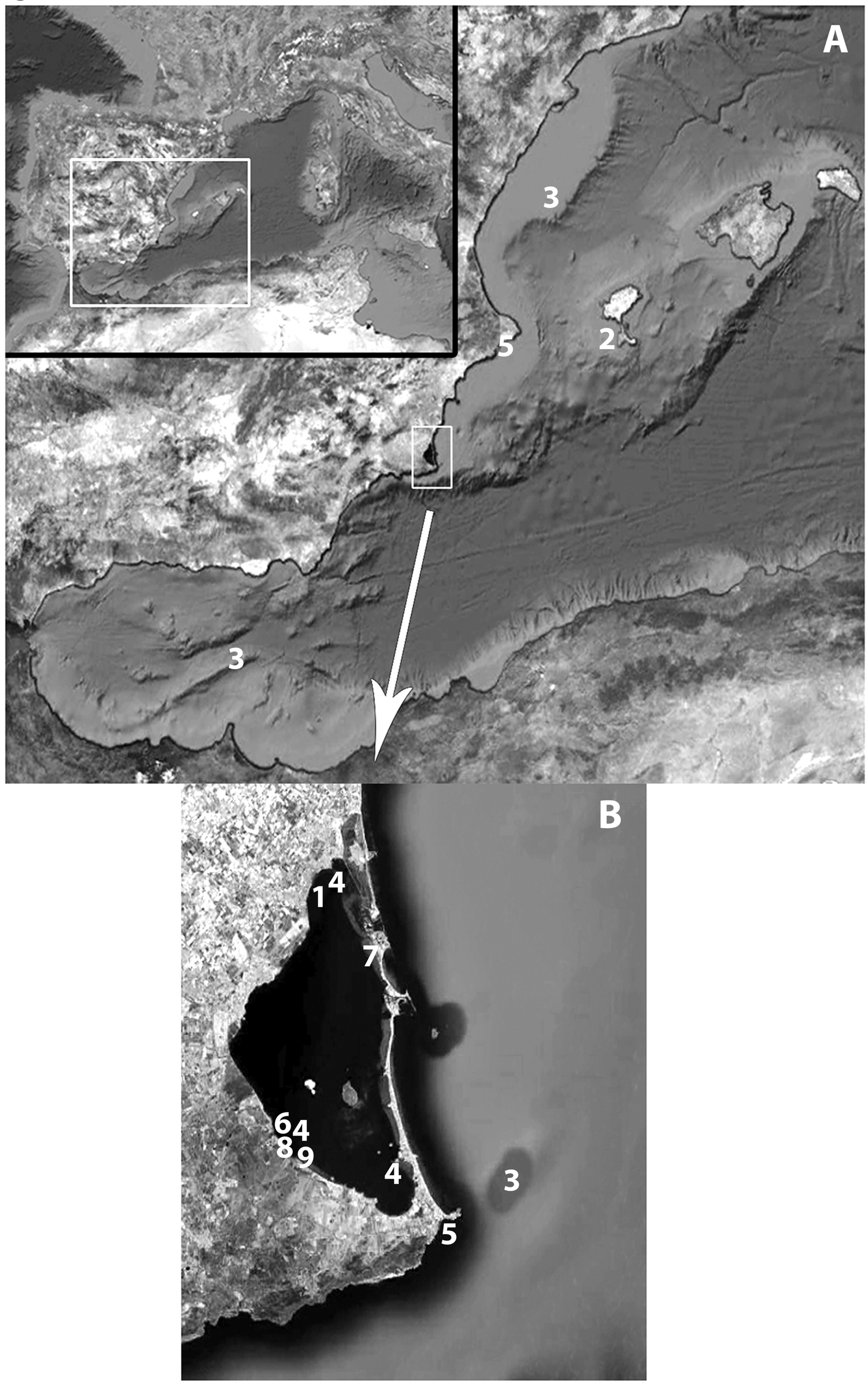
Material examined. One mature specimen from Cabo de Palos (Murcia, Spain) and two mature specimens from littoral areas near Calpe (Alicante, Spain) were studied. One mature specimen from Calpe was sectioned sagittally and mounted on 50 slides ( MNCN 4.01/130 - MNCN 4.01/180). The specimen from Cabo de Palos and the second specimen from Calpe were stored in alcohol 70%.
Localities: Cala Racó, Calpe, Alicante (38º38.07’N, 0º04.46’E) Spain; Cabo Palos, Murcia, Spain (37º37.39’N, 0º42.25’W); Fig. 1 View FIGURE 1 (5).
Other material. Planocera pellucida (Mertens, 1833) Oersted, 1844 Zoologisches Institut und Zoologisches Museum der Universität Hamburg, slides: V 13167 View Materials / 1- V View Materials 13167/18 (Atlantic Ocean, pelagic); V 131171 / 1- V View Materials 13171/ 15 (Atlantic Ocean, pelagic); V 13169 View Materials / 1- V View Materials 13169/37 (Atlantic Ocean); V 13177 View Materials / 1- V View Materials 13177/11 (Atlantic Ocean) and 5 specimens in alcohol: V13131 View Materials ( Cape Verde Islands).
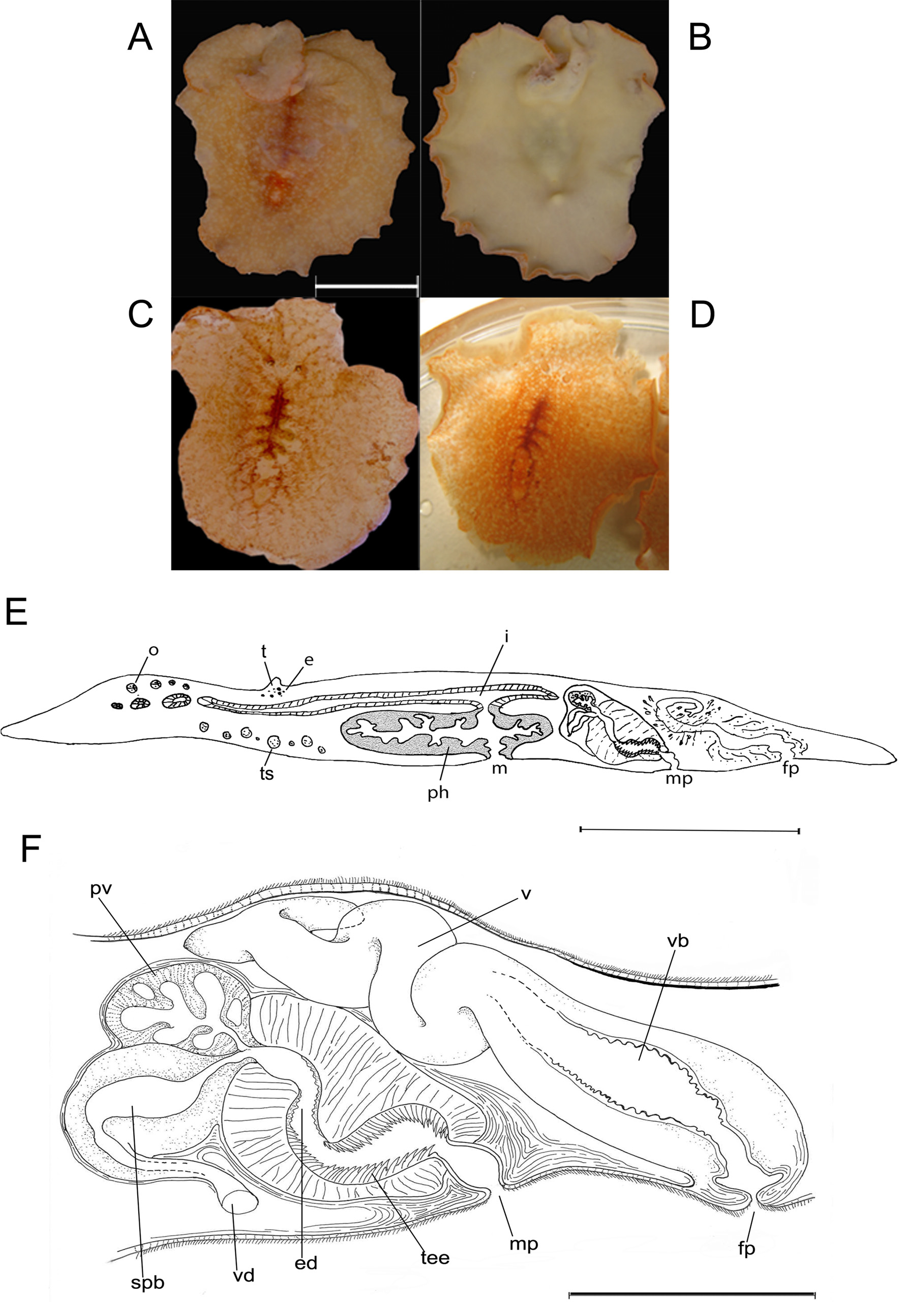
Description. The specimen ( Fig. 9 View FIGURE 9 A–D) has a broadly oval, almost round, form with slightly ruffled margins, 20 mm long by 15 mm wide (measurements from fixed specimen). The ground colour is yellow-orange, with a reddish net-like accumulation of pigment granules at the body midline along the intestine branches. The terminal ramifications on the dorsal side and the margin of the body sometimes reflect a white colour. The body appearance is fleshy, but very transparent and frail in appearance. Two conspicuous conical tentacles located near the brain, but far from the margins. Numerous tentacular eyes at the base of each tentacle. Several small cerebral eyes present anterior as well as posterior to the tentacles, being more abundant behind the tentacles. A broad ruffled pharynx occupies the central region of the body ( Fig. 9 View FIGURE 9 E).
In general, the organisation of the copulatory complex ( Fig. 9 View FIGURE 9 F) agrees with the detailed descriptions of Lang (1884). Nevertheless, some additional details can be discerned in our specimens. For example, the vasa deferentia dilate and become the spermiducal bulbs before entering proximally into the muscular cirrus bulb. The prostatic vesicle opens together with the spermiducal bulbs at the distal end of the male bulb. The prostatic vesicle is lined with a glandular epithelium forming the characteristic tubular chambers. The ejaculatory duct is wide and short. The cirrus is composed of distal spines and teeth that progressively increase in size towards the genital atrium. The rather short male genital atrium is bottle-shaped. The entire male complex is surrounded by thick muscle layers. The female apparatus does not show remarkable differences from Lang's (1884) description of the species. Briefly, it consist of a muscular external vagina or vagina bulbosa, surrounded by muscle layers, which is lined with a delicate cuboidal, glandular epithelium at the distal end; in some sections, this epithelium shows fringe-like extensions. The internal vagina first runs anteriorly and then posteriorly. Numerous cement glands open in the distal section of the internal vagina. Proximally, the two oviducts join the internal vagina and Lang’s vesicle is either entirely lacking or only present as a vestigial vesicle.
Discussion. The genus Planocera Blainville, 1828 shows a wide, almost cosmopolitan, distribution ( Fig. 8 View FIGURE 8 ). From the Mediterranean basin, three species have been reported: Planocera folia Grube, 1840 , Planocera graffi Lang, 1879 and Planocera ceratommata ( Palombi, 1936) .
Planocera graffi View in CoL was first described for the Gulf of Naples ( Lang 1879, 1884), and also reported for the islands of Sao Vicente ( Cape Verde) ( Laidlaw 1903) and the Catalan coast ( Novell 2003).
Planocera folia View in CoL was originally described for Palermo by Grube (1840), and later cited for Barwick Bay, USA (Apalachee Bay?) by Johnston (1865). Unfortunately, the internal anatomy of P. folia View in CoL is unknown, thus making it impossible to compare with other species of the genus. Ludwig von Graff (1904) last cited P. folia View in CoL , merely as the record of Grube (1840); therefore, the last real record of P. folia View in CoL appears to be in 1865 by George Johnston. For the Mediterranean, the only record is the original description of Grube (1840).
Planocera ceratommata View in CoL was originally described by Palombi (1936) for Cape Town ( South Africa), but more recently has been cited by Novell (2003) for the Catalan coast.
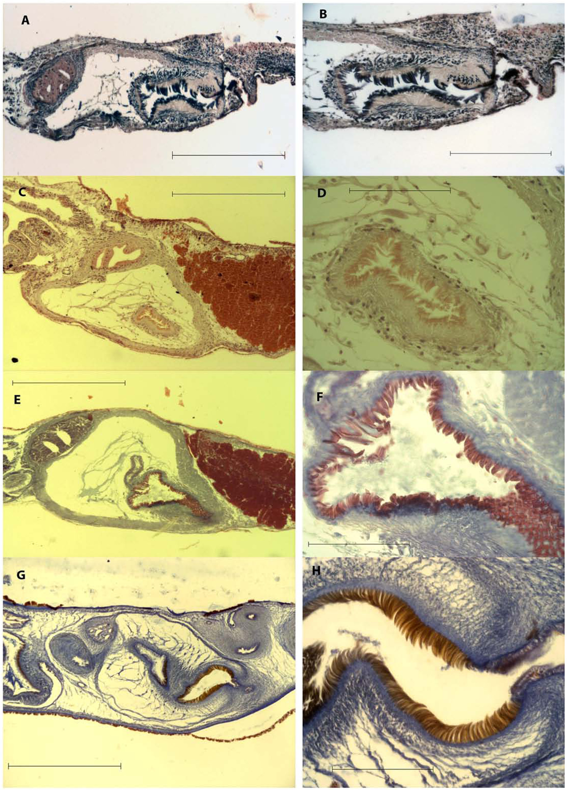
Although Lang (1879, 1884) could not compare Planocera graffi View in CoL with P. pellucida (Mertens, 1833) View in CoL as the description of P. pellucida View in CoL does not include internal anatomical features, he did comment on the great similarity between P. g r a f f i and P. pelagica (Moseley, 1877) View in CoL , which only differ in the shape of the ejaculatory duct (tortuous in P. pelagica View in CoL ). Years later, Faubel (1983) synonymised P. pelagica View in CoL with P. pellucida (Mertens, 1833) View in CoL .
Bock (1913) and Faubel (1983) described P. pellucida View in CoL as oval, tapering posteriorly and P. graffi View in CoL as rounded, sometimes broader than long. However, Prudhoe (1985) reported that P. pellucida View in CoL could show both body shapes, slightly elongated or rounded. This variation in body shape is, in our opinion and after a careful reading of the original description, the only reported difference between both species. An anatomical comparison between both species was necessary. Therefore, specimens of P. pellucida View in CoL from the invertebrate collection if the Zoological Museum of Hamburg, captured in the Atlantic shores, were compared with our specimens from the Mediterranean coasts. The results of this comparative study ( Fig. 10 View FIGURE 10 ) are the following: the cirrus of the male copulatory organ shows the same shape and type of spines in all specimens, the disposition and localisation of the prostatic and seminal vesicle is the same and the vasa deferentia show identical trajectories. It is evident that there are no significant morphological differences between the two species and a synonymization is justified. Therefore, we propose that P. graffi View in CoL is synonymous with P. pellucida View in CoL with the consequent expansion of the distribution of P. pellucida View in CoL within the Mediterranean Sea.
With respect to the third Planocera View in CoL species cited for the Mediterranean shores, P. ceratommata ( Palombi, 1936) View in CoL , this species is actually the most cited species of the genus Planocera View in CoL within the Mediterranean Sea, although it was originally described for Still Bay, Cape Town ( South Africa) ( Palombi 1936).
Unfortunately, the original material of P. ceratommata View in CoL is missing, but numerous similarities between this species and P. pellucida View in CoL suggest a possible case of synonymy; although, some differences must also be considered, that not allow a synomization. The most significant variation between P. ceratomata from South Africa and our material lies in the arrangement of the eyes. In P. pellucida View in CoL , the tentacular eyes surround the tentacles in an orderly (stereotypical) manner, whereas in P. ceratommata View in CoL the arrangement of the eyes is irregular ( Palombi, 1936; fig. 17). A similar pattern is also observed for the cerebral eyes. Another difference concerns the organisation of the prostatic vesicle. According to Palombi (1936), the copulatory organ “É formato de una grossa vescicola glandulare granulosa avvolta di una rica fascci muscolare costituita di fibri longitudinale e circolari” (“It is formed by a large glandular prostatic vesicle involved by well developed bundles of muscles formed by longitudinal and circular fibers"); this description, together with the figure ( Palombi 1936, fig. 18), illustrate a prostatic vesicle that is different from the vesicle in P. pellucida View in CoL described by Bock (1913). Nevertheless, we assume that many of the records of P. ceratommata View in CoL for the Mediterranean in fact concern P. pellucida View in CoL .

| MNCN |
Museo Nacional de Ciencias Naturales |
No known copyright restrictions apply. See Agosti, D., Egloff, W., 2009. Taxonomic information exchange and copyright: the Plazi approach. BMC Research Notes 2009, 2:53 for further explanation.
|
Kingdom |
|
|
Phylum |
|
|
Class |
|
|
Order |
|
|
Family |
|
|
Genus |
Planocera graffi Lang, 1879
| Marquina, Daniel, Osca, David, Rodríguez, Jorge, Fernández-Despiau, Estrella & Noreña, Carolina 2014 |
P. ceratommata (
| Palombi 1936 |
P. pelagica
| Moseley 1877 |
P. pellucida
| Mertens 1833 |
P. pellucida
| Mertens 1833 |