
Paramicrolaimus damodarani, Jacob, Jini, Jaleel, Abdul & Vijayan, Anil Kumar, 2015
|
publication ID |
https://doi.org/ 10.11646/zootaxa.3904.4.5 |
|
publication LSID |
lsid:zoobank.org:pub:3999945E-A42C-4963-A479-CA56B27C3950 |
|
DOI |
https://doi.org/10.5281/zenodo.5618591 |
|
persistent identifier |
https://treatment.plazi.org/id/BE015000-FF85-FFE1-9EE5-FDD2FD20F824 |
|
treatment provided by |
Plazi |
|
scientific name |
Paramicrolaimus damodarani |
| status |
|
Description of Paramicrolaimus damodarani sp. nov.
( Figs 2–4 View FIGURE 2 View FIGURE 3 View FIGURE 4 )
Material examined. Holotype, two paratype males and two juveniles [Slide No.IO/SS/NEM/00021; Deposited at FORV Referral Centre, Centre for Marine Living Resources and Ecology, Kochi, Kerala, India.] collected from continental margin of south eastern Arabian Sea.
Type locality. Holotype and paratype males: Continental margin of south eastern Arabian Sea—off Kannur, 11° 45’ 02” N, 74° 41’ 47” E, 95 m, 11.02.2012 (FORVSS 295). Two juveniles: South eastern Arabian Sea—off Cape Comorin, 7° 09’ 12” N, 77° 19’ 14” E, 207 m, 21.04.2005 (FORVSS 233). Sediment texture was silt with low percentages of clay, bottom temperature 26.6°C, bottom salinity 35.65 psu, bottom dissolved oxygen concentration 2.98 ml/l.
Description. Holotype (male): Body cylindrical, long and thread like. Total body length 1280 µm, a =51.2, b =9.14, c =20.32. Body diameter 20 µm at the level of posterior cephalic setae, with maximum 25 µm at mid body and 25 µm at anus. Cuticle thick, striated; striation in the cephalic region weak, distinct at mid body and caudal region. Hypodermal gland cells present. Head without striation, slightly constricted at the level of amphids. Labial sensillae barely visible. Cephalic setae in two separate circles (6+4), with similar lengths (13–14 µm). Somatic setae present in cephalic (5 µm) and caudal (7 µm) regions. Buccal cavity irregular, with deep and narrow anterior part and posterior part with sclerotized walls, two teeth present as dorsal and right subventral projections. Amphids wide (11 µm), thick walled, transversely oval-shaped with a small dorsal limb, located 19 µm away from the anterior end. Oesophagus 140 Μm long with swollen anterior end, middle part thin and cylindrical; posterior end swollen resembling a slightly elongated or weak oesophageal bulb (35 Μm long and 10 Μm wide). Males diorchic. Spicules paired, equal in size, 28 µm long, strongly arcuate, proximally cephalate with a distinct ventral, raised keel-like structure at mid-length. Gubernaculum parallel to spicule, simple in shape with lateral wing in the middle part. Seven distally expanded, cuticularised, protruding, precloacal supplements, each with distal thorn-like structures at their tips. First anterior and last posterior precloacal supplements slightly smaller than those between. Tail conoid, attenuated, ventrally coiled; 63 µm in length. Short, broad terminal spinneret present, strongly cuticularised, with long terminal setae arising from it dorsally.
Females and Juveniles. Females not found; juveniles resemble males in general morphology ( Table 1 View TABLE 1 ).
Diagnosis. Cuticle finely striated. Conspicuous amphids with thick wall, transversely oval-shaped with a small dorsal limb. Spicules paired, strongly arcuate, proximally cephalate with a distinct central keel. Gubernaculum simple, plate-like, with a lateral wing in the middle part. Seven cuticularised, protruding, ventrally placed precloacal supplements. Tail conoid with a cuticularised terminal spinneret.
Relationships. The general shape of the body and the spicular apparatus, position and number of cephalic setae, and position and shape of the amphid place the present specimens in Paramicrolaimus Wieser, 1954 ( Figs 2–4 View FIGURE 2 View FIGURE 3 View FIGURE 4 ; Tables 1 View TABLE 1 & 2 View TABLE 2 ). They are most similar to Paramicrolaimus mirus Tchesunov, 1988 in the general shape of the body, buccal cavity, oesophagus, size and shape of amphid and shape of gubernaculum. Paramicrolaimus damodarani sp. nov. strongly differs from P. mirus in body length (1.28 mm vs 4.06 mm), length of cephalic setae (6+8 µm vs 13+14–15 µm), a -value (105–106 vs 51–52.4), b - value (21.7–21.8 vs 8.8–9.1), c -value (28–40.5 vs 18.8–20.3), number of precloacal supplements (7 vs 9), shape and size of spicular apparatus (28 µm vs 23 µm), and in having a terminal spinneret which is absent in P. mirus ( Figs 2–4 View FIGURE 2 View FIGURE 3 View FIGURE 4 , Tables 1 View TABLE 1 & 2 View TABLE 2 ). Specimens of P. mirus from the Yellow Sea ( Huang & Zhang, 2005) showed larger measurements in all morphological characters compared with P. damodarani sp. nov. ( Table 2 View TABLE 2 ). Also, the spicule of P. mirus has a velum ( Huang & Zhang 2005) whereas P. damodarani sp. nov. has a central keel at mid-length. The gubernaculum in both the species were plate-shaped with a lateral wing in middle part but is of different size (19 µm vs 18 µm). The spinneret of P. damodarani sp. nov. has strongly cuticularised walls and the setae in the caudal region are longer ( Figs 2 View FIGURE 2 , 3 View FIGURE 3 ) than in P. mirus .
Paramicrolaimus damodarani sp. nov. can be differentiated from P. spirulifer in being smaller (1.28 mm vs 4.43 mm) in all morphological measurements in addition to the number of precloacal suppliments (7 vs 6) and the shape of the gubernaculum ( Figure 2–4 View FIGURE 2 View FIGURE 3 View FIGURE 4 ; Table 2 View TABLE 2 ). While Wieser, 1959 reported 6 precloacal supplements in P. spirulifer, Jensen (1978) counted 10 in the redescription of the species. The gubernaculum in P. spirulifer is weakly sclerotized and apparently surrounds the distal parts of the spicules but in P. damodarani sp. nov. it is plate-shaped with a lateral wing in the middle part.
Etymology. The species is named in honour of Prof. R. Damodaran, with deep gratitude and in appreciation of his invaluable contributions to benthic studies in India.
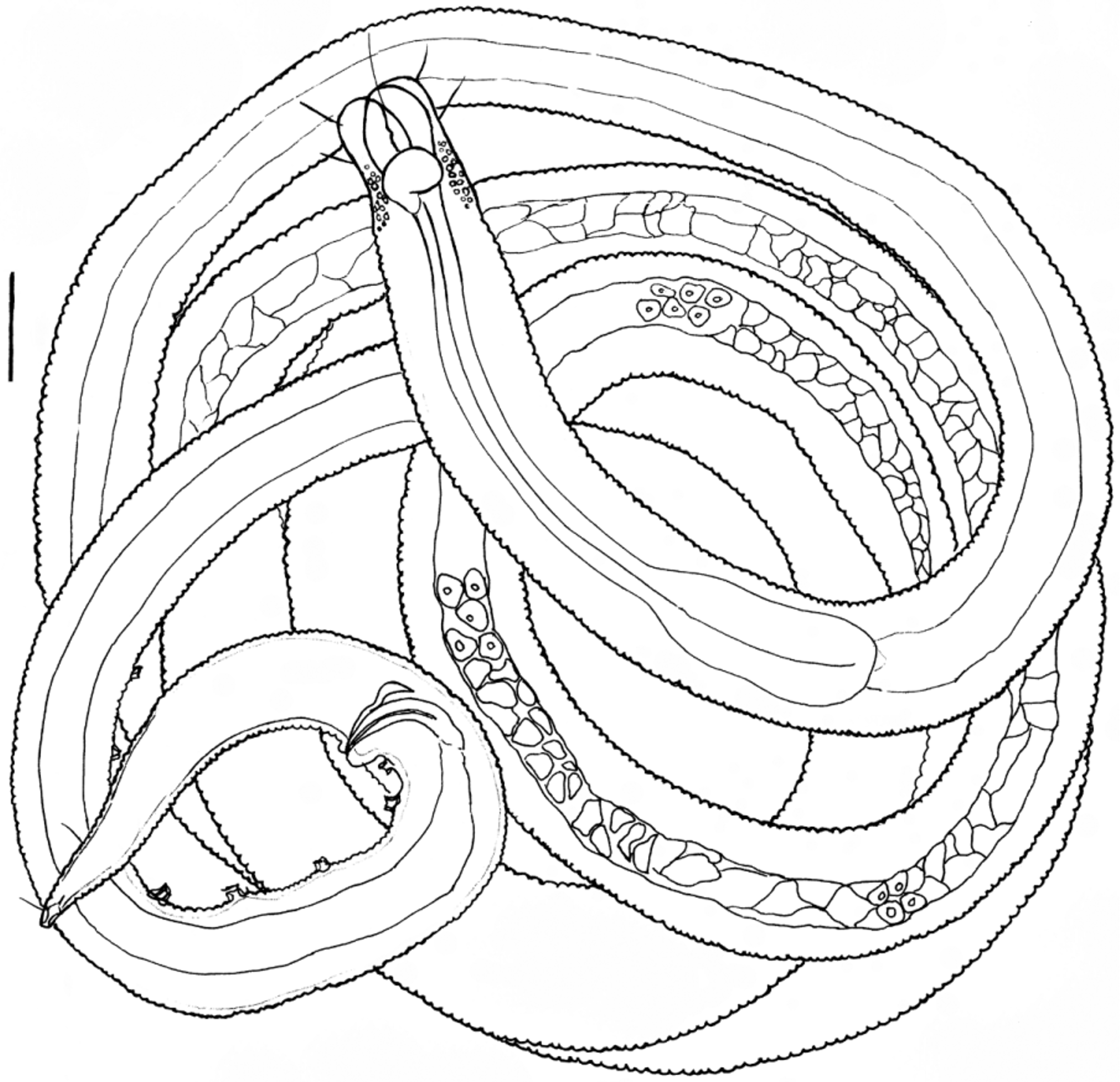
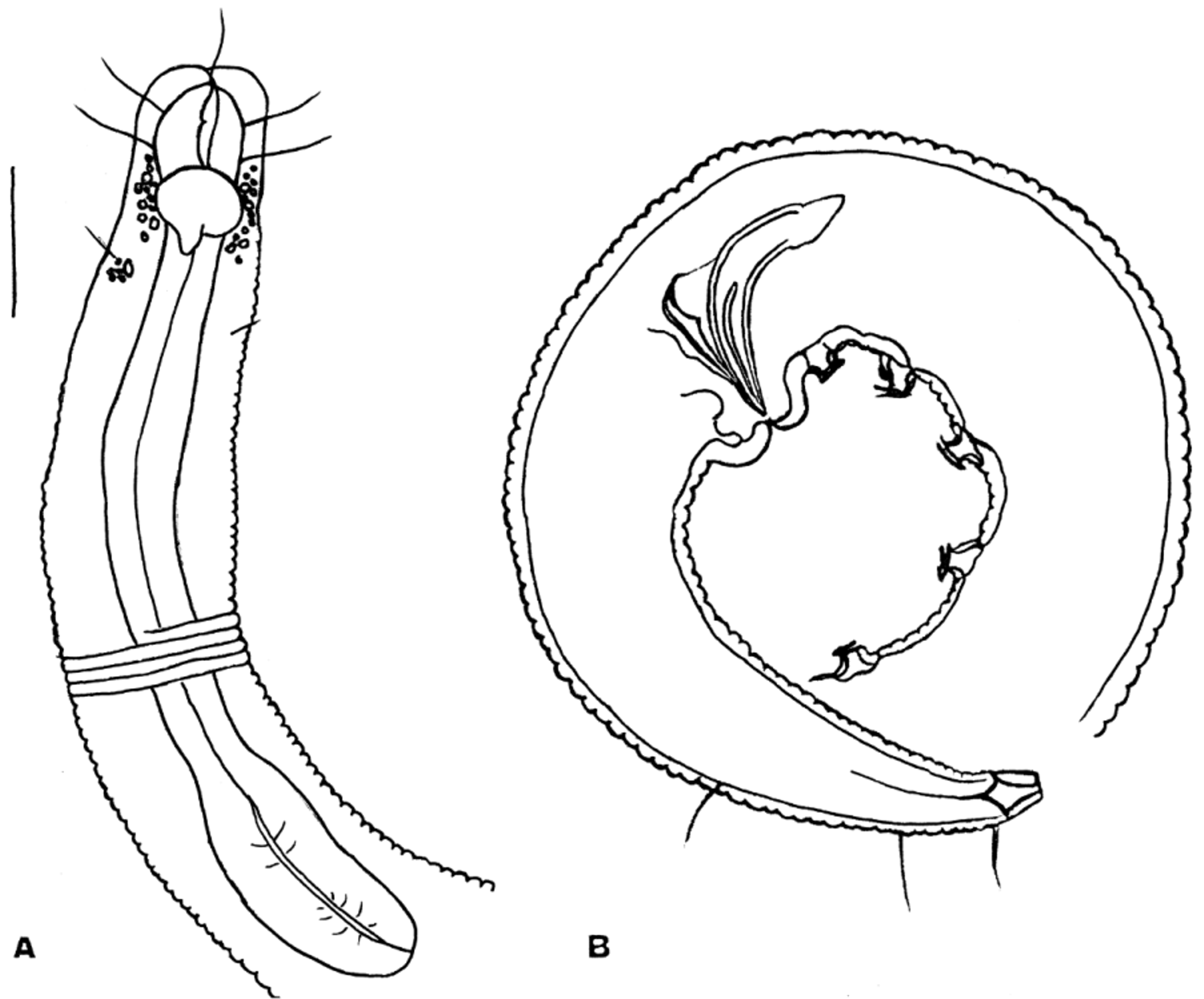
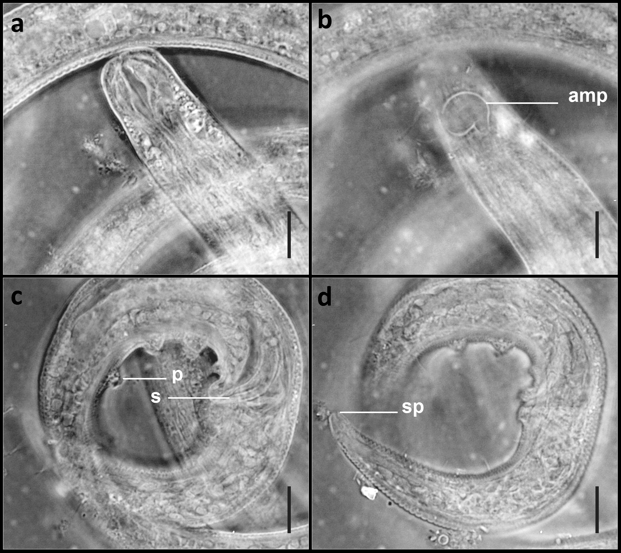
TABLE 1. Morphometry of Paramicrolaimus damodarani sp. nov.
| Material examined | Holotype ♂ | Paratype ♂1 | Paratype ♂2 | Juvenile 1 | Juvenile 2 |
|---|---|---|---|---|---|
| L (µm) | 1280 | 1310 | 1225 | 1230 | 1285 |
| mbd (µm) | 25 | 25 | 24 | 23 | 25 |
| a | 51.2 | 52.4 | 51.0 | 53.5 | 51.4 |
| b | 9.1 | 9.0 | 8.8 | 8.7 | 9.0 |
| c | 20.3 | 20.2 | 18.8 | 18.9 | 19.8 |
| hd (µm) | 20 | 20 | 20 | 20 | 20 |
| cs (µm) | 13+14 | 13+15 | 13+14 | 13+14 | 13+14 |
| ss (µm) | 5–7 | 5–7 | 5–7 | 5–7 | 5–7 |
| aw (µm) | 11 | 12 | 11 | 11 | 11 |
| Distance from anterior end (µm) | 19 | 20 | 19 | 19 | 19 |
| Oesophagus length (µm) | 140 | 145 | 140 | 142 | 142 |
| Spicule length as arc (S) (µm) | 28 | 29 | 28 | – | – |
| L. of gubernaculum (µm) | 19 | 19 | 19 | – | – |
| No. of supplements | 7 | 7 | 7 | – | – |
| t (µm) | 63 | 65 | 65 | 65 | 65 |
No known copyright restrictions apply. See Agosti, D., Egloff, W., 2009. Taxonomic information exchange and copyright: the Plazi approach. BMC Research Notes 2009, 2:53 for further explanation.
|
Kingdom |
|
|
Phylum |
|
|
Class |
|
|
Order |
|
|
Family |
|
|
Genus |
Paramicrolaimus damodarani
| Jacob, Jini, Jaleel, Abdul & Vijayan, Anil Kumar 2015 |
P. spirulifer
| Jensen 1978 |