
Zoothamnium xuianum, Sun, Ping, Ji, Daode & Song, Weibo, 2005
|
publication ID |
https://doi.org/ 10.5281/zenodo.170316 |
|
DOI |
https://doi.org/10.5281/zenodo.5662212 |
|
persistent identifier |
https://treatment.plazi.org/id/BF1C87AE-FFFE-FFE0-FE9B-F9DDFBB03E7D |
|
treatment provided by |
Plazi |
|
scientific name |
Zoothamnium xuianum |
| status |
|
Description of Zoothamnium xuianum n. sp. (Figs. 1 & 2; Table 1 View TABLE 1 )
Diagnosis: Marine Zoothamnium with alternately branched stalk. Zooids measuring about 45 × 30 µm with unfolded peristomial lip. Contractile vacuole apically located; macronucleus Cshaped and transversely orientated; more than 50 transverse silverlines between anterior end and aboral trochal band and on average 14 between aboral trochal band and scopula; three rows of peniculus 3 about equal length and parallel to each other, with the outer two rows closely set.
Type specimens: One holo– and one paratype slides (registration number: 0 4040701, 04040702) with protargol and silver nitrate impregnated cells are deposited in the Laboratory of Protozoology, Ocean University of China.
FIGURE 1. Morphology of Zoothamnium xuianum n. sp. from live cells (A–E), and after protargol (F–H) impregnations. (A) General view of a typical zooid. (B) Zooid with different body shape. (C) Extended and contracted zooids. (D) Detail of stalk, with thecoplasmic granules in spasmoneme. (E) Colony form. (F, G) Comparison of detailed arrangement of three peniculi between Qingdao population and Weihai population, double arrowhead to show detail of the outer two rows in peniculus 3. (H) Oral infraciliature, arrow marks epistomial membrane, double arrowhead shows short kinety fragment, arrowhead notes the separated upper end of outer row in peniculus 2. G = germinal kinety; H = haplokinety; P1–3 = peniculus 1–3; Po = polykinety. Scale bars in (A) = 20 µm, in (E) 200 µm.
Dedication: We dedicate this species to Prof. Dr. Muqi Xu, a diligent protozoologist of the Institute of Zoology, Chinese Academy of Sciences, who has made a lot of contribution to the ecology of ciliated protozoa.
Description: Zooids not differentiating into micro and macrozooid, fully extended individuals cylindroid to slenderly campanulate, 30–50 × 20–40 µm in vivo, length up to 2 times width, with common ratio 3:2 ( Table 1 View TABLE 1 ). Maximum width of body at peristomial border and not constricted below it. Peristomial disk flat, moderately elevated (Figs. 1A, 1B; 2D). Cell insensitive to stimuli, contracted zooid usually fistshaped (Figs. 1C; 2G). Pellicle completely smooth when observed at low magnification, and very fine striations can be detected only using powerful objectives (400× or higher).
Abbreviations: CV = coefficient of variation in %, Max = maximum, Mean = arithmetic mean, Min = minimum, n = number of individuals examined, SD = standard deviation, SE = standard error of mean.
Cytoplasm moderately dense and finely granular, usually containing several 3–8 µm sized granules (Figs. 1A, 1B; 2A, 2D). Food vacuoles often contain diatoms or small flagellates. One large, apically located contractile vacuole (Fig. 2E, arrow) rather inactive, and contracting at a rate of ca. 4 min. Macronucleus usually C–shaped and horizontally orientated, surrounding vestibulum at upper 1/3 of body. Micronucleus not observed.
Colony regularly alternatively branched (Figs. 1E; 2B, 2C), with up to 50 zooids in a large colony. Stalk up to 800 µm long (Figs. 1E; 2B), diameter about 7 µm in main branch and about 5 µm in distal part. Its smooth surface usually has some bacteria attached (Fig. 2F). The spasmoneme is 2 µm in diameter, with a string of dark grey thecoplasmic granules (0.4–0.6 µm in diameter) (Figs. 1D; 2F, arrowheads).
Structure of oral apparatus typical. The haplokinety (H) and polykinety (Po) describe about 1.5 turns around the peristomial disc before entering the infundibulum where they make a further turn (Fig. 1H). Near distal end of haplo– and polykinety, one short kinety fragment always recognizable (Figs. 1H, double arrowhead; 2I, arrow).
FIGURE 2. Photomicrographs of Zoothamnium xuianum n. sp. from live cells (A–G), after protargol (H, I) and silver nitrate (J) impregnations. (A) A typical zooid. (B) Colony form. (C) Zooids at low magnification. (D, E) Zooids of different body shape, arrow in figure E to mark the contractile vacuole. (F) Stalk and spasmoneme, arrowheads to show thecoplasmic granules. (G) To show the contracted ovalshaped zooids. (H) Detail of oral peniculi. (I) Apical view, arrow to show the oral distal fragment and double arrowhead to mark the epistomial membrane. (J) silverline system. P1–3 = peniculus 1–3. Scale bars in (A, D, E) = 20 µm, in (B) 200 µm.
Infraciliature of oral peniculi as follows: rows of peniculus 1 (P1) equal in length, extending to cytopharynx. P2 terminates distinctly above and between P1 and P3 (Figs. 1F, 1H; 2H); at adoral end of P2, the outer row apparently separated from the other two (Figs. 1F, arrow; 1H, arrowhead). Three rows of P3 parallel for entire length, ending together at approximately the same level. Germinal kinety (G) parallel to the haplokinety within upper half of infundibulum (Fig. 1H). Epistomial membrane (EM) located near the opening of the infundibulum (Figs. 1H, arrow; 2I, double arrowhead). Trochal band consisting of barren dikinety that encircles cell in aboral region.
Silverline system typical of Zoothamnium pattern (Fig. 2J). Striations closeset and many conspicuous pellicular pores associated with silverlines. More than 50 silverlines between anterior end and aboral trochal band, 12–17 between aboral trochal band and scopula.
Comparison and discussion
Zoothamnium xuianum n. sp. is identified by its alternately branching form, small zooid size, unfolded peristomial lip and closely spaced pellicular striations. Both populations collected bear all the above characters; thus, morphological and morphometrical differences between these two populations revealed the extent of the variation within the species ( Table 1 View TABLE 1 ; Figs. 1F; 1G).
Considering colony size and alternately branched stalk, singlelayered peristomial lip and marine habitat, four species should be compared with the current organism: Zoothamnium chlamydis Hu & Song, 2001 ; Z. plumula Kahl, 1933 ; Z. alternans Claparède & Lachmann, 1858 and Z. sinense Song, 1991 .
Our species bears a strong resemblance to Zoothamnium chlamydis in body shape, size and appearance of peristomial lip, ( Figs. 5 View FIGURE 5 A, 5B). However, the latter can be distinguished from the former by having macrozooids (vs. all zooids about equal size). Furthermore, the number of pellicular striations is quite different (27–47, 19–29 vs. more than 50, 12–17) ( Hu & Song 2001).
Zoothamnium xuianum is somewhat similar to Z. plumula sensu Song, 2002 ( Figs. 5 View FIGURE 5 C, 5D) in branching style and habitat. However, Z. xuianum can be clearly characterized by smaller body size (30–50 vs. 50–100 µm), absence of enlarged zooids (vs. present) and lower number of striations between aboral trochal band and scopula (12–17 vs. 24–28) ( Song et al. 2002).
Kahl (1933, 1935) redescribed three different forms of Zoothamnium alternans , Z. alternans sensu Claparède & Lachmann (1858) ( Fig. 5 View FIGURE 5 F), Z. alternans sensu Greeff (1870) ( Fig. 5 View FIGURE 5 E) and Z. alternans sensu Kent (1881) ( Fig. 5 View FIGURE 5 G). All above forms are distinct from Z. xuianum by possession of specialized zooids.
Zoothamnium sinense Song, 1991 ( Figs. 5 View FIGURE 5 H, 5I) can be separated from the new one by distinctly short and plump body shape (vs. elongated and slender in Z. xuianum ) and more silverlines from aboral trochal band to scopula (17–23 vs. 12–17).
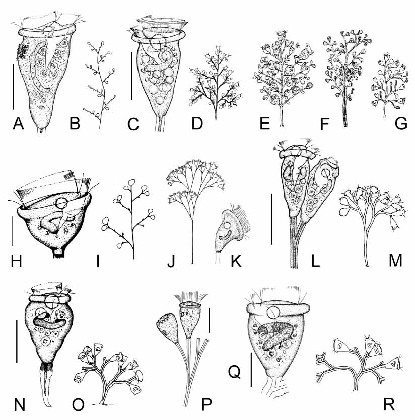
TABLE 1. Morphometric data on Zoothamnium xuianum n. sp. (Weihai population, first line; Qingdao population, second line) and Z. paraentzii (third line). All measurements in µm. , data not available.
| Character | Min | Max | Mean | SD | SE | CV n |
|---|---|---|---|---|---|---|
| Body length in vivo | 44 32 56 | 48 50 60 | 45.7 44.5 59.0 | 2.06 5.63 1.85 | 0.59 1.99 0.65 | 4.5 12 13.0 8 3.1 8 |
| Body width in vivo | 24 26 32 | 36 32 40 | 29.1 29.7 38.0 | 4.01 1.98 3.70 | 1.16 0.70 1.31 | 13.8 12 7.0 8 9.7 8 |
| Number of silverlines from peristome to aboral trochal band | 50 75 | 83 | 69.6 79 | 2.97 2.76 | 1.05 1.13 | 4.3 8 3.5 6 |
| Number of silverlines from aboral trochal band to scopula | 12 28 | 17 33 | 14.4 30.9 | 1.51 1.86 | 0.53 0.70 | 10.5 8 6.0 7 |
No known copyright restrictions apply. See Agosti, D., Egloff, W., 2009. Taxonomic information exchange and copyright: the Plazi approach. BMC Research Notes 2009, 2:53 for further explanation.
|
Kingdom |
|
|
Phylum |
|
|
Class |
|
|
Order |
|
|
Family |
|
|
Genus |
Zoothamnium xuianum
| Sun, Ping, Ji, Daode & Song, Weibo 2005 |
Z. plumula sensu
| Song 2002 |
Z. alternans sensu
| Kent 1881 |
Z. alternans sensu
| Greeff 1870 |
Z. alternans sensu Claparède & Lachmann (1858)
| Claparede & Lachmann 1858 |